info@buzaevclinic.ru
  • Home
  • Focused Ultrasound
    • MRI-guideded focused ultrasound treatment
      • Parkinson’s Disease
      • Essential Tremor
      • Dystonia
      • Neuropathic Pain
    • Treatment Prices
    • Patient Stories
  • Patients
    • Make an appointment
      • I have a tremor!
      • WhatsApp
      • Online Appointment
      • 88003008462
      • Telemedicine
    • Services and Prices
    • Treatment and Diagnostics
    • Preparing for the Examination
    • Questions and Answers
      • Video
    • Support Group
    • Public Offer
  • Doctors
    • I want to join your team
    • Refer the Patient
    • MRgFUS Monograph
    • Video with Results
    • Results of Our Treatment in Research Articles
    • Want to Learn from You
  • Research and Education
    • Our research
    • Directory of Diseases
    • Education
    • Education Login
  • About Us
    • Mission and Vision
    • Team
    • News
    • Address, opening hours
    • Legal Section
  • Menu Menu

MRI of the knee joint
You are here: Home1 / Therapeutic and Diagnostic Areas of the V.S. Buzaev International Medical Centre2 / Expert MRI Diagnostics3 / MRI of the knee joint

MRI of the knee joint

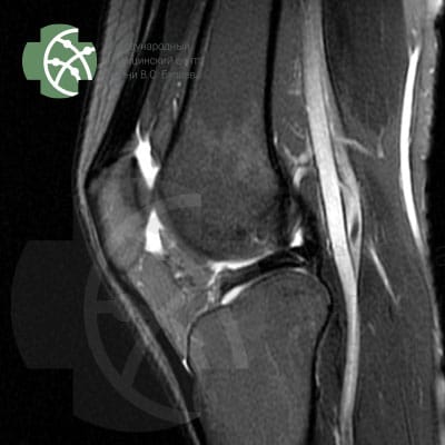
MRI of the knee joint. Program T2 FS Sagittal 3 mm.
MRI of the Knee Joint. Program T2 FS Sagittal 3 mm.
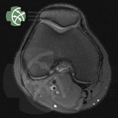
MRI of the Knee Joint. Program PD FS Axial 3 mm.
MRI of the knee joint. Program PD FS Axial 3 mm.
MRI of the Knee Joint. Program PD FS Coronal 3 mm
MRI of the knee joint. Program PD FS Coronal 3 mm

Magnetic resonance imaging of the knee joint without contrast agent

Rating:      

Reviews 2GIS Yandex Google

What you will receive

Medical report with recommendations
We will record the MRI data on DVD-disk. On the disk envelope, we will indicate QR-CODE, which allows you to download the study description 24 hours after the examination.

Who needs this service?
Indications for MRI of the knee joint (determined by the trauma doctor during an in-person consultation):

MRI examination is performed after consultation with an orthopedic doctor. The most common indications for MRI of the knee joint:

  • traumatic injuries
  • preparation for surgery
  • signs of joint inflammation;
  • suspicion of dystrophic changes
  • congenital developmental anomalies
  • the need to exclude tumors, metastases;
  • questionable data obtained during ultrasound, CT, or X-ray.

Who cannot receive such a service? Contraindications for MRI of the knee joint (applicable to the MRI machine in our clinic):

Absolute contraindications for MRI of the knee joint:

  • The presence of “embedded” metallic elements, implants, shrapnel, and electronic stimulators in the body that cannot be removed
  • Early pregnancy in women
  • Chronic diseases of the urinary system, kidney ailments accompanied by reduced excretory functionality. The restriction applies to the procedure in contrast mode
  • The presence of allergic reactions in a patient to a specific contrast agent

Conditional contraindications:

  • Weight over 170 kg
  • shock, extremely severe condition of a person
  • CNS disorders accompanied by uncontrolled movements

How we do it

MRI of the knee can be performed in a lying position, feet towards the aperture of the machine, which allows successful examination even for people with claustrophobia (fear of enclosed and tight spaces)

MRI does not require anesthesia or special preparation.
Before the procedure, the MRI-technician will check you with a special device to ensure there are no metals in your body or clothing that react to the magnetic field. Then, you will be placed on the MRI table and instructed. You will be given a button in your hand, which can instantly stop the examination if pressed. Then, the knee joint examination protocol, consisting of seven programs, will be initiated.

If necessary to clarify the obtained data, additional research programs may be connected after agreement with you.

What is included in the service?

The research protocol in different clinics can vary significantly. MRI is always a compromise between the time spent on the examination and its quality.
We use all recommended programs, ensuring the highest quality of research, allowing for a detailed examination of the knee area without missing pathological changes.
An important aspect is the research step – thickness “sections “, we obtain as a result of the study. The smaller the step and the more slices, the more detailed we can assess the condition of the tissues.

Our MRI knee joint protocol includes the following sequences:

Program / slice thickness

PD FS saggital 3 mm
T2 saggiati 3 mm
T2 FS saggital 3 mm
PD FS coronal 3 mm
T1 coronal 3 mm
PD FS axial 3 mm
MERGE saggital 3 mm


 How long will it take?

The MRI protocol of the knee joint in our clinic, including 7 different research programs, will take about 40 minutes.

How else can this be done?

Assessing the condition of the knee and knee joint can be done based on symptoms during an examination by a trauma orthopedist. This specialist should see the patient, make a preliminary diagnosis, and after reviewing the examination results, provide a final diagnosis and prescribe appropriate treatment.
In addition to MRI, CT (computed tomography) and ultrasound can be performed.
But it should be understood that CT uses ionizing radiation, the dose of which needs to be strictly controlled; while ultrasound provides much less informative results.

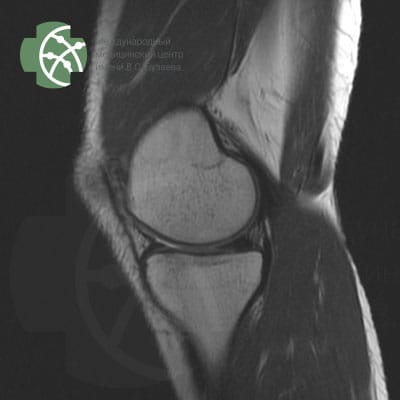
MRI of the knee area - joint and surrounding tissues. Program PD FS sagittal 3 mm.
MRI of the knee area – joint and surrounding tissues. Program PD FS sagittal 3 mm.
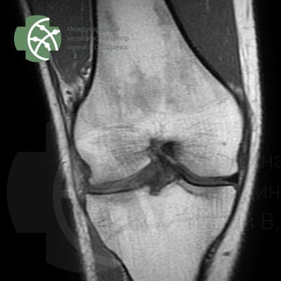
MRI of the knee area - joint and surrounding tissues. Program T1 coronal 3 mm.
MRI of the knee area – joint and surrounding tissues. Program T1 coronal 3 mm.
MRI of the knee area - joint and surrounding tissues. T2 FS sagittal 3 mm program
MRI of the knee area – joint and surrounding tissues Program T2 FS sagittal 3 mm

You might need

  • Consultation with a Trauma and Orthopedic Specialist
  • Second Expert Opinion (description of MRI studies conducted in other clinics)
  • Center for Extrapyramidal and Cognitive Disorders
  • Center for Head and Facial Pain
  • Endocrinology
  • Expert MRI Diagnostics
    • MRI of the brain
    • MRI of the heart
    • MRI of the knee joint
    • MRI of the spine
    • Whole-body MRI
  • Gastroenterology and Endoscopy
  • Heart and Vascular Treatment. Intelligent Cardiology
    • 24-hour Ambulatory Blood Pressure Monitoring (ABPM) in Ufa
  • Innovative Non-Invasive Neurosurgery with Focused Ultrasound
  • Pediatrics and Child Vaccination
  • Treatment of Tremor with MRI-guided Focused Ultrasound (MRgFUS)
    • Surgery for Parkinson’s Disease Will Eliminate Tremor and Stiffness
    • Surgery for Tremor
  • Urology and Andrology

Book an Appointment

  • WhatsApp
  • +78003008462
  • Online
  • info@buzaevclinic.ru

Attention!

The information provided is for reference only. There are contraindications. Specialist consultation is required. This is a machine translation of the original site buzaevclinic.ru and is currently being revised.

This article is subject to copyright.

(c) 2025 Exclusive rights belong to the International Medical Center named after V.S. Buzaev.

You may reprint or quote for non-commercial purposes provided that a proper reference to this original source is included, and you may not alter or distort the text, images, or audio in any way.

License: CC-BY-NC-ND
Creative Common licence-by-nc-nd-buzaevclinic

Do you know?

Tremor and rigidity can be reduced or even completely eliminated

Contents

  • Book an Appointment
  • Leave a review
  • Preparation for Research
  • MRgFUS CT Protocol
  • CT for MRI-guided focused ultrasound
  • Tax deduction
  • Company Details
  • Archive
  • Parkinson’s community
  • Original (Русский)
  • For Arab Patients
  • Turkish
  • O’zbek

Working Hours (GMT+5)

Monday09:00 - 21:00
Tuesday09:00 - 21:00
Wednesday09:00 - 21:00
Thursday09:00 - 21:00
Friday09:00 - 21:00
Saturday09:00 - 17:00
Sunday09:00 - 17:00
©2020 V.S. Buzaev Medical Center®. There are contraindications. Specialist consultation is required. Use of website materials is permitted only with authorization and proper source citation.
Scroll to top Scroll to top Scroll to top