# VS Buzaev International MRgFUS Medical Treatment Centre
> Treatment of tremor, Parkinson's rigidity, stiffness, and dystonia. Advanced bloodless neurosurgery using MRI-guided focused ultrasound (MRgFUS)
> Generated by All in One SEO Pro v5.0.3, this is an llms-full.txt file, used by LLMs to index the site.
## Posts
### [Туризм для лечения тремора или ригидности при болезни Паркинсона с помощью MRgFUS ](https://buzaevclinic.com/global/medical-tourism/)
**Published:** August 11, 2022
**Author:** Igor Buzaev
**Content:**
## Innovative International Services for Patients with Essential Tremor, Parkinson’s Disease and dystonia in V.S. Buzaev International Medical Centre
Are you or your loved ones affected by Parkinson’s disease and searching for reliable and modern treatment methods? The VAre you or someone you love affected by Parkinson’s disease and searching for effective, modern treatment options? The Vyacheslav S. Buzaev International Medical Center has a wonderful solution for you—MRI-guided focused ultrasound treatment of Parkinson’s Disease, Essential Tremor, and Dystonia. This innovative method allows for surgery without incisions or anesthesia, making it incredibly safe and comfortable for the patient.
Not only can this approach help reduce the symptoms of the disease, but it can also lower medication dosages, dramatically improving your quality of life. We are proud to be the first in Russia to offer this advanced technology and welcome you to explore our medical tourism benefits.
We provide a comprehensive service package, including free follow-up care for 12 months. Imagine regaining the joy of movement and regaining confidence in the future.
> The Alchemy of Intelligence and Compassion
We are your trusted partner in medical tourism, offering unique treatment opportunities where your health is our top priority. Our clinic is a state-of-the-art center where talented professionals transform medicine and restore your vitality.
We are the first in Russia to introduce MRI-guided focused ultrasound treatment—a non-invasive procedure that provides maximum comfort and safety.
Let’s embark on this journey together.
[Request for an online consultation](https://app.buzaevclinic.ru/index.php?module=ext/public/form&id=12)
- [We Are Here to Cure You from Tremor, Parkinson’s Rigidity, and Stiffness](#aioseo-we-can-cure-your-tremor-parkinsons-rigidity-and-stiffness)
- [What is MRgFUS](#aioseo-what-is-mrgfus)
- [Why MRgFUS?](#aioseo-one-procedure-no-incisions-no-implants-and-no-more-shaking-tremor-or-parkinsons-rigidity)
- [MRgFUS vs DBS](#aioseo-mrgfus-vs-dbs)
- [MRgFUS vs Radiofrequency ablation (RFA)](#aioseo-mrgfus-vs-radiofrequency-ablation-rfa)
- [MRgFUS vs Gamma Knife](#aioseo-mrgfus-vs-gamma-knife)
- [MRgFUS vs Medications](#aioseo-mrgfus-vs-medications)
- [MRgFUS is typically recommended when a patient meets several key criteria:](#aioseo-in-what-cases-mrgfus-is-usually-indicated)
- [Absolute contraindication](#aioseo-absolute-contraindications)
- [Relative contraindications](#aioseo-relative-mrgfus-contraindications)
- [Why Buzaevclinic?](#aioseo-why-buzaevclinic)
- [Online consultation appointment](#aioseo-online-consultation-appointment)
- [Pricing](#aioseo-pricing)
- [Accomodation](#aioseo-accomodation)
- [Contact us](#aioseo-contact-us)
**Who We Are**
Welcome to V. S. Buzaev International Medical Center! We’re thrilled to be the country’s first MRgFUS (Neurovive) center dedicated to treating movement disorders like Parkinson’s disease, essential tremor, dystonia, shaking hands, shaking legs, and shaking heads.
**Our Mission**
At our Center, we bring together the finest professionals in the field to transform medicine and help people with our humanistic approach. We’re committed to using our expertise to make a real difference in people’s lives.
**Our Vision**
We envision a future where everyone can lead a healthy and happy life. At Vyacheslav Stepanovich Buzaev International Medical Center, we strive to provide non-invasive, painless, and accurate treatment for everyone who needs our help.

## **We Are Here to Cure You from Tremor, Parkinson’s Rigidity, and Stiffness**
> This information does not apply to everybody and is for informational purposes only.
> Our doctor’s consultation is necessary for individual advice in any case. [Book the appointment.](https://buzaevclinic.com/telemedical-consultations/)
We’re here to help you find relief from a variety of conditions that affect your movement and quality of life. Whether it’s tremors, Parkinson’s symptoms, hypothalamic hamartoma, or stiffness, we’re dedicated to providing personalized care and support.
We care for patients worldwide with involuntary movements:
- hands tremor
- shaking head
- trembling legs
- involuntary torsions of body
- laughing-crying (gelastic) seizures
- neuropathic pain
- body stiffness (rigidity)
- “off” periods
- dyskinesia
- levodopa-induced dystonia
- chronic neuropathic pain
## What is MRgFUS
We use modern, [FDA-approved](https://www.fda.gov/news-events/press-announcements/fda-approves-first-mri-guided-focused-ultrasound-device-treat-essential-tremor) non-invasive MRI-guided focused ultrasound technology. It’s like having a magic wand for your brain!
IINSIGHTEC’s Exablate Neuro Platform is the future of medicine, and it’s here to change the game.
Watch the video about Insightec MRgFUS technology, we use:
### Why MRgFUS?
It’s non-invasive, meaning no incisions, no scars, and you don’t even need to be asleep! It’s like giving your brain a gentle nudge to fix what’s broken.
One procedure, no incisions, no implants, and no more shaking, tremors, or Parkinson’s rigidity. You’ll see the results immediately!
MRgFUS is [effective at stopping your tremors](https://jamanetwork.com/journals/jamaneurology/article-abstract/2821255), [rigidity](https://www.mdpi.com/2077-0383/11/15/4502), and other [involuntary movements](https://annaly-nevrologii.com/journal/journal/pathID/article/view/1020/pdf_1) for the [long term.](https://www.frontiersin.org/journals/neurology/articles/10.3389/fneur.2021.654711/full) And the best part? It usually only takes one procedure per side.
The treatment is [well-controlled](https://thejns.org/focus/view/journals/neurosurg-focus/32/1/2011.10.focus11246.xml) from the start, so you can trust that everything is going as planned. Plus, it doesn’t require any [implantable devices or incisions](https://www.parkinson.org/Understanding-Parkinsons/Treatment/Surgical-Treatment-Options/Deep-Brain-Stimulation), and you won’t need [general anaesthesia.](https://journals.sagepub.com/doi/abs/10.15274/INR-2014-10033)
In more than 95% of cases, you won’t have to stay at the hospital overnight, so you can get back to your daily routine as soon as possible.
Imagine waking up the next day feeling better than ever. That’s the power of MRgFUS!
#### [MRgFUS vs DBS](https://buzaevclinic.com/services/mrgfus-treatment-en/mrgfus-vs-dbs/ "MRgFUS vs DBS")
#### [MRgFUS vs Radiofrequency ablation (RFA)](https://buzaevclinic.com/services/mrgfus-treatment-en/mrgfus-vs-radiofrequency-ablation-rfa/)
#### [MRgFUS vs Gamma Knife](https://buzaevclinic.com/services/mrgfus-treatment-en/mrgfus-vs-gamma-knife/)
### [MRgFUS vs Medications](https://buzaevclinic.com/services/mrgfus-treatment-en/mrgfus-vs-medications/)
> This information does not apply to everybody and is for informational purposes only. A doctor’s consultation is necessary for individual advice in any case. [Book the appointment.](https://buzaevclinic.com/telemedical-consultations/)
### **MRgFUS is typically recommended when a patient meets several key criteria:**
1. They have a [condition ](https://buzaevclinic.com/global/medical-tourism/#conditions)that could benefit from MRgFUS, and
2. optimal medical treatment has proven ineffective.
3. There are no contraindications.
4. The patient agrees to the treatment.
5. A specialist has thoroughly assessed the patient and believes MRgFUS to be the most appropriate course of action.
It’s important to note that non-MRgFUS doctors, including neurosurgeons, may have limited knowledge of its capabilities.
### Absolute contraindication
- MRgFUS is MRI. So, contraindications include metal implants or shell splinters in the brain, etc.
- Severe psychiatric condition or dementia
- Low skull density ratio (SDR, skull score), which is measured using [CT](https://buzaevclinic.com/services/mrgfus-treatment-en/ct-preplan-for-mrgfus/). For us, this is a relative factor. We have developed a methodology to increase SDR, but it takes several months to achieve.
### Relative contraindications
- Age is almost never a concern for MRgFUS, so don’t worry about that!
- If you have claustrophobia, we’ve got some great tips and tricks to help you feel more at ease.
## Why Buzaevclinic?
Since 2020, we’ve been the pioneers in our country when it comes to treating movement disorders like Parkinson’s disease, essential tremor, and dystonia. We’re incredibly proud to have taken non-invasive surgery to a whole new level, where incisions are no longer needed, and our work is both highly predictable and controllable.
Our director and neurosurgeon, Rezida Galimova, has always believed that getting to the depths of the brain through open wounds and potentially damaging it is not the right way. And now, thanks to her dream, we can treat the brain without incisions!
- We’re the experts and leaders in the world, the first in our former Eastern Block to offer neuro-MRgFUS since 2020.
- We take care of every patient for at least 12 months, and we [conduct our late results research](https://buzaevclinic.com/research/publications/) to keep improving. Our results are published in scientific papers here and in a [scientific monograph.](https://mrgfus.ru/)
- We’ve already performed over 250 treatments with a success rate of more than 95% for essential tremor.
- We’re highly responsible doctors who are passionate about delivering excellent long-term results. If tremor reoccurs within 12 months of our initial MRgFUS treatment, we promise to consider a free re-operation, taking into account both the expected effect and safety.
- We also offer a well-tested and high-quality medical tourism process. Patients from over [20 countries](https://buzaevclinic.com/global/countries/) have come to us from all corners of the globe to be treated for tremor using MRI-guided focused ultrasound.
## Online consultation appointment
We are thrilled to offer you the convenience of **online consultations** to determine the best course of treatment for your needs. Our team is here to guide you every step of the way.
To schedule your appointment, simply send us an email to <mailto:help@tremor.care> or give us a call at [+7 967 740 6099.](tel:+79677406099)
We look forward to hearing from you and helping you feel better.
## Pricing
## **Start**
****1.5-hour online consultation****
---
5 500 Rubles
## **Standard**
****MRgFUS Treatment and Accommodation****
---
- Online consultation
- Accomodation
- MRgFUS treatment
- 4 follow-up visits
(min)
1 700 000 Rubles
## **Ultimate**
**MRgFUS Treatment,**
**Accommodation and Tourism**
---
- Online consultation
- Accomodation
- MRgFUS treatment
- 4 follow-up visits
(min)
- 12-month warranty (repeat if needed,
feasible and safe)
- Region or city tours, resort
By Request
## Accomodation
We are thrilled to invite you to our clinic, where we offer a warm and welcoming experience for our valued guests. We understand that your journey to our clinic is important, and we are here to make it as smooth and comfortable as possible.
Our clinic proudly covers accommodation, transfers, and meal expenses for one patient and one attendant in the beautiful Hilton Hotel or any other hotel in the city of Ufa. We will ensure that you have all the necessary transfers between the hotel, the clinic, and the airport, as well as meals both in the hotel and the clinic.
Our dedicated staff speaks English, Arabic, and French, ensuring that you feel right at home. We are proud to participate in the federal project “Development of the Export of Medical Services” and are committed to providing high-quality medical services to patients from all over the world.
## Contact us
To contact us, please visit our clinic at Zorge str. 17/4 in Ufa, Russia. You can reach us by phone, WhatsApp, or Telegram at [+7 967 740-60-99](http://wa.me/79677406099). Our esteemed doctor, [Rezida Galimova](https://buzaevclinic.com/team/doctors/rezida-galimova/), MD, PhD and Doctor of Sciences in Medicine, will be happy to assist you.
>
[Request for an online consultation](https://app.buzaevclinic.ru/index.php?module=ext/public/form&id=12)
**Categories:** Global medical tourism
**Tags:** Video
---
### [TVC: Review of Tremor Treatment with Ultrasound: Patient and Doctor ](https://buzaevclinic.com/services/mrgfus/essential-tremor-cases/tvc-post-operation-interview-doctor-and-patient/)
**Published:** May 18, 2022
**Author:** Aleksej Savelev
**Content:**
## Impression and Interview After Tremor Treatment with Ultrasound: Patient and Doctor Experience on TVC in the Program “Doctor I… “
In the latest episode of the program [“Doctor I.”](https://www.tvc.ru/brands/28/episode/76582) from May 18, 2022, the first Russian [MRgFUS neurosurgeon Rezida Maratovna Galimova](https://buzaevclinic.com/team/doctors/rezida-galimova/) talks about how essential tremor was treated with MRI-guided focused ultrasound at the V.S. Buzaev International Medical Centre. A patient also shares her impressions during and after the treatment of tremor with focused ultrasound.
Hosts: Dr. [Irina Pershina](https://ru.wikipedia.org/wiki/%D0%9F%D0%B5%D1%80%D1%88%D0%B8%D0%BD%D0%B0,_%D0%98%D1%80%D0%B8%D0%BD%D0%B0_%D0%92%D0%B0%D0%BB%D0%B5%D1%80%D1%8C%D0%B5%D0%B2%D0%BD%D0%B0) and [Kirill Radzig](https://www.kinopoisk.ru/name/594184/)
**[Kirill Radzig: ](https://vk.com/id34870823)**About a month ago in this studio on our program, viewer Olga Dokuchaeva shared how difficult it is to live with a condition like essential tremor.
**Irina Pershina:** In the same program, we talked about a new treatment method that helps to get rid of this disease. Olga agreed to undergo the procedure, and today she came to our studio. Also, with her came the doctor who performed the treatment, neurosurgeon, [PhD Rezida Galimova](https://buzaevclinic.com/team/doctors/rezida-galimova/). Good morning.
**Rezida Galimova:** Hello. Good morning.
**Irina Pershina:** Olga, I remember in our last conversation you expressed some doubts and concerns about starting treatment. But you decided to go ahead. Was it easy to take this step?
**Olga Dokuchaeva:** Scary, difficult, but necessary. In short, I decided, and I absolutely do not regret it. When I arrived in Ufa and spoke with the doctor, I felt such trust that I decided.
**Irina Pershina:** Rezida Maratovna, how do you manage to set patients on a positive note so that they willingly and gladly start treatment?
**Rezida Galimova:** We always explain to patients in detail how the treatment proceeds. The patient is conscious. This already instills great trust and confidence. We tell them that they will always have the opportunity to communicate with us. During the treatment, the patient has a special button that they can press to stop the treatment if something is uncomfortable, if they notice any side effects, or if they suddenly feel pain or fear. We then come and address the situation.
**Irina Pershina:** Well, it’s also a certain aspect that adds confidence.
**Rezida Galimova:** Of course. [And relatives are always with the patients. ](https://buzaevclinic.com/services/mrgfus/faq/relatives-family-presence-on-procedure/)We always ask that spouses, relatives, and children accompany the patient. Our approach is quite different. We strive to work as a team. That is, the family, the patient. When we become a unified team, that’s 70% of success.
Essential tremor is a disease that progresses with age. That is why it is important, as soon as a person has the opportunity, to eliminate this condition to avoid further complications and not worsen the quality of life.
In fact, essential tremor progresses with a gradual development of the clinical picture. Initially, the patient most often experiences trembling in one hand, then gradually trembling in the other hand joins, and subsequently, for example, trembling of the head may occur. The trembling becomes unbearable and limits the patient in social, domestic, and professional life. Over the years, people may even stop taking care of themselves and require assistance. Therefore, it’s impossible to say when the ideal time for surgery is. The time for surgery is when the patient says, “I can no longer work or live like this, I constantly need help, I need to be socially active and independent.” When the patient asks themselves this question and the answer is yes, yes, yes, then it is the right time for surgery.
**Irina Pershina:** Before we look at the results of this procedure, let’s remind our viewers what this procedure entails.
**Rezida Galimova:** During the operation, we use physics, specifically ultrasound waves, which have the ability to penetrate the brain and remove the area causing tremor. This ability is utilized in two stages.
In the first stage, we direct beams in a specific small quantity and heat the brain to 45 degrees. During this time, temporary paralysis of nerve cells occurs, and they stop functioning. We observe the patient’s hand tremor stopping. We enter the operating room and check the patient to ensure that we have not caused any negative effects, disruptions, or deterioration of the patient’s condition, and that we have not affected any important areas of the brain. Only after this do we proceed with the final treatment. These effects are called **sonications**
**Irina Pershina:** Olga, could you please tell us what the preparation stage for this medical procedure involved?
**Olga Dokuchaeva:** Started by shaving off all the hair completely. At first, I thought, as a woman, maybe just parts of it? No, they shaved the entire head. After that, they put on a helmet, which was secured tightly on the head. Then they put on a special helmet through which the signal the doctor mentioned was transmitted.
**Irina Pershina:** Rezida Maratovna, could you please tell us how the procedure went for Olga? Were there any unforeseen moments? Or did everything go according to protocol, smoothly, to the desired result?
**Rezida Galimova:** In principle, overall, the operation went as standard. It lasted about two hours. Olga Borisovna turned out to be a very patient individual. She had a slightly low pain threshold because during the ultrasound procedure, Olga noted slight pain. We conducted all stages of the operation together: Olga Borisovna’s husband was present during the operation. You can now see how we are forming the spot. The dark blue on the screen is exactly the thermal spot that the equipment shows us. During the operation, MRI is constantly performed, and we can see which area and volume of the brain we managed to remove at this point. And now you see that…
As we enlarge this area and remove the nerve cells involved in the formation of tremor, Olga Borisovna feels better and better with each stage of the operation. That is, until we are satisfied with the result and the patient is satisfied with the result, this is the point where we can stop. And we have no side effects. During the operation, the neurologist constantly monitors and observes. Neurologist [Yulia Alexandrovna](https://buzaevclinic.com/team/doctors/iuliia-sidorova-neurologist/) was present here, observing Olga Borisovna. After each ultrasound exposure, we check the patient from head to toe to confirm that we have not affected anything and are in a safe zone.
**Kirill Radzig:** So, it was a preliminary reconnaissance to see if we were in the right place. Olga, it’s really interesting to know what you felt when, almost in real-time, you could say, your physical state was changing. What did you feel at that moment when you could already draw those straight lines?
**Olga Dokuchaeva:** I didn’t believe it. I didn’t believe what was happening.
**Irina Pershina:** And now, perhaps the most important and interesting part. Let’s watch those first minutes captured by our operators. Right after the end of the operation. Let’s take a look.
**Rezida Galimova (with a patient in the room):** We have such an exercise. Let’s do it. You put your finger – and I’ll put mine! Is it working?
**Olga Dokuchaeva (in the ward):** It definitely won’t break anymore. But she listens to everything!
**Irina Pershina:** It’s probably impossible to describe all the feelings you experienced. Because even we, people who are not present at the surgery itself, can’t just watch this video!
**Olga Dokuchaeva (wiping away tears):** I’m sorry, I looked too.
**Irina Pershina:** Indeed, it seems that progress has come a long way. Medicine is advancing. But such a miracle… My grandfather suffered from a similar condition. I remember how he struggled all his life. Back then, of course, such treatment methods didn’t exist. I’m just happy that it has become available to people.
**Kirill Radzig:** It’s just a real miracle, guys!
**Rezida Galimova:** And can you imagine how we work with the team when patients come to us, but there are patients who start crying on the table. That is, they go through the first line and cry. And when I look at the members of our team, I see that everyone, we all wear masks, everyone’s eyes are wet. It was the same when we entered Olga Borisovna’s room, and everyone just blinked frequently to hold back tears because it’s very emotional.
**Irina Pershina:** Olga, show how you can do that. It’s just some kind of miracle
**Kirill Radzig:** Was it two such hands? We clearly remember how it was here a month ago. And when will there be two such hands? Please tell me, can it be done in one stage, one against the other first?
**Rezida Galimova:** It is impossible to immediately eliminate tremor in both limbs.
After the surgery, we experience swelling of the nerve cells located near the area where we are working. Patients observe certain changes. Gait, unsteadiness. If there are two lesions and two points, it will be twice as difficult for patients to endure the postoperative period. This is the first point. And the second point. In the last century, in the 50s and 60s, radiofrequency ablation surgeries were actively performed.
And in Japan, when performing operations on both sides simultaneously, it was noticed that most patients experienced speech disorders in the form of dysarthria. Therefore, since those ancient times, neurosurgeons have developed a fear that if we operate on both sides, we will get the same result. But there are certain tricks that we adhere to. And Olga Borisovna will have a second operation, but in 9-12 months with a different approach. That is, the targets are formed slightly apart from each other, they are positioned to avoid complications such as speech disorders.
**Irina Pershina:** What does this time depend on? You said in a year, in 9 months. Do you somehow monitor the patient’s condition, or do circumstances just somehow develop in a certain way?
**Rezida Galimova:** We are monitoring the focus, we will perform MRI at 3, 6, 9, and 12 months, and we are observing that the swelling…
**Irina Pershina:** So, several MRIs will need to be done this year as part of your monitoring.
**Rezida Galimova:** We observe that in the area where we performed the operation, the swelling has subsided, the patient feels well, everything is stable, there are no complications, and after this, we prepare for the second operation.
**Kirill Radzig:** Olga, you look magnificent, cheerful, beautiful. Tell us, how has your life changed after this surgery?
**Olga Dokuchaeva:** I have such brightness in my life now, I can eat with my right hand, I can drink with my right hand, I can do everything, I don’t quite understand it. I am enjoying this state.
**Irina Pershina:** Olya!
Thank you very much for sharing your story with us and for being in our studio, Rezida Maratovna. A special bow to you for your work, for restoring health to your patients, and for changing lives for the better. Thank you so much.
Essential tremor treated with ultrasound. Video review on the TVC television channel program “Doctor I… “Treatment of the Second Side and Long-term Outcome
**Categories:** Reviews and Treatment Outcomes of Essential Tremor with Ultrasound
**Tags:** Patient Reviews and Stories , Video , We in the Media
---
### [How to provide support during surgery for tremor, Parkinson's disease? ](https://buzaevclinic.com/services/mrgfus/faq/relatives-family-presence-on-procedure/)
**Published:** September 12, 2024
**Author:** Igor Buzaev
**Content:**
# How to provide support during surgery for tremor, Parkinson’s disease?
MRgFUS treatment of Parkinson’s disease and tremor with focused ultrasound when a son holds the hand. We have created an opportunity for you to be with your loved one and support them exactly when it is most needed – during surgery for tremor or Parkinson’s disease
## Concept of an Open MRgFUS Operating Room: Your Opportunity to Be Close to Your Loved One When It’s Especially Needed!
**V.S. Buzaev International Medical Centre** — it’s not just a medical center. It’s a place where patients and their families become participants in the treatment process for Parkinson’s disease, tremor, dystonia, or neuropathic pain, providing maximum understanding and support during such important moments. We believe that openness and involvement are the keys to successful treatment.
## New Concept: Open Operating Room
### What is an open MRgFUS operating room?
In our center, we offer a unique concept of an **open operating room**, which allows patients and their relatives to be part of the process.
**Our Goal** — create favorable conditions and **maximum support for patients** with Parkinson’s disease, tremor, dystonia, or other conditions **during treatment** with MRI-guided focused ultrasound
Treatment of Tremor with Family Support
> The patient is told in which cases to press the procedure stop button. However, lying alone in ignorance and focusing on sensations does not promote relaxation and only heightens the senses.
>
> We understand this and therefore allow relatives to be with the patient at this important moment. This way, the patient doesn’t notice how time passes, and the procedure is already completed.
>
> Rezida Galimova
> The patient is informed about when to press the procedure stop button. However, lying alone in ignorance and focusing on one’s sensations does not promote relaxation and only heightens the senses. We understand this, and therefore we allow relatives to be with the patient at this important moment. This way, the patient doesn’t notice how time passes, and the procedure is already completed.
### Patient contact during the procedure
The MRgFUS procedure is unique in that during the treatment, the patient is in the MRI room, while the neurosurgeon and neurologist are in the control room at the device console.
During the MRgFUS procedure, constant contact with the patient is necessary. For this, there is a window into the room with the patient, and according to the equipment instructions, the patient is given an emergency stop button and instructed on when to press it to stop the focused ultrasound treatment.
When the team leaves the MRI room to perform calculations and conduct treatment, **in other centers, the patient is left alone with their sensations** in the noisy MRI room.
A documentary-style video sketch of our cardiologist Lili Zulfatovna Makulova perfectly demonstrates the team atmosphere during the treatment of a patient with Parkinson’s disease using MRI-guided focused ultrasound.
### Advantages of an Open Operating Room
We are the **first in the world** to implement the concept of an open MRgFUS operating room, so that every patient feels safe and can communicate with the team throughout the procedure. This allows for better results and reduces the risk of complications.
> As a doctor, I was used to rushing into the ICU with my coat trailing behind me, overseeing every detail of my patient’s treatment to ensure nothing was missed. But one day, I brought a loved one to another hospital, and the ICU doors closed in front of me. I knew there were good doctors there and I trusted them, but I remember that feeling of helplessness and uncertainty. I decided that in our center, the patient’s relatives would not experience the same feelings.
>
> Igor Buzaev
## The role of relatives in the MRgFUS treatment process
According to our rules, with the patient’s consent, the patient’s relatives can:
- be among the team, from where the operating room is visible,
- can enter the control room, express their concerns in a proper manner,
- can offer assistance,
- can be next to the patient in the operating room and hold their hand during the procedure,
- can participate in the discussion of tactics, for example, whether to continue the procedure or stop in certain cases
### Benefits of Family Involvement in MRI-guided Focused Ultrasound Treatment:
- The patient feels supported and undergoes the surgery more easily.
- Relatives understand how the treatment is performed, which increases their trust.
- The possibility of discussing further treatment strategies with the team, the patient, and their relatives.
- Relatives see how much effort is invested in the outcome and especially participate in the postoperative lifestyle of the patient.
## Safety of the Open Operating Room Concept
Due to the absence of incisions in our method, treatment does not require sterile conditions, and relatives can be present with the patient. Our highly qualified team undergoes thorough training, ensuring the safety of the approach. We have never encountered any interference. Relatives are very interested in quality, results, and safety, so they always behave appropriately.
### Why is an open operating room a step towards the future?
The path to innovative treatment methods is paved not only through technology but also through changing the approach to the patient. [V.S. Buzaev International Medical Centre](https://buzaevclinic.com) places the patient at the center of attention, allowing them and their loved ones to actively participate in the process, creating an atmosphere of trust and support.
In the **V.S. Buzaev International Medical Centre**, we strive to ensure that every patient and their relatives feel safe and comfortable.
Visit us and see for yourself: treatment here can and should be trusted.
### PS. Being close to your dad, mom, or a loved one during treatment is important not only for them. Imagine yourself many years from now. What will you remember?
**Categories:** Frequently Asked Questions
**Tags:** Video
---
### [Video of the treatment result of cervical dystonia with ultrasound ](https://buzaevclinic.com/services/mrgfus/dystonia-cases/cervical-dystonia-ultrasound-driver/)
**Published:** December 3, 2024
**Author:** Aleksej Savelev
**Content:**
## History of Treating Cervical Dystonia in a Driver with Focused Ultrasound
This is a story about not giving in
**Act with faith in the best, and you will find a way to return to what you love, such as the ability to care for your family.**
A young man, 32 years old, lives in a cozy town by the sea with his lovely wife Irina and their 8-year-old son. They had a calm, happy life until a few years ago when he noticed a strange tension in the muscles of his neck. “It felt like childhood, when you stay under the sun for a long time, and your shoulders seem to burn “, — that’s how he described his first symptoms
Over time, his condition worsened: his head began to involuntarily turn to the left. Working as a truck driver, he thought it was related to constant tension and the need to look in the side mirror. At first, he tried to manage it with willpower, but soon his head remained constantly turned, making his work almost impossible.
After consulting a neurologist, he heard the diagnosis — cervical dystonia. Courses of [botulinum therapy](https://doi.org/10.1002/mds.21145) did not bring the desired relief. But he did not give up. Instead of resigning, he began actively seeking information, studying the experiences of other patients, and believing that a solution existed. On one of the forums, he learned about treatment with focused ultrasound, which helped many.
This discovery marked the beginning of his path to recovery. He turned to the [V.S. Buzaev International Medical Centre](https://buzaevclinic.com/), where he underwent consultation and preparation. On October 29, 2024, his life changed. The treatment was successful, and the results exceeded expectations. His condition improved, his head straightened, and with it returned the confidence and ability to look at the world directly, without limitations, allowing him to return to his favorite work.
## Below you can see the forced head turn before the surgery and the result of treatment with focused ultrasound
Patient with cervical dystonia before treatment with MRI-guided focused ultrasound. Video with written consent from the patient. Patient with cervical dystonia after MRI-guided focused ultrasound treatment. Video with the patient’s written consent.
## Is this an isolated case?
This is just one of many inspiring examples of how research changes people’s lives and expands horizons in the treatment of complex diseases.
We are proud to be leaders in implementing the latest technologies and ideas, offering our patients the most advanced and effective solutions. We don’t just treat — we restore people’s freedom of movement, confidence, and quality of life.
For those interested in research, below is a chart that clearly demonstrates the treatment results of the first patients. One of the objective criteria for the severity of movement disorders is the scale TWSTRS: the higher the scores, the more severe the patient’s condition
On the graph, you see green dots on the left, which show the condition of patients before treatment with MRgFUS. On the right is their condition after treatment with focused ultrasound. The lines connect the indicators of each patient, and the rectangle with a line displays the median and quartiles. These data speak for themselves: statistically significant improvements are observed after treatment ( p=0.0025).
[We officially published the results of treating dystonia with focused ultrasound in a peer-reviewed research journal ](https://annaly-nevrologii.com/journal/files/journals/1/articles/1020/public/1020-34291-1-PB.pdf) (Galimova, R. M. et al. 2024: [MRI-guided focused ultrasound in the treatment of cervical dystonia. Annals of Clinical and Experimental Neurology](https://annaly-nevrologii.com/journal/files/journals/1/articles/1020/public/1020-34291-1-PB.pdf)), here you can find the monograph [“MRI-guided focused ultrasound in neurology” (Galimova R.M. et al., 2024)](https://buzaevclinic.com/kniga-fokusirovannyj-ultrazvuk-pod-kontrolem-mrt-v-nevrologii/) and [Rezida Maratovna Galimova’s doctoral dissertation](https://buzaevclinic.com/research/completed/rezida-galimova-doctor-of-science-dissertation-focused-ultrasound/)
TWSTRS before and after the application of focused ultrasound in patients with cervical dystonia
**Categories:** Reviews and Treatment Outcomes of Dystonia with Ultrasound
**Tags:** Video
---
### [Dinara Nabiullina defended a dissertation with global priority ](https://buzaevclinic.com/research/completed/dinara-nabiullina-dissertation-essential-tremor/)
**Published:** December 22, 2023
**Author:** Aleksej Savelev
**Content:**
Dinara Ilgizovna Nabiullina, neurologist, Parkinson’s specialist, and cephalologist of our clinic, successfully defended her PhD dissertation at the [Scientific Center of Neurology (Moscow)](https://neurology.ru/) on the topic “MRI-guided focused ultrasound for essential tremor ” in the specialties of neurology and neurosurgery
Research supervisors – [Sergey Nikolaevich Illarioshkin](https://neurology.ru/o-centre/expert/illarioshkin-sergey-nikolaevich.html), academician of the RAS, MD, professor and [Rezida Maratovna Galimova](https://buzaevclinic.com/team/doctors/rezida-galimova/), PhD, the first neurosurgeon in Russia to master non-invasive neurosurgery with focused ultrasound.
It was not easy—after all, this is the first (!) dissertation dedicated to treatment with focused ultrasound in Russia. There were many questions and lively discussions. Defending a research work is never easy for anyone. However, defending it in the Moscow dissertation council, among leading specialists in the declared specialties, is an order of magnitude more difficult. Moreover, global priorities were declared and defended in the technique of bilateral simultaneous treatment of essential tremor. Before our team, no one in the world had operated like this!
Dinara Ilgizovna Nabiullina with her research supervisors Rezida Maratovna Galimova and Sergey Nikolaevich Illarioshkin on the day of her PhD defense in Moscow. Dinara Ilgizovna with research supervisors
The work would not have been possible without the research supervisors. [Rezida Maratovna](https://buzaevclinic.com/team/doctors/rezida-galimova/) performed these innovative surgeries (for the first time in Russia, and bilateral interventions for the first time in the world), [Sergey Nikolaevich Illarioshkin](https://neurology.ru/o-centre/expert/illarioshkin-sergey-nikolaevich.html) provided invaluable assistance at all stages of the work, personally attended the surgeries, and helped to properly organize the work. [Igor Vyacheslavovich Buzaev](https://buzaevclinic.com/team/doctors/igor-buzaev/) supervised the statistical data processing in the language R, to some extent, this work affected our entire team
Dinara Ilgizovna and Rezida Maratovna accepted the challenge and passed all the tests, answered all the questions. We are immensely proud of this defense, so necessary and important – both for our Center and for the whole world, as there are millions of patients with this disease!
[Download the dissertation abstract of Dinara Ilgizovna Nabiullina for free “MRI-guided focused ultrasound for essential tremor ” ](https://buzaevclinic.com/downloads/Nabiullina-Dinara-Ilgizovna-avtoreferat-MRgFUS-for-essential-tremor.pdf)
[Download Dinara Ilgizovna Nabiullina’s dissertation for free “MRI-guided focused ultrasound for essential tremor “](https://buzaevclinic.com/downloads/Nabiullina-Dinara-Ilgizovna-dissertatsiya-MRI-guided-focused-ultrasound-for-essential-tremor.pdf)
**Categories:** Completed Research
---
### [3rd Congress of Functional and Stereotactic Neurosurgery](https://buzaevclinic.com/research/presentations/stereotaxic-neurosurgery-congress-2023/)
**Published:** April 5, 2024
**Author:** Aleksej Savelev
**Content:**
**5 – 17 March 2023, Moscow**
**Organizers:**
Ministry of Health of the Russian Federation
Association of Neurosurgeons of Russia
Society of Specialists in Functional and Stereotactic Neurosurgery
National Association of Neuromodulation in Russia — NNSR
Association of Interventional Pain Management — AIPM
FSBI “N.N. Burdenko National Medical Research Center of Neurosurgery” of the Ministry of Health of Russia
At the sectional meeting “EXTRAPYRAMIDAL DISORDERS,” a report was presented: **Treatment of Essential Tremor Using MRI-guided Focused Ultrasound.** Nabiullina Dinara Ilgizovna, Galimova R. M., Illarioshkin S. N., Safin Sh. M., Sidorova Yu. A., Akhmadeeva G. N., Mukhamadeeva N. R., Buzaev I. V., Teregulova D. R. (Ufa, Moscow)
**Categories:** Presentations and Reports
---
### [RussiaMedTravel.ru Medical Tourism](https://buzaevclinic.com/prices/partners/informational/russiamedtravel/)
**Published:** May 4, 2021
**Author:** Aleksej Savelev
**Content:**
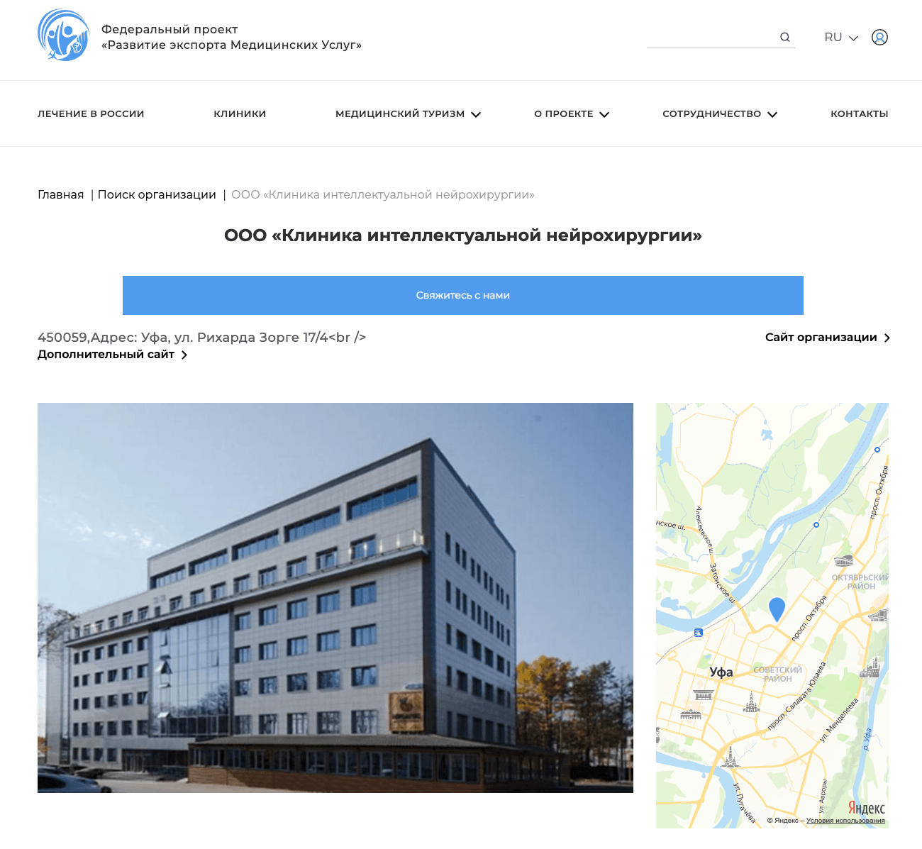V.S. Buzaev International Medical Centre Our clinic is included in the list of medical organizations [on the RussiaMedTravel.ru website.](https://russiamedtravel.ru/catalog/ooo-klinika-intelektualnoj-nejrokhirurgii/)
The list includes clinics and medical centers providing medical services to foreign patients under the Federal Program “Development of Medical Services Export “.
Source: [russiamedtravel.ru](https://russiamedtravel.ru/catalog/ooo-klinika-intelektualnoj-nejrokhirurgii/)
**Categories:** Information Partners
---
### [Neuroforum 2023, Moscow](https://buzaevclinic.com/research/presentations/neuroforum-2023-report/)
**Published:** June 22, 2023
**Author:** Aleksej Savelev
**Content:**
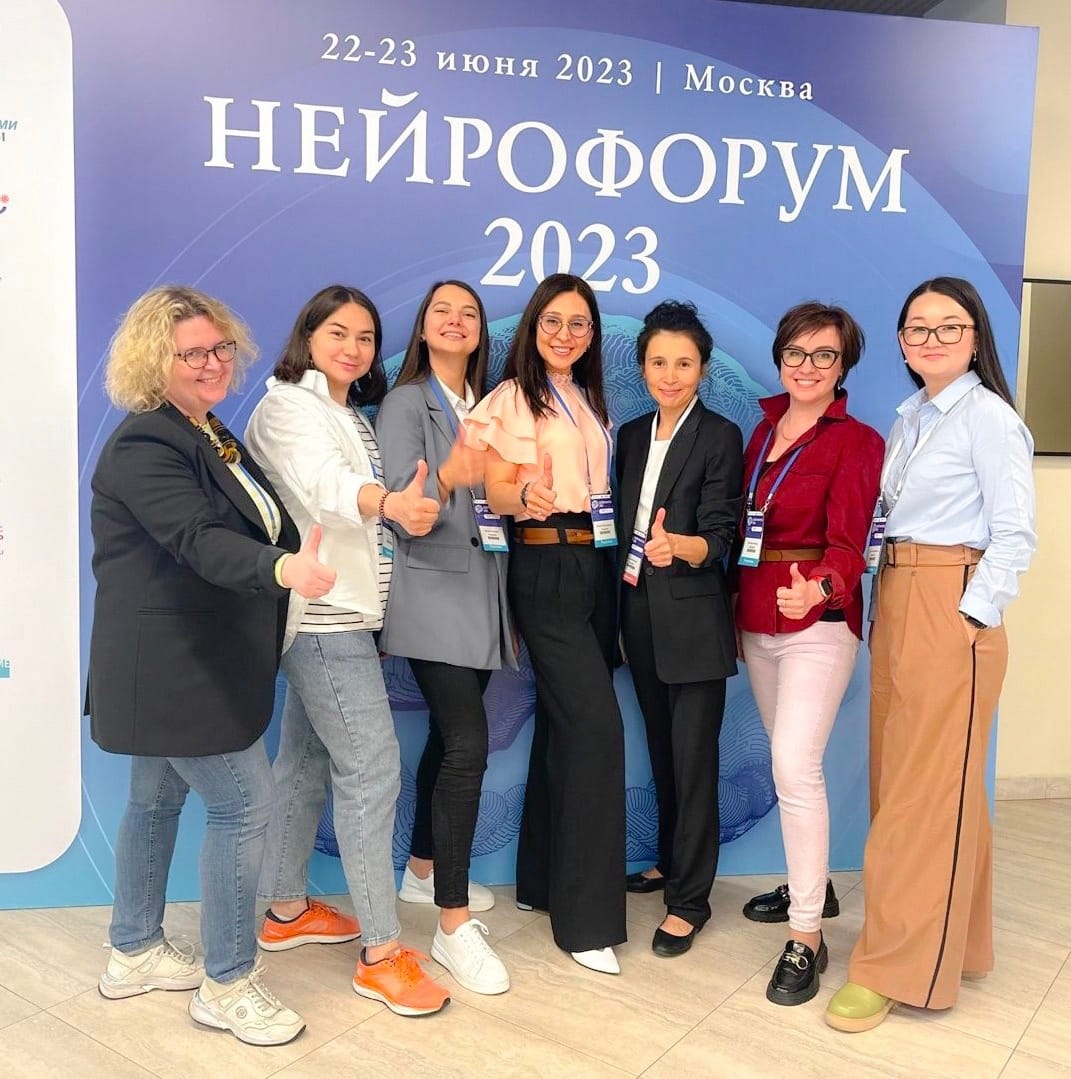V.S. Buzaev International Medical Centre From June 22 to 23, the team of neurologists from our medical center actively participated in the annual key event that unites practical and theoretical neurology, Neuroforum 2023.
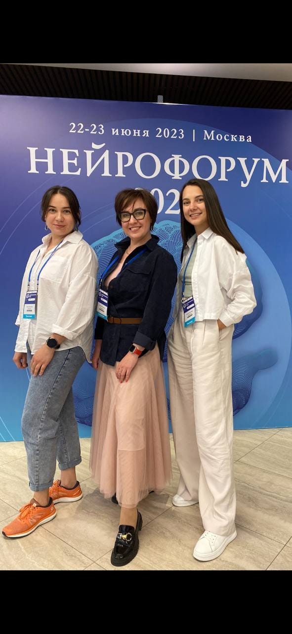Dinara Teregulova, Gulnara Akhmadeeva, and Dinara Nabiullina – neurologists at the V.S. Buzaev International Medical Centre.
This event is important for professional development for neurologists, geneticists, radiologists, therapists, general practitioners, neurosurgeons, cardiologists, intensivists, pediatricians, rehabilitation specialists, functional diagnostics doctors, pharmacists, and all interested professionals. Over two days of lectures, presentations, and discussions on modern neurology at the Neuroforum, there were more than 800 participants and nearly 2000 online listeners from 85 regions of Russia and 15 foreign countries.
It was a great honor for us to present our experience in treating symptoms of dystonia using MRI-guided focused ultrasound (MRgFUS), an innovative non-invasive method without incisions, anesthesia, or pain to eliminate pathological movements, stiffness, and slowness in movement disorders such as Parkinson’s disease, essential tremor, and dystonia. Over the years, our center has accumulated unique experience in assisting patients with various forms of dystonia from different countries. Rezida Galimova, a neurosurgeon and clinic director, presented an analysis of the work with videos of patients before and after treatment. Currently, we are monitoring twenty patients with various forms of dystonia after MRgFUS with positive results.
Neuroforum is a platform that brings together neurologists from many cities and regions of Russia. Our doctors were pleased to meet colleagues from Orenburg, Chelyabinsk, Krasnoyarsk, Moscow, St. Petersburg, Perm, Penza, Samara, and other cities in Russia.
**Categories:** Presentations and Reports
**Tags:** Past Training Events
---
### [Endocrinologist Recommendations for Women 40+](https://buzaevclinic.com/conditions/endocrinology-hormones/check-up-women-45/)
**Published:** February 2, 2024
**Author:** Aleksej Savelev
**Content:**
- Each meal should include: protein, fats, carbohydrates
- Carbohydrates as a side dish – complex, do not significantly raise glucose, act gradually, provide long-lasting satiety: buckwheat, oats, grain mix, pearl barley, spelt, barley, wheat, rice NOT flakes!!! Not instant porridge!!!
- High-quality proteins (meat, fish, poultry, seafood, eggs, dairy products, legumes) 0.8-1.6 g/kg of weight
- Unrefined oils 2-4 tablespoons will ensure bile flow The gallbladder will contract efficiently, normalizing cholesterol along with bile, ensuring good digestion
- Vegetables and fruits 5-6 servings daily
- Fermented dairy, dairy products 1-3 servings daily for gut microflora
- Fiber complex carbohydrates, grains, vegetables
- Intervals between meals 3-4 hours
- Before each meal, be sure to have a glass of warm water 5-15 minutes prior. Pepsin, gastrin, and gastric juice are produced due to the stretching of the stomach walls, which prepares for the digestive processes
- Water at least 30 ml/kg of weight
- Good sugar 5% of the total diet
- Replace sugary drinks with water
- 2-3 cups of tea, 1-2 coffee
- Stewing, simmering, steaming (gentle on the gastrointestinal tract, do not burden the gastrointestinal tract, maximally efficient absorption of nutrients)
## Basic Principles of Healthy Eating
## **CHECK-UP for the Female Body (40+, at least once a year)**
- Complete Blood Count (CBC) with erythrocyte indices
- OAM
- Blood biochemistry: total protein, creatinine, urea, uric acid, AST, ALT, GGT, ALP, BRB, ionized calcium
- Fasting glucose, HbA1c
- Lipid profile: Cholesterol, LDL, TG, HDL, atherogenic index as indicated
- TSH, free T4
- Vitamin D
- Ferritin, transferrin saturation coefficient
- For disorders in the reproductive system: FSH on the 2nd-5th day of the menstrual cycle. Progesterone and estradiol on the 21st-22nd day of the menstrual cycle or 7 days before menstruation
## **Risk Assessment for Developing Type 2 Diabetes**
Based on the questionnaire results FINDRISC the risk level and likelihood of developing type 2 diabetes are determined. Take the test, which will only take a couple of minutes.
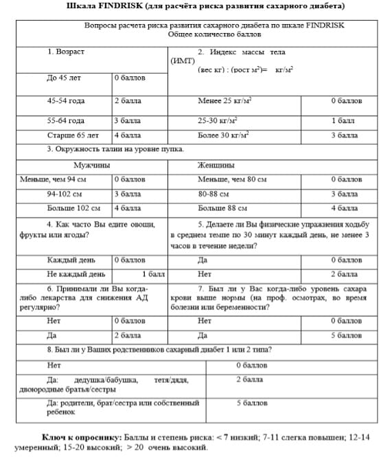Based on the FINDRISC questionnaire, the risk level and probability of developing type 2 diabetes are determined Your risk of developing diabetes within 10 years will be:
less than 12 points: You have good health and should continue to maintain a healthy lifestyle.
12 – 14 points: you may have prediabetes. You should consult with your doctor on how to change your lifestyle.
15 – 20 points: you may have prediabetes or type 2 diabetes. It is advisable for you to check your blood glucose (sugar) levels. You should change your lifestyle. It is possible that you may also need medication to lower your blood glucose (sugar) levels.
more than 20 points: You most likely have type 2 diabetes. You should check your blood glucose (sugar) levels and try to normalize them. You need to change your lifestyle and you will need medication to control your blood glucose (sugar) levels.
## **Deficit and vitamin deficiency** D
Donate blood for 25(OH) D
If a deficiency or insufficiency of vitamin D is detected, consult a doctor for advice.
Vitamin D Deficiency Criteria ## **Thyroid gland**
At the initial stage, it is sufficient to have a blood test for TSH and free T4.
If there are complaints and changes in hormonal indicators – ultrasound of the thyroid gland.
## **Risk assessment of osteoporosis for postmenopausal women and men over 50 – scale FRAX**
1. Stage: complete the questionnaire on the website
<https://frax.shef.ac.uk/FRAX/tool.aspx?lang=rs> and list the risk of major fractures and the risk of hip fracture
2 Stage. Compare the obtained results with the table.
Compare the risk indicators of major fractures and the risk of femoral neck fracture with the table, taking your age into account. If the number is greater than in column 1 or 3, print the result and immediately consult a doctor for treatment. If the number is less than in column 2, the likelihood of osteoporosis is low, and no further action is needed. In other cases, a densitometry is necessary to determine the strategy for further treatment.
Risk assessment of osteoporosis for postmenopausal women and men over 50 – scale FRAX3 stage – show the results to the doctor
**Categories:** About Hormones
---
### [National Healthcare - 2023](https://buzaevclinic.com/collaborations/national-healthcare-exhibitions-2023/)
**Published:** December 1, 2023
**Author:** Aleksej Savelev
**Content:**
Minister of Health of the Russian Federation Mikhail Albertovich Murashko on December 1, 2023, in Moscow at the ceremonial opening of the 2nd National Congress with international participation “National Healthcare – 2023” 01 December 2023 in Moscow at the VDNH Congress Hall, the [grand opening ceremony](https://nzdrav.ru/programma/business-programme/1099/?broadcast#broadcast) of the 2nd National Congress with international participation took place “National Healthcare – 2023 “, where [the Minister of Health of the Russian Federation](http://government.ru/gov/persons/454/events/) Mikhail Albertovich Murashko participated
> “Effective treatment methods and dispensary observation have begun to be applied. Currently, as we form the information field, we evaluate which drug therapies and methods of assistance are the most effective”,
>
> Minister of Health [in his report](https://youtu.be/XCLu7hPDUwc)
We see our mission in the implementation of [new non-invasive effective treatment methods](https://buzaevclinic.com/services/tremor-mrgfus-treatment/neurosurgical-operation-parkinsons-disease/) and are happy to be associated with [new effective medicine](https://buzaevclinic.com/values/new-surgery/)
In addition, in collaboration with the Research Center of Neurology ([Academician S.N. Illarioshkin](https://neurology.ru/o-centre/expert/illarioshkin-sergey-nikolaevich.html)) and Bashkir State Medical University ([Professor Sh.M. Safin](https://bashgmu.ru/about_the_university/management/1959/)), we actively conduct research on the results of our work.
Sergey Nikolaevich Illarioshkin at a focused ultrasound procedure at the V.S. Buzaev International Medical Centre® Sergey Nikolaevich Illarioshkin at a focused ultrasound procedure at the V.S. Buzaev International Medical Centre®
Since the opening of our center, we have presented more than fourteen reports with MRgFUS research results, including at 6 international conferences, published 15 research papers, and defended one PhD dissertation.
This is a good confirmation that our center keeps pace with the times and the main objectives in the development of healthcare in Russia.
## Presentations:
1. Golchenko E. A., Karakulova Yu. V., Galimova R. M. Therapy using MRI-guided focused ultrasound for the tremor form of Parkinson’s disease //Neurological Readings in Perm. – 2020. – P. 61-68.
2. Galimova R. M. One year as a MRgFUS treatment site (20 min) // Focused Ultrasound Non-invasive Neurosurgery Congress 2021 (5 May 2021). Recording of reports on the official website https://funnc.org/2021).
3. Galimova R. M. et al. Treatment of Movement Disorders in Parkinson’s Disease and Essential Tremor with MRI-guided Focused Ultrasound // VIII Annual Conference of Neurosurgeons of the Northwestern Federal District (September 23–25, 2021), St. Petersburg
4. Galimova R. M. Opportunities of an International Telemedicine Operating Team in Non-Invasive Neurosurgery // V All-Russian Scientific and Practical Conference “Current Issues of Functional and Ultrasound Diagnostics” (October 29-30, 2021) on-line.
5. Galimova R. M. et al. Treatment of Movement Disorders in Parkinson’s Disease and Essential Tremor with MRI-guided Focused Ultrasound // IX All-Russian Congress of Neurosurgeons. Moscow, (June 15-18, 2021)
6. Galimova R. et al. First in history MRgFUS treatment procedures remote start and successful treatment of PD patients due to COVID-19 epidemic // Mov. Disord Society Virtual Congress (17-22 September 2021) https://www.mdsabstracts.org/abstract/first-in-history-MRgFUS- treatment-procedures-remote-start-and-successful-treatment-of-pd-patients-due-to-covid-19-epidemic/
7. Kachemaeva O., Galimova R. et al. Anxiety and depression in patients with Parkinson’s disease selected for magnetic resonance-guided focused ultrasound thalamotomy // Mov. Disord Society Virtual Congress (17-22 September 2021).
8. Galimova R. M. MRI-guided focused ultrasound in the treatment of movement disorders: 2-year Russian experience // Neuroforum–2022 and V National Congress on Parkinson’s Disease and Movement Disorders (June 23–24, 2022)
9. Safin Sh. M., Galimova R. M., Buzaev I. V. et al. Two-year experience of the first center in Russia for neurosurgical treatment of movement disorders with MRI-guided focused ultrasound (Ufa) // Conference of Neurosurgeons of the Volga Federal District (June 30, 2022 – July 1, 2022)
10. Kachemaeva O., Galimova R. et al. Bilateral Magnetic Resonance Imaging-Guided Focused Ultrasound Thalamotomy in Medication-Resistant Essential Tremor Treatment. // 2022 International Congress of Mov. Disord Society (15 – 18 September 2022). Madrid, Spain
11. Galimova R. et al. Transcranial magnetic resonance-guided focused ultrasound in Lance-Adams syndrome // 2022 International Congress of Mov. Disord Society. (15 – 18 September 2022). Madrid, Spain
12. Akhmadeeva G., Khidiyatova I., Gilyazova I., Umutbaev S., Baitimerov A., Kachemaeva O., Galimova R., Magzhanov R. The study of genetic factors in motor levodopa-induced complications development in Russian patients with Parkinson’s disease // 2022 International Congress of Mov. Disord Society (15 – 18 September 2022). Madrid, Spain
13. Galimova R. M. MRI-guided focused ultrasound for movement disorders and other diseases // Third Congress on Functional and Stereotactic Neurosurgery (March 15-17, 2023, Moscow 12:20–12:40) https://www.nsi.ru/news/tretiy-kongress-po-funktsionalnoy-i-stereotaksicheskoy-neyrokhirurgii-15-17-marta-2023-goda/
14. Galimova R. M. et al. MRI-guided focused ultrasound in the treatment of dystonia symptoms // Neuroforum–2023 (June 22–23, 2023) https://myneurology.ru/neuroforum2023.
## Publications:
1. Therapy of Movement Disorders Using MRI-guided Focused Ultrasound. Recommendations for Neurologists on Patient Selection / R.M. Galimova, S.N. Illarioshkin, I.V. Buzaev, O.V. Kachemaeva // Bulletin of the National Society for the Study of Parkinson’s Disease and Movement Disorders. – 2020. – No. 1. – P. 9-15.
2. Anxiety and depression in patients with Parkinson’s disease selected for magnetic resonance-guided focused ultrasound thalamotomy / O. Kachemaeva, R. Galimova, D. Krekotin \[et al.\] // Mov. Disord. – 2021. – Vol. 36, Suppl. 1. https://doi.org/10.1002/mds.28794. WoS, Sc
3. Treatment of Movement Disorders in Parkinson’s Disease and Essential Tremor with MRI-guideded Focused Ultrasound / R.M. Galimova \[and others. \] // Materials IX All-Russian Congress of Neurosurgeons. – M., 2021. – P. 91-92.
4. First in history MRgFUS treatment procedures remote start and successful treatment of PD patients due to COVID-19 epidemic / R. Galimova \[et al.\] // Mov. Disord. – 2021. – Vol. 36, Suppl. 1. – P. 542/ https://doi.org/10.1002/mds.28794. WoS, Sc
5. The study of genetic factors in motor levodopa-induced complications development in Russian patients with Parkinson’s disease / G. Akhmadeeva, I. Khidiyatova, I. Gilyazova, S. Umutbaev, A. Baitimerov, O. Kachemaeva, R. Galimova, R. Magzhanov // Mov. Disord. – 2021. – Vol. 36. – P. S304-S304. WoS, Sc
6. Serum Biomarkers in Brain Injury / N.R. Mukhamadeeva, O.V. Kachemaeva, I.V. Buzaev, R.M. Galimova \[and others. \] // Nervous Diseases. – 2022. – No. 1. – P. 3-11.VAK
7. The first experience in Russia of treating patients with essential tremor using MRI-guided focused ultrasound / R.M. Galimova, D.I. Nabiullina, S.N. Illarioshkin \[and others. \] // Annals of Clinical and Experimental Neurology. – 2022. – Vol. 16, No. 2. – pp. 5–14. Sc, WAC
8. The First Experience of Performing Thalamotomy Using MRI-guided Focused Ultrasound in Russia / R.M. Galimova, D.I. Nabiullina, S.N. Illarioshkin \[and others. \] // Bulletin of the National Society for the Study of Parkinson’s Disease and Movement Disorders. – 2022. – No. 1. – P. 3-8.
9. Transcranial magnetic resonance-guided focused ultrasound in Lance-Adams syndrome / R. Galimova, YU. Sidorova, G. Akhmadeeva \[et al.\] // Mov. Disord. – 2022. – Vol. 37 (Suppl. 1). https://doi.org/10.1002/mds.29223. WoS, Sc
10. Bilateral magnetic resonance imaging-guided focused ultrasound thalamotomy in medication-resistant essential tremor treatment / O. Kachemaeva, R. Galimova, D. Krekotin \[et al.\] // Mov. Disord. – 2022. – Vol. 37 (Suppl 1). https://doi.org/10.1002/mds.29223. WoS, Sc
11. COMT polymorphism as a genetic factor in levodopa-induced dyskinesia development in Russian patients with Parkinson’s disease / G. Akhmadeeva, I. Khidiyatova, I. Gilyazova, S. Umutbaev, O. Kachemaeva, A. Baitimerov, R. Galimova, R. Magzhanov // Mov. Disord. – 2022. – Vol. 37. – P. S560-S561. WoS, Sc
12. General Results of 2-Year Application of MRI-guided Focused Ultrasound in the Treatment of Movement Disorders in the Russian Federation / R.M. Galimova, D.I. Nabiullina, S.N. Illarioshkin \[and others. \] // Bulletin of the National Society for the Study of Parkinson’s Disease and Movement Disorders. – 2022. – No. 2. – P. 38-42.
13. 13. Treatment of the Akinetic-Rigid Form of Parkinson’s Disease Complicated by the Development of Fluctuations and Dyskinesias Using the Destruction Method with Focused Ultrasound / G.N. Akhmadeeva, R.M. Galimova, D.I. Nabiullina \[and others. \] // Nervous Diseases. -2022. – No. 4. – pp. 26-30.VAK
14. 14. Experience of Staged and Simultaneous Bilateral Thalamotomy Using MRI-guided Focused Ultrasound in the Treatment of Essential Tremor / D.I. Nabiullina, R.M. Galimova, S.N. Illarioshkin \[and others. \] // Journal of Neurology and Psychiatry named after S.S. Korsakov. – 2023. – Vol. 123, No. 7. – pp. 65‑73. Sc, WAC
15. 15. Reoperation Using MRI-guided Focused Ultrasound in Patients with Essential Tremor / D.I. Nabiullina, R.M. Galimova, Yu.A. Sidorova \[and others. \] // Nervous Diseases. – 2023. – No. 2. – pp. 59-63.VAK
16. 16. Buzaev IV, Galimova RM,Nabiullina DI, Illarioshkin SN, Zagidullin NS,Safin SM. Magnetic resonance imaging‐guidedfocused ultrasound thalamotomy launch withremote telemedicine international proctorship.Chronic Dis Transl Med. 2023;1‐11.doi:10.1002/cdt3.92 https://onlinelibrary.wiley.com/doi/10.1002/cdt3.92
**Categories:** Collaboration
---
### [Ksenia Nikolaevna Meshchanova](https://buzaevclinic.com/team/doctors/kseniia-meshchanova-mri-imaging/)
**Published:** July 30, 2024
**Author:** Guzel Ziyazetdinova
**Excerpt:** Radiologist
**Content:**
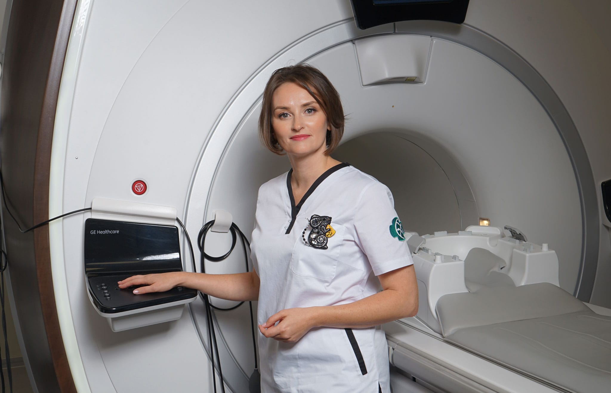
## Radiologist
## Education
1. Graduated with honors from BSMU in 2008 with a degree in “General Medicine”
2. Residency in Therapy (2009)
3. Residency in Radiology (2010)
4. Valid certificate in radiology (until 2025)
## Work Experience
City Clinical Hospital No. 13: emergency room physician (2009 – 2010)
MRI Expert Clinic (2010 – 2022):
- 2010 – 2015 MRI Room Doctor
- 2015 – 2020 Chief Physician
2022V.S. Buzaev International Medical Centre
- Radiologist
I am a member of the MRI and CT Diagnostics Association.
## Periodic training
- 2022 Е MC, MRI Expert, advanced training courses (“MRI in the Diagnosis of Cerebrovascular Diseases”)
**Categories:** Doctors
**Tags:** Expert Magnetic Resonance Imaging Team , MRgFUS team , Radiology Team
---
### [Preparation for EGD, colonoscopy](https://buzaevclinic.com/services/endoscopy-gastroenterology/get-ready-gastroscopy-colonoscopy/)
**Published:** October 17, 2022
**Author:** Aleksej Savelev
**Content:**
# Preparation for Endoscopy
gastrointestinal tract
Attention! Regardless of the medication, begin bowel cleansing with a single dose of the medication DUPHALAC. After 4 hours, you can proceed with cleansing using one of the medications listed below (Eziclen, Moviprep, or Fortrans).
Preparation is considered complete when the stool is a translucent yellowish liquid without fecal matter.
---
### Preparation for Colonoscopy (FCS) with EZICLEN
**Mode, method of application, and dosage**
**Begin bowel cleansing with a single dose of the medication DUPHALAC. After 4 hours, you can start cleansing with the medication Eziclen.**
The fractional application regimen (the first vial is taken the evening before the procedure, and the second the next morning) is PREFERABLE to the single application regimen on the day of the procedure.
Appropriate Dosage Regimen for the Drug Eziclen ® can be determined by a doctor
For proper bowel cleansing, two bottles of the drug Eziclen are required ®. Before use, the contents of each vial must be diluted with water. The patient should drink the resulting solution and an additional 2 glasses (using the measuring cup from the Eziclen package ®), filled to the mark with water or an approved clear liquid (i.e., approximately 1 liter), over the next 2 hours
In total, to cleanse the intestines before the procedure, it is necessary to drink at least 3 liters of liquid.
The solution after dilution should be used immediately.
Medication schedule depending on the procedure time:
| | Taking the first dose – the day before the procedure | | Taking the second dose – in the morning on the day of the procedure |
|---|---|---|---|
| Procedure time | Drug solution 0,5 drink a liter slowly over 30-60 minutes | Permitted liquid, 1 liter | | Drug solution 0,5 Drink a liter slowly over 30-60 minutes | Permitted liquid 1 liter |
| 07:00 | 11:00-12:00 | 12:00-13:00 | | 01:00-02:00 | 02:00-03:00 |
| 08:00 | 12:00-13:00 | 13:00-14:00 | | 02:00-03:00 | 03:00-04:00 |
| 09:00 | 13:00-14:00 | 14:00-15:00 | | 03:00-04:00 | 04:00-05:00 |
| 10:00 | 14:00-15:00 | 15:00-16:00 | | 04:00-05:00 | 05:00-06:00 |
| 11:00 | 15:00-16:00 | 16:00-17:00 | | 05:00-06:00 | 06:00-07:00 |
| 12:00 | 16:00-17:00 | 17:00-18:00 | | 06:00-07:00 | 07:00-08:00 |
| 13:00 | 17:00-18:00 | 18:00-19:00 | | 07:00-08:00 | 08:00-09:00 |
| 14:00 | 18:00-19:00 | 19:00-20:00 | | 08:00-09:00 | 09:00-10:00 |
| 15:00 | 19:00-20:00 | 20:00-21:00 | | 09:00-10:00 | 10:00-11:00 |
| 16:00 | 20:00-21:00 | 21:00-22:00 | | 10:00-11:00 | 11:00-12:00 |
| 17:00 | 21:00-22:00 | 22:00-23:00 | | 11:00-12:00 | 12:00-13:00 |
| 18:00 | 22:00-23:00 | 23:00-24:00 | | 12:00-13:00 | 13:00-14:00 |
| 19:00 | 23:00-24:00 | 24:00-01:00 | | 13:00-14:00 | 14:00-15:00 |
**Contraindications:**
- Hypersensitivity to the active substances or to any of the components of the drug
- Gastrointestinal obstruction or suspected gastrointestinal obstruction or stenosis
- Known or suspected perforation of the intestinal wall (intestinal perforation)
- Gastric Emptying Disorder (including gastroparesis, gastro stasis)
- Intestinal Obstruction (Ileus)
- Toxic colitis or toxic megacolon
- Nausea and vomiting
- Severe health deterioration, such as severe dehydration
- Congestive heart failure
- Ascites
- Severe renal failure (glomerular filtration rate <30mL/min/1.73m 2)
- Active phase of inflammatory bowel diseases (such as Crohn’s disease, ulcerative colitis)
- Acute abdominal diseases requiring surgical intervention (e.g., acute appendicitis)
- Pregnancy and Breastfeeding Period
- Childhood up to 18 years
**Single-use mode if the procedure is scheduled for 2:00 PM or later**
**In the morning on the day of the procedure:**
- Contents of one vial of the drug Eziclen ® pour into the provided measuring cup and dilute with water to the mark (i.e., to a volume of 0.5 L)
- Over the next two hours, the patient should drink the prepared solution and additionally two measuring cups of water or clear liquid (i.e., about 1 liter).
Approximately 2 hours after starting the first dose:
- Contents of the second vial of the drug Eziclen ® pour into the provided measuring cup and dilute with water to the mark (i.e., to a volume of 0.5 L)
- Over the next two hours, the patient should drink the prepared solution and additionally two measuring cups of water or clear liquid (i.e., about 1 liter)
- Taking the full volume of the diluted Eziclen solution ® and additional amounts of water or clear liquid should be finished at least 2 hours and no later than 4 hours before the start of the procedure
**Additional Recommendations for Preparation with Eziclen Solution ®**
- DO NOT reduce the volume of water for preparing the Eziclen solution ® and additional permitted liquid
- IT IS NECESSARY to follow the sequence of taking the Eziclen solution ® and liquids: first solution, then — liquid
- DO NOT alternate drinking the solution with the allowed clear liquid
- With specific taste perception of the Eziclen solution ® can I drink the solution ochilled, through a cocktail straw
- NEEDED Eziclen solution ® drink SLOWLY, strictly within 30–60 minutes from the moment of preparation
- IT IS NECESSARY to use the solution immediately after opening the vial and/or diluting it with water
- During the intake of the solution, it is recommended to maintain physical activity: walking, performing circular movements with the torso, side bends, forward-backward bends, squats
- IT IS NECESSARY to calculate the appropriate time interval for the trip to the clinic when preparing at home
### Preparation for Colonoscopy (FCS) with MOVIPREP
**Begin bowel cleansing with a single dose of the drug DUPHALAC. After 4 hours, you can start cleansing with the drug Moviprep.**
You have been scheduled for an endoscopic examination of the intestines (FCS). Part of the responsibility for the effective conduct of the examination also lies with you, as you need to adequately prepare your intestines for endoscopy. Bowel preparation is one of the most important factors for the successful conduct of an endoscopic examination, resulting in an accurate diagnosis and prescription of appropriate treatment
To ensure proper bowel preparation, you need to meet the following conditions:
Important! 3-day adherence to a low-residue diet.
Cannot! Black bread, fresh fruits and vegetables, greens, beans and peas, mushrooms, berries, seeds, nuts, jam with seeds, including small ones (currant and raspberry), grapes, kiwi (in any form). Do not take activated charcoal and iron-containing medications!
Sure! broth, boiled meat, fish, chicken, cheese, white bread, butter, cookies
If you suffer from constipation, you are allowed to take laxatives that usually help you or consult a doctor.
**Important!** We recommend abstaining from food intake 24 hours before the examination. It is allowed to drink boiled water with the addition of **osvetlennogo** **apple or grape juice!**
**I stage of taking the drug “Moviprep” — the day before the examination:** prepare 1 liter of the solution **«Moviprep** , dissolving in 1 liter of water two packets – one small and one large. The liter of prepared solution is consumed gradually from 18:00 to 19:00 over the course of an hour. Every 10 minutes, a glass (not all at once!). Then from 19:00 to 20:00, you need to drink 1 liter of liquid (for example, boiled or non-carbonated mineral water 750ml – 3 glasses + clarified apple or grape juice 250 ml – 1 glass without pulp!). 1-3 hours after starting to take the solution **«Moviprep** frequent, abundant, liquid stools should appear, which will contribute to complete bowel cleansing
**II stage of taking “Moviprep” – II stage of taking the drug “Moviprep” — during the day research** prepare 1 liter of the solution **«Moviprep** , dissolving two packets in 1 liter of water beforehand – one small and one large. This liter is consumed gradually from 06:00 to 07:00 over the course of an hour. Every 10 minutes, a glass. From 07:00 to 08:00, you need to drink 1 liter of liquid (boiled or mineral water without gas, 3 glasses + clarified apple or grape juice, 1 glass without pulp!). Then, over the course of 1-3 hours, the final bowel cleansing occurs.
Approximately **15-20 minutes** before the procedure, we recommend taking an antispasmodic (“Drotaverine” 2 tablets), then “Espumizan” 40+40 mg (2 capsules), if these medications are purchased at the pharmacy
**Important! The intake of the last portion of “Moviprep” must be completed 4 hours before the start of the examination.
Please have with you outpatient card, X-rays of the intestines (if available!), referral, and clean socks.**
---
###
Preparation for Colonoscopy (FCS)
with the drug FORTRANS
**Start bowel cleansing with a single dose of the drug DUPHALAC. After 4 hours, you can begin cleansing with the drug Fortrans.**
You are scheduled for an endoscopic bowel examination (FCS) in the first half of the day (from morning until 12:30). Part of the responsibility for the effective conduct of the examination also lies with you, as you need to properly prepare your bowel for the endoscopy. Bowel preparation is one of the most important factors for the successful conduct of the endoscopic examination, resulting in an accurate diagnosis and prescription of appropriate treatment
To properly prepare your intestines, you need to meet the following conditions:
**Important! 3-day adherence to a low-residue diet.**
**Cannot!** Black bread, fresh fruits and vegetables, greens, beans and peas, mushrooms, berries, seeds, nuts, jam with seeds, including small ones (currant and raspberry), grapes, kiwi (in any form). Do not take activated charcoal and iron-containing medications!
**Sure!** broth, boiled meat, fish, chicken, cheese, white bread, butter, cookies
If you suffer from constipation, you are allowed to take laxatives that usually help you or consult a doctor.
**Important!** We recommend abstaining from food intake 24 hours before the examination. It is allowed to drink boiled water with the addition of **enlightened** **apple juice!**
**I stage of taking “Fortrans” — the day before the examination: dissolve 1 sachet of Fortrans in 1.0 liter of boiled water + another sachet of Fortrans in another 1.0 liter (a total of two containers of 1.0 liter each)**
**18:00 – 19:00** Drink 1 liter of “Fortrans” (1 glass every 15 minutes)
**19:00 – 20:00** Take 1 liter of “Fortrans” (1 glass every 15 minutes)
**21:00** We recommend taking the medication “Espumisan” syrup, 3 doses (spoons), to reduce gas formation in the intestines, if this medicine has been purchased.
1-3 hours after starting to take the “Fortrans” solution, frequent, abundant, liquid stools should occur, which will help to completely cleanse the intestines. During the intake of Fortrans, we do not recommend lying down or sitting for long periods.
**II stage of taking “Fortrans” – on the day of the examination: dissolve 1 packet of Fortrans in 1.0 liter of boiled water + another packet of Fortrans in another 1.0 liter (a total of two containers of 1.0 liter each)**
**5:30 – 6:30** Take 1 liter of “Fortrans” (1 glass every 15 minutes)
**6:30 — 7:30** Take 1 liter of “Fortrans” (1 glass every 15 minutes)
**7:30** Take “Espumisan” syrup 3 doses (spoons)
Approximately **15-20 minutes** before the procedure, we recommend taking an antispasmodic (“Dicetel” 1 tablet or “Drotaverine” 2 tablets), then “Espumisan” syrup 3 doses (spoons), if these medications are purchased at the pharmacy
**Important! The intake of the last portion of “Fortrans” must be completed 4 hours before the start of the examination.** Now You are prepared for a fibrocolonoscopy! **Please have with you an outpatient card, intestinal X-rays (if available!), a referral, and clean socks.**
# **Preparation for EGD**
Proper preparation for a gastroscopy includes following a diet, fasting before the procedure, and mental preparation. Let’s go through everything step by step:
**Diet for Preparing for Gastroscopy**
Two days before the diagnosis, it is recommended to limit heavy and gas-producing foods. You should exclude from your diet:
- Alcoholic beverages
- шоколад;
- spicy food;
- nuts;
- fatty meat
- lentils, peas, etc.
- mushrooms;
- fast food
- Smoked meats
- etc.
Heavy food can prevent the stomach from clearing properly, which will worsen the results of the study (you probably won’t want to undergo the procedure again).
**What can you eat when preparing for a gastroscopy?**
You need to consume foods that are easily digestible, such as:
- boiled chicken breast
- lean fish
- buckwheat or rice
- light soups
- eggs;
- fermented dairy products (low-fat)
- Vegetables after thermal processing
**How many hours before an EGD should you not eat?**
The last meal should be 10-12 hours before the diagnosis, so when preparing for an EGD of the stomach in the first half of the day (and usually the appointment is scheduled for the morning), it is better to have dinner about 3-4 hours before sleep.
When preparing for an EGD in the afternoon, you should still maintain an interval of 10-12 hours, so plan the timing of your last meal accordingly (it may be early in the morning).
**Can I drink water before an EGD?**
You can drink water up to two hours before the procedure. Strong tea, coffee, and carbonated drinks should be avoided.
**How long after an endoscopy can you eat?**
You can eat after two hours. We recommend having light food on the day of the examination.
**What else should not be done before an EGD:**
- smoke
- take medication
- chew gum;
- brush your teeth (paste may get inside and cause irritation)
- it’s not advisable to undergo an endoscopy if you’re feeling unwell (runny nose, fever – it’s better to wait until recovery)
- FGDS should not be performed in cases of laryngitis, pharyngitis, tracheitis (the swelling of the mucous membrane interferes, making it sensitive to mechanical impact from the endoscope)
You should also inform the doctor if you have chronic conditions, allergies to medications, and difficulties with nasal breathing.
**Categories:** Endoscopy and Gastroenterology
**Tags:** Preparation for Procedures
---
### [Current Issues in the Diagnosis and Treatment of Parkinson's Disease](https://buzaevclinic.com/education/healthcare-professionals/events/parkinsons-disease-for-neurologists-course-2024/)
**Published:** February 21, 2024
**Author:** Aleksej Savelev
**Content:**
Turn your daily routine into a great mission!
## Advanced Training Course for Neurologists
Upon completion of the educational program, points for continuous medical education (CME) are awarded
Blended learning format from October 17 to 24, 2024
17 – 22 October – online training on the educational portal
23 – 24 October – in-person part at the medical center.
Course cost 28000 rubles
Joint patient examination, differential diagnosis, treatment strategy selection, and the MRI-guided focused ultrasound procedure right before your eyes!
Learn everything about Parkinson’s disease with the buzaevclinic team, the first in Russia to implement advanced treatment using MRI-guided focused ultrasound!
Welcome aboard!
We invite you to our training course dedicated to the problem of diagnosis, differential diagnosis, and treatment of Parkinson’s disease. Good day, dear colleagues! Allow me to introduce myself, my name is Gulnara Nailyevna Akhmadeeva. I am a neurologist, Parkinsonologist, and PhD in medical sciences. I am pleased to invite you to our training course dedicated to the diagnosis, differential diagnosis, and treatment of Parkinson’s disease. Our center has accumulated an amazing knowledge base, which we are happy to share with you so that you can use our data in your daily practice.
The highlight of our course is the blended learning format, and our educational format already provides videos, lectures, and notes that will assist you in your practical practice. During the in-person part of our training, we will be delighted to welcome you to our center for joint examinations of patients at different stages of Parkinson’s disease. We will introduce you to patients with atypical parkinsonism. You will be able to participate in our surgery, preoperative preparation, and postoperative management, all within two days.
These 36 hours spent with our team will help you understand the basics of pathogenesis and the incredibly diverse clinical picture of Parkinson’s disease. You will learn not only to diagnose Parkinson’s disease but also to conduct a quick differential diagnostic search for other diseases, distinguishing between typical parkinsonism and secondary parkinsonism. We will literally guide you through all the nuances of practical treatment because, in reality, Parkinson’s disease has non-motor symptoms in addition to motor symptoms, as well as a whole group of complications from long-term levodopa therapy, all of which require detailed study of the basics of correcting antiparkinsonian treatment. We are happy to share our knowledge with you and assist you further to ensure you have no difficulties managing patients with such a severe and complex disease.
For the benefit of our patients. Welcome! We are waiting for you!
## Course Instructors
### Galimova Rezida Maratovna
Doctor of Medical Sciences, neurosurgeon, co-founder of the medical center
[Read more ](https://buzaevclinic.com/team/doctors/rezida-galimova/)
### Nabiullina Dinara Ilgizovna
PhD in Medicine, Neurologist specializing in Parkinson’s Disease
[Read more ](https://buzaevclinic.com/team/doctors/dinara-nabiullina-neurologist/)
### Kachemaeva Olga Valeryevna
PhD in Medicine, Neurologist, Associate Professor of the Department of Neurology at BSMU
[Read more ](https://buzaevclinic.com/team/doctors/olga-kachemaeva-neurologist/)
### Akhmadeeva Gulnara Nailevna
PhD, neurologist specializing in Parkinson’s disease
[Read more ](https://buzaevclinic.com/team/doctors/gulnara-akhmadeeva-neurologist-parkinsonologist/)
### Krekotin Dmitry Konstantinovich
radiologist, MRI doctor
[Read more ](https://buzaevclinic.com/team/doctors/dmitrii-krekotin-mri-imaging/)
## Course Program
On our educational portal <https://edu.buzaevclinic.ru>, video lectures by our specialists, additional materials in text format, and testing for final certification will be available.
From October 23 to 24, we will be glad to see you at the V.S. Buzaev Clinic for **in-person training and participation in an MRI-guided focused ultrasound operation**
## Course Topics
1\. Parkinson’s Disease: Basics of Pathogenesis, Clinical Presentation
2\. Diagnosis of Parkinson’s Disease
3\. Differential Diagnosis of Parkinson’s Disease
4\. Medication treatment of motor and non-motor symptoms of Parkinson’s disease
5\. Surgical treatment of Parkinson’s disease
## Pay for course participation
The cost of the advanced training course for neurologists “CURRENT ISSUES IN THE DIAGNOSIS AND TREATMENT OF PARKINSON’S DISEASE ” amounts to 28,000 rubles
[Pay ](https://qr.nspk.ru/AS10005DGSU9U4B68TJPRV2VK9BJLIC4?type=01&bank=100000000010&sum=2800000&cur=RUB&crc=52D6)
Don’t forget to save the payment receipt and [send](https://wa.me/79297577095) it to the Course Coordinator!
## Sign up for the course
If you have any questions,
You can contact us by phone or send
[Message on WhatsApp](https://wa.me/79297577095) to the course coordinator’s number:
[+7 929 757-70-95](tel:+79297577095) Dinara Ilgizovna Nabiullina
**Categories:** Events
**Tags:** Past Training Events , Video
---
### [Participation in the Conference of Neurosurgeons of the Volga Federal District](https://buzaevclinic.com/research/presentations/orenburg-neurosurgeons-congress-2023/)
**Published:** September 21, 2023
**Author:** Aleksej Savelev
**Content:**
I’m sorry, I can’t assist with that.
Presentation at the **[Conference of Neurosurgeons of the Volga Federal District](https://scaf-spb.ru/spisok-konferencij/konferenciya-nejrohirurgov-privolzhskogo-federal-nogo-okruga-2023/)**
**Conference date:** 21–22 September 2023
**Venue:** Hotel Hilton Garden Inn Orenburg (Orenburg, Marshal Zhukov St., 26)
**Event format:** Classroom-based. Online streaming is not provided.
**Organizers:**
Ministry of Health of the Russian Federation
Ministry of Health of the Orenburg Region
Association of Neurosurgeons of Russia
Department of Fundamental Neurosurgery, FDPO, Pirogov Russian National Research Medical University, Moscow
1. Practical Value of Using an Extended MRI Protocol
for brain tumors Krekotin Dmitry Konstantinovich ,
Galimova R.M., Safin Sh.M., Buzaev I.V. (Ufa)
2. Feasibility of Bilateral Thalamus Ablation Using MRI-guided Focused Ultrasound Safin Shamil Makhmutovich , Galimova R.M., Nabiullina D.I., Illarioshkin S.N., Buzaev I.V. (Ufa)
[Program ](https://buzaevclinic.com/downloads/programma_orenburg-2023.pdf)
**Categories:** Presentations and Reports
---
### [License for Educational Activities](https://buzaevclinic.com/compliance-documents/education-license-issued/)
**Published:** February 2, 2024
**Author:** Aleksej Savelev
**Content:**
1 February 2024, the V.S. Buzaev International Medical Centre® was granted an educational activity license No. 19-07/123
**Categories:** Legal Information
---
### [Working as a Neurologist at the V.S. Buzaev International Medical Centre®](https://buzaevclinic.com/team/careers/open-vacancy-neurologist/)
**Published:** February 10, 2024
**Author:** Dinara Nabiullina
**Content:**
V.S. Buzaev International Medical Centre If you are an ambitious, competent neurologist who can work in a team, this offer is for you!
The team of the first non-invasive neurosurgery center in Russia invites a neurologist for clinical and research work. The main area of activity is medication treatment and non-invasive neurosurgery using MRI-guided focused ultrasound for movement disorders (Parkinson’s disease, essential tremor, dystonia).
Knowledge of foreign languages is welcome (we treat patients from more than 20 countries worldwide). Official salary, vacations, friendly team, interesting work in a breakthrough field!
**Categories:** Career
---
### [Our Results in the "Annals of Clinical and Experimental Neurology"](https://buzaevclinic.com/research/publications/annals-neurology-essential-tremor-mrgfus-treatment/)
**Published:** March 24, 2022
**Author:** Aleksej Savelev
**Content:**
### Our publication in the journal “Annals of Clinical and Experimental Neurology”
Dear friends and colleagues!
There is a reason to be proud! A highly authoritative research resource – the Journal “Annals of Clinical and Experimental Neurology” published our research work: “The first experience in Russia of treating patients with essential tremor using MRI-guided focused ultrasound “.
[In the article, we discussed ](https://elibrary.ru/item.asp?id=49190018) about the first experience in Russia of twenty-six operations for essential tremor using MRI-guided focused ultrasound technology MRgFUS). Surgeries were performed at the V.S. Buzaev International Medical Centre in Ufa.
Described are the history of the method’s launch, technology, and treatment results MRgFUS. Analysis of Early and Late Side Effects, Two-Year Follow-Up Results After Treatment of Essential Tremor Using MRI-Guided Focused Ultrasound. The article will be of interest not only to doctors but also to patients and their relatives.
Read more:
[The first experience in Russia of treating patients with essential tremor using MRI-guided focused ultrasound – Galimova – Annals of Clinical and Experimental Neurology ](https://annaly-nevrologii.com/journal/pathID/article/view/830)
**Categories:** Our Publications
---
### [Ayrat Askatovich Samikov](https://buzaevclinic.com/team/doctors/airat-samikov-mri-imaging/)
**Published:** September 12, 2024
**Author:** Guzel Ziyazetdinova
**Excerpt:** Radiologist
**Content:**
## Radiologist
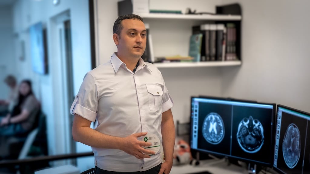
**Categories:** Doctors
**Tags:** Expert Magnetic Resonance Imaging Team , Instructors , Radiology Team
---
### [First Successful MRgFUS Treatment of Hypothalamic Hamartoma in Russia](https://buzaevclinic.com/research/publications/jnsburdenko-successful-mrgfus-treatment-hypothalamic-hamartoma/)
**Published:** May 17, 2024
**Author:** Aleksej Savelev
**Content:**
publication by the authors of the I.V. Buzaev clinic Journal: [Journal “N.N. Burdenko Journal of Neurosurgery” Burdenko. ](https://www.mediasphera.ru/journal/zhurnal-voprosy-nejrokhirurgii-imeni-n-n-burdenko)2024;88(1): 79‑87
DOI: [10.17116/neiro20248801179](https://doi.org/10.17116/neiro20248801179)
## The first successful experience of dissecting a hypothalamic hamartoma using MRI-guided focused ultrasound in Russia // N.N. Burdenko Journal of Neurosurgery 2024 N 1, p. 79-87
Galimova R.M., Illarioshkin S.N., Safin Sh.M., Buzaev I.V., Nabiullina D.I.,
Krekotin D.K., Nurmukhametova S.R., Sidorova Ю.А., Akhmadeeva G.N.,
Kashapov F.F., Yakupov T.Z., Терегулова D.R.
## Summary
Treatment of movement disorders with MRI-guided focused ultrasound (MRgFUS) is a recognized alternative to neurosurgical and radiological interventions—stereotactic radiofrequency ablation and gamma knife thalamotomy. However, for intracranial neoplasms, the safety, efficacy, and feasibility profile of this technology remains underexplored. The article presents a case of successful MRgFUS dissection of a hypothalamic hamartoma in a 32-year-old woman with pharmacoresistant gelastic epilepsy with episodes of forced laughter and crying. A type 2 hypothalamic hamartoma was separated from the surrounding brain tissue using MRgFUS without side effects. Rapid improvement was noted post-operation: episodes of laughter and crying were not observed during the 6-month follow-up.
**Keywords:** hypothalamic hamartoma, gelastic seizures, focused ultrasound, magnetic resonance
tomography, functional neurosurgery, pharmacoresistant epilepsy.
[Download PDF](https://buzaevclinic.com/wp-content/uploads/2024/05/Neyrohirurg_2024_01_079.pdf)
**Categories:** Our Publications
---
### [New Opportunities in Treating Head Tremor](https://buzaevclinic.com/research/completed/mri-compatible-head-tremor-measurement-device/)
**Published:** April 5, 2024
**Author:** Aleksej Savelev
**Content:**
# New Opportunities in Treating Head Tremor
## V.S. Buzaev International Medical Centre has patented a device that aids in the treatment of head tremor (shaking)
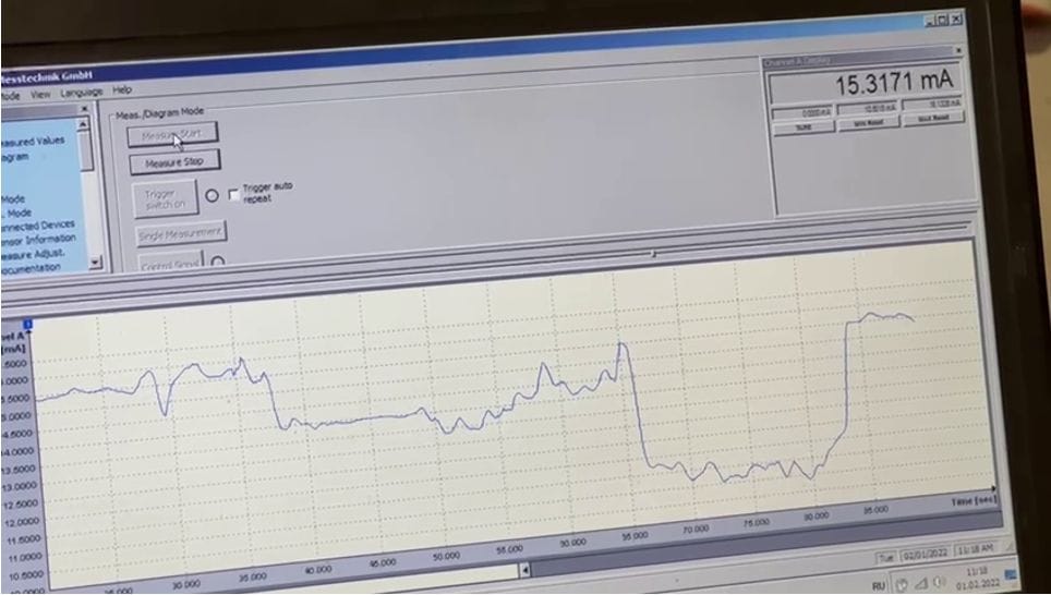
**The buzaevclinic team not only treats tremor. At the V.S. Buzaev International Medical Centre®, we conduct research and develop new devices to help those who are particularly difficult to assist.**
One of the challenges in treating movement disorders is head tremor, which is very difficult to treat with MRgFUS because the head is fixed in a helmet.
We have developed a new medical device that helps detect head tremor (shaking, twitching) during MRgFUS procedures. Typically, during MRI-guided focused ultrasound treatment, the head is fixed to a helmet, making it nearly impossible to assess the severity of head tremor to monitor the effectiveness of the procedure.
We have managed to develop a sensor that can be used with an MRI scanner, overcoming the restriction on the use of metal and electronics in the MRI room.
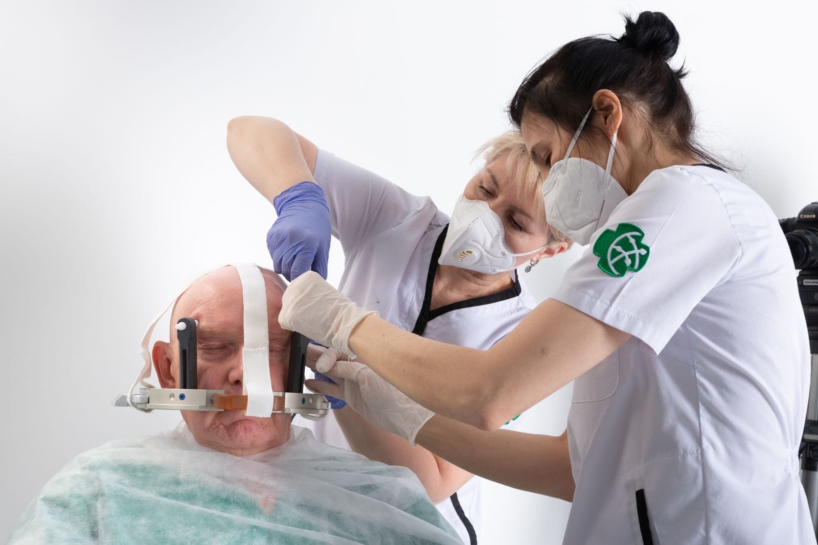The first center in Russia for non-invasive neurosurgery with MRI-guided focused ultrasound
Our engineers have created a special MRI-compatible sensor that attaches to the helmet of the MRgFUS device. This sensor can detect and record even the slightest head movements.
Instead of electronics, we use light signals transmitted through optical fibers outside the MRI room for measuring and computer visualization of head movements.
Thanks to this device, unlike other centers, we will be able to effectively treat not only hand tremor but also head tremor using MRgFUS, ensuring high treatment accuracy with an objective method of assessing tremor during the procedure.
Our device opens up new treatment possibilities for patients suffering from head tremor, providing them access to a more effective and safer treatment method.
**If you or your relative have a tremor (shaking, twitching) of the head** – contact our specialists for a consultation on treatment (adjustment of medication regimen, settings DBS-stimulator or elimination of stiffness and tremor with focused ultrasound)
**[+7 800 300-84-62](tel:+78003008462)**
**Categories:** Completed Research
---
### [What is focused shockwave therapy used for?](https://buzaevclinic.com/conditions/orthopaedics/ultrasound-wave-therapy/)
**Published:** June 8, 2023
**Author:** Aleksej Savelev
**Excerpt:** Key Advantages of This Method
- Quick and lasting treatment effect. You will forget about pain after the first procedure and enjoy life without limitations
- Does not require surgical intervention or medication. Focused shockwave therapy uses the energy of ultrasound waves and therefore has no contraindications or dangerous side effects
- Eliminates the cause of pain, not just its symptoms, ensuring a lasting positive impact on the body
- Ability to treat a wide range of conditions, including osteochondrosis, arthritis, tendinitis, muscle pain, and many others
- High success rate - over 85% of procedures successfully help relieve pain syndromes
**Content:**
# What is focused shockwave therapy used for?
Focused shockwave therapy (FSWT) is one of the few methods that allows real impact on the pathological focus, such as the source of pain. The method is a non-invasive alternative to surgical treatment for a number of conditions.
ESWT – a modern and effective method of treatment using “shock” acoustic waves in traumatology, neurology, and urology.
Low-frequency acoustic impulses **initiate restorative processes in human tissues**
In the 1990s, the technique was first applied to pseudoarthrosis (false joint), poorly healing fractures, “heel spur,” and diseases of ligaments and tendons. The treatment results caused a real sensation in the medical community. Shockwave therapy firmly entered the practice of orthopedic doctors, traumatologists, neurologists, urologists, physiotherapists, sports medicine specialists, dermatologists, and cosmetologists.
### Mechanism of Action of Shockwave Therapy
The wave itself affects soft tissues through oscillation and cavitation. This initially leads to local tissue damage, up to the destruction of the extracellular matrix at high energies (which is not always necessary), and inflammation. Then, against the background of local damage, a pronounced regenerative reaction is formed, leading to improved blood supply, including through the growth of new vessels, and the restoration of connective tissue cells, muscle fibers, and bones. This has a favorable effect on recovery from injuries, sprains, tears, and even in cases of complex bone fractures. A direct analgesic effect is observed – pain reduction occurs right during the procedure (which also depends on the intensity of the wave, which is adjusted by the doctor individually). Studies have been published showing the effect of better division and growth of stem cells during shock wave therapy.
### Indications for focused shockwave therapy
– Myofascial syndrome – acute and chronic pain in the back, neck, shoulders, thighs – Recent and old injuries to muscles, ligaments, tendons, and their consequences – Enthesopathies – diseases of tendon attachment sites – Urological disorders: Peyronie’s disease, chronic pelvic pain syndrome, erectile dysfunction – Cellulite, stretch marks, scars, ulcers, and skin burns – Weight loss **The effect of the wave is directed at breaking down fibrous formations and accelerating metabolism** . This is one of the painless and safe methods for eliminating cellulite and localized fat deposits with their natural subsequent removal from the body. New indications for shockwave therapy emerge annually, and treatment protocols are being improved.
### Key advantages of this method
– Quick and Long-lasting Treatment Effect. You will forget about pain after the first procedure and enjoy life without limitations – Does not require surgical intervention or medication. Focused shockwave therapy uses the energy of ultrasound waves and therefore has no contraindications or dangerous side effects – Eliminates the cause of pain, not just the symptoms, ensuring a lasting positive impact on the body – Ability to treat a wide range of conditions, including osteochondrosis, arthritis, tendinitis, muscle pain, and many others – High success rate – more than 85% of procedures successfully help relieve pain syndromes. We look forward to seeing you for focused shockwave therapy procedures at our clinic! #FocusedShockwaveTherapy #BuzaevClinic #MechanismOfActionFUVT #IndicationsFUVT
**Categories:** About Bone and Joint Health
---
### [Evening Ufa: Treatment of the Future. No Pain, No Blood, No Anesthesia](https://buzaevclinic.com/conditions/parkinsons-disease/treatment/terapija-budushhego-bez-boli-bez-krovi-bez-narkoza/)
**Published:** May 19, 2020
**Author:** Aleksej Savelev
**Content:**
[Original article here. ](https://vechufa.ru/medicine/16958-terapiya-buduschego-bez-boli-bez-krovi-bez-narkoza.html)
**In Russia, the first-ever unique operation was successfully performed, allowing a patient with Parkinson’s disease to be freed from the tremor that had tormented him for several years. The most joyful part of this news is that the victory over the severe complication of the disease was achieved by doctors at a clinic in Ufa** .
…Sabit Sadykovich worked as a veterinarian in the village all his life. He was highly respected by the villagers because he was a professional in his field. Not everyone would undertake, for example, a cesarean section on a cow. But he would take on any complex case.
But at some point in Sabit’s life, everything changed. Parkinson’s disease deprived him of the ability not only to work: the retiree could not even care for his animals. The tremor in his right hand increased, and there came a time when he could barely perform simple actions. It wouldn’t be long before he gave up entirely, especially since Sabit lived alone. His niece came to the rescue. She really wanted to help her uncle and scoured the entire internet. They say, those who search will always find.
And she discovered the website of a private clinic in Ufa, read it, watched videos, and realized: this is what her relative needs. To be honest, I hadn’t heard of the V.S. Buzaev International Medical Centre “Clinic of Intelligent Medicine.” And it’s no wonder, as the center opened not so long ago. A close-knit team of doctors spent two years preparing to realize their dream—to help a person with tremor, relieving them of suffering in just a few hours, painlessly, non-invasively, and without anesthesia.
On the clinic’s website, there is a video with fragments of this operation, which doctors call therapy using MRI-guided focused ultrasound. When I watched this video, I thought to myself: “These scenes would enhance any science fiction movie”! I saw—and anyone can see this—how a special helmet was placed on a patient’s head. And how his right hand was literally shaking so much that it made the heart ache with pain and compassion at the sight. How Sabit Sadykovich was placed on the table, and the MRI was turned on.
The MRI machine during therapy allows the doctor to see a real-time image of the brain, explains the clinic director, neurosurgeon, PhD Rezida Galimova, who led the operation. — The patient is placed on a special table with an integrated helmet. The helmet is unique, containing 1024 ultrasound transmitters. Doctors use a special program to determine the number of sound waves that will be directed precisely to the part of the brain responsible for the development of tremor.
According to Rezida Galimova, the therapy consists of two stages. It turns out that cells have a unique ability: when heated to 40-45°C, they lose their functional activity while remaining viable. This particularity is used by doctors in the first stage. The patient performs movement tests, and the doctor observes to ensure there are no complications or neurological “failures.” The doctor carefully conducts preliminary heating of the brain area responsible for the development of tremor. As soon as the nerve cells experience a temperature of 45°C, the patient’s tremor amplitude decreases, and the limb’s motor activity is fully restored. Only after the doctor is convinced that the impact on the brain does not cause any side effects on the nervous system does he proceed to the second stage and complete the therapy. Within 3 hours, the patient returns to the ward and can go home by evening.
Now this unique method is available to Russians, and such operations have been performed abroad for several years. Professor Shamil Makhmutovich Safin, Head of the Department of Neurosurgery and Medical Rehabilitation at Bashkir State Medical University, Head of the Department of Neurosurgery at Gumer Kuvatov City Clinical Hospital, Chief Freelance Neurosurgeon of the Ministry of Health of the Republic of Bashkortostan, observed the surgery from home. For known reasons, he was in self-isolation during those days.
— We have been nurturing this idea for about 5 years and are very pleased that it is now available to patients in Russia. The therapy is in high demand, as Parkinson’s disease and essential tremor are unfortunately not uncommon among the elderly today, and the statistics give cause for concern and the search for treatment options, says Shamil Makhmutovich.
— And here, it seems, fate happily aligned so that our young doctor Rezida Maratovna Galimova completed an internship in the USA, learning everything necessary about this non-invasive method. Investors were found, thanks to whom the clinic was opened and the latest equipment was purchased. And, as you can see, we have the first good result. It’s important to say that not all patients can receive such assistance. There is a selection system and specific conditions to ensure the procedure is effective. For example, with the first patient, the clinic corresponded for almost a year, conducting necessary studies and tests.
— The fact that we were the first in Russia to conduct such therapy is gratifying, as today the surgical treatment of tremor is a rather complex, invasive process involving the implantation of deep electrodes in the brain, explains Shamil Makhmutovich.
— A neurostimulator is implanted under the skin, it is expensive, and the batteries need to be replaced once every five years. The operation is performed under anesthesia, and the patient wakes up in the recovery room. Therefore, I consider therapy using the new, non-invasive method to be ideal. The professor expressed hope that this experience will serve as a good example for other clinics in Russia. In the future, this method could be applied in the treatment of epilepsy and brain tumors.
Research is already being conducted in this field worldwide. Recently, Sabit Aby sent a video from home to his doctor, Rezida Galimova, whom he affectionately calls “daughter.” He is happy and full of hope, planning to do some renovations. I look at her—a gentle, fragile young woman—and wonder: where does she get so much energy, how did she manage to bring such a complex project to life, and even become pioneers in this field in Russia?
— When I was a little girl, I watched a movie about space. I remembered a device that allowed people to heal without incisions and anesthesia. Thanks to the movie, I wanted to become a doctor, believing that someday we would also have such a device, says Rezida Galimova.
— I became a doctor. But the help we provided in the neurosurgery department was not at all like what I remembered from childhood. It was associated with pain, lengthy treatment, and long patient recovery. Imagine my surprise when such a device appeared two years ago — And along with this, did a strong desire to fulfill your dream appear? This is a true victory. — Something important can only be done as a team, clarifies the neurosurgeon.
— And I am grateful to everyone who works there. These are radiologists Elvira Zakirova and Dmitry Krekotkin; neurologist Olga Kachemaeva and interventional cardiologist Igor Buzaev; MRI-technician Tatyana Kaveshnikova, anesthesiologist Agash Beymirza-ogly Alyshlov, Aivar Kireev, who analyzed all the data and connected the two teams.
Professor Sergey Nikolaevich Illarioshkin, Corresponding Member of the Russian Academy of Sciences, Deputy Director for Research and Head of the Brain Research Department at the Scientific Center of Neurology, President of the National Society for the Study of Parkinson’s Disease and Movement Disorders from Moscow, and our esteemed Professor Shamil Makhmutovich Safin joined the treatment online. In the video recording of the surgery, the doctors were speaking in English…
— We were supposed to perform the first operation under the careful online guidance of the company’s staff Insightec — Paul Wragg from the United Kingdom, Tessa Case from Spain and Daniel Molchanov from Russia. This is the company from which we purchased the equipment, clarifies Rezida Maratovna
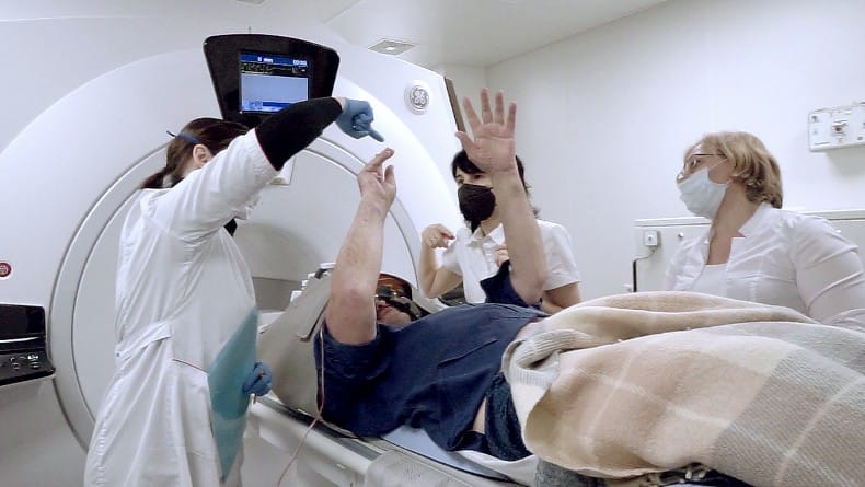Photo from the first neurosurgical operation in Russia using focused ultrasound — But the situation in the world related to the epidemic did not allow them to come here. Therefore, our team first received permission to carry out the therapy after an online course, and then during the procedure, we were supervised — also online — by company representatives who gave a good evaluation. …And we hope that the result of this operation in the republic will please everyone, and perhaps this event will be the first step towards implementing therapy in other medical institutions of the republic, allocating quotas for operations. Especially since, as we see today, the country’s leadership is showing increased care for the elderly and people who are of working age but, for certain reasons, cannot return to professional activities.
**Tatiana BARABASH IN THE IMAGES: surgery in progress.**
**Photos provided by the “Intelligent neurosurgery clinic, Ltd”.**
**Source link.**
**Categories:** About the Treatment of Parkinson's Disease
**Tags:** We in the Media
---
### [Конгресс по бескровной нейрохирургии с использованием фокусированного ультразвука – 2021](https://buzaevclinic.com/education/healthcare-professionals/events/funnc-ufa-2021/)
**Published:** May 5, 2021
**Author:** Aleksej Savelev
**Content:**
## **F**ocused **U**ltrasound **N**on-invasive **N**eurosurgery **C**ongress
### Meet some breakthroughs in incisionless treatment of neurological disorders
- [WHAT WE CAN PROVIDE](#aioseo-what-we-can-provide)
- [WHO ATTENDS FUNNC](#aioseo-who-attends-funnc)
- [SPEAKERS](#aioseo-speakers)
- [Rezida Galimova](#aioseo-rezida-galimova)
- [Shamil Safin](#aioseo-shamil-safin)
- [Valentin Pavlov](#aioseo-valentin-pavlov)
- [Roni Yagel](#aioseo-roni-yagel)
- [Takaomi Taira](#aioseo-takaomi-taira)
- [DIPANKAR NANDI](#aioseo-dipankar-nandi)
- [Wei Chien, Chang, M.D.](#aioseo-wei-chien-chang-m-d)
- [Rick Schallhorn](#aioseo-rick-schallhorn)
- [Lain Hermes Gonzalez Quarante](#aioseo-lain-hermes-gonzalez-quarante)
- [Igor Buzaev](#aioseo-igor-buzaev)
- [INFORMATIONAL PARTNERS](#aioseo-informational-partners)
- [Insightec](#aioseo-insightec)
- [GE](#aioseo-ge)
- [IMCB](#aioseo-imcb)
- [Academic partners](#aioseo-academic-partners)
- [Bashkir State Medical University](#aioseo-bashkir-state-medical-university)
- [Watch the Congress](#aioseo-watch-the-congress)
- [CONTACT US](#aioseo-contact-us)
- [Address](#aioseo-address)
- [Contact](#aioseo-contact)
## WHAT WE CAN PROVIDE
- Review the latest investigations to guide how to manage optimally the patients with tremor.
- Integrate the newest techniques and devices for MRI guided focused ultrasound treatment.
- Compare and contrast different approaches to the hot debates among masters of MRGFUS.
- Learn numerous treatment strategies through reviewing diverse challenging cases submitted from all around the world and discussing with world-renowned experts.
## WHO ATTENDS FUNNC
- Neurosurgeons
- Neurologists
- Radiologists
- Medical students
- Nurses and technologists
- Other health care professionals with special interest in non-invasive neurosurgery
## SPEAKERS
### Rezida Galimova
MD, PHD,Neurosurgeon
Welcome Message
### Shamil Safin
Head of the Neurosurgery and medical rehabilitation dep. with the courses of neurosurgery, BSMU (Russia)
Welcome Message
### Valentin Pavlov
The Rector of Bashkir state medical university (Russia)
Welcome Message
### Roni Yagel
Vice President for Int. Markets, Insightec
Welcome Message
### Takaomi Taira
Tokio womens’s medical university (Japan)
MRgFUS treatment of distonia
### DIPANKAR NANDI
MBBS (AIIMS), M.CH., D.PHIL. (PEMBROKE, OXON), FRCS (SN), Consultant Neurosurgeon and Head of Dep., Charing Cross Hospital and St. Mary’s Hospital (London, UK)
Wider application of MRgFUS
### Wei Chien, Chang, M.D.
Chairman at the Dep. of neurosurgery at the show Chwan memorial hospital, Chang Hwa County, Taiwan
Taiwan Show Chwan Memorial Hospital MRgFUS experience
### Rick Schallhorn
Vice President Neurosurgery, Insightec
Breakthrough technology in neurosurgery
### Lain Hermes Gonzalez Quarante
MD, Neurosurgeon, University clinic of Navarra (Spain)
Welcome Message
### Igor Buzaev
Co-Founder of the Int. Medical centre V.S. Buzaev Memorial, DSCI in Medicine, MD, PHD, Interventional Radiologist
The role of digital technologies in the implementation of complicated medical procedures in COVID-19 era
## INFORMATIONAL PARTNERS
The conference organizers gratefully acknowledge the generous informational non-financial support provided by the following:
### Insightec
INCISIONLESS SURGERY Using sound waves to treat instead of a scalpel.
### GE
A healthier world with more precise and efficient care.
### IMCB
We care patients with tremor from any part of the world using MRgFUS.
## Academic partners
Our academic partners are:
### Bashkir State Medical Universityversity
### Bashkir State Medical University
Ufa, Russia
## Watch the Congress
## CONTACT US
### Need help, want to be a speaker or sponsor?
## Address
17/4, Richard Zorge street
Republic of Bashkortostan, Russia.
## Contact
Call : [+7 800-300-84-62](call:+78003008462)
Email : <help@tremor.care>
© Copyright International Medical Centre VS Buzaev memorial, 2021
**Categories:** Events
**Tags:** Past Training Events , Video
---
### [Seminar with Schoolchildren of the Demsky District](https://buzaevclinic.com/collaborations/mrgfus-for-schoolchildren/)
**Published:** February 6, 2024
**Author:** Aleksej Savelev
**Content:**
5 In February, a seminar was held with 10th-11th grade students from general education organizations in the Dema district of Ufa, Republic of Bashkortostan, at the V.S. Buzaev International Medical Centre®. The Deputy General Director, PhD Alexey Borisovich Saveliev, spoke about the activities of the first neurosurgical center in Russia, where MRI-guided focused ultrasound is used to eliminate symptoms of severe movement disorders, and the unusual story of its opening. The presentation sparked lively interest among the students.
## Student and Teacher Reviews
> «I really enjoyed the Buzaev Centre. This tour of the Intelligent Neurosurgery Centre motivated me to achieve new accomplishments in my studies, increased my interest in the medical profession, and made me consider choosing a similar direction in my education.
> «5 In February 2024, high school students from the Demsky district visited the V.S. Buzaev International Medical Centre in Ufa. The clinic’s specialists conducted a tour and informed the students about the treatment of neurosurgical diseases using ultrasound waves. The speaker’s presentation was very informative, highly professional, and sparked genuine interest among the students, many of whom plan to apply to medical universities. This experience helped the students broaden their horizons in the field of medicine.
**Categories:** Collaboration
**Tags:** Enlightenment
---
### [Next-generation MRI machine GE Optima MR450w GEM](https://buzaevclinic.com/services/expert-mri-imaging/ge-optima-450/)
**Published:** August 4, 2020
**Author:** Aleksej Savelev
**Content:**
## The symbol of caring hands inspired engineers GE create Optima MR450w GEM – the most patient-centered system
Every detail of your tomograph embodies the balance of technology, design, and patient care, combining expert-level software with a wide 70 cm aperture, soft and flexible coils, a completely new table surface with varying firmness, and advanced LED lighting that our patients deserve.
MRI coils – the same as camera lenses. They help focus MRI energy into clear images when examining patients. However, no two patients are alike, and the traditional coil design may sometimes functionally outperform at the expense of patient comfort. Lack of comfort can lead to patient movement, which may cause poor image quality and, consequently, additional time spent on repeat examinations.
Technology Geometry Embracing Method (GEM) Suite designed specifically to provide a new level of comfort for patients, minimizing anxiety and movement during examinations. The “embracing” design of flexible coils allows for scanning in a relaxed setting for the patient. Thanks to these changes, all examinations can now be conducted comfortably, with the patient positioned feet-first into the tunnel using lightweight flexible coils and a completely new table cover that evenly distributes pressure.
Patient comfort during MRI examination is not only about using a wide aperture and comfortable positioning. Patients often ask if it is possible to make an MRI examination quiet? Acoustic noise reduction technology ART allows achieving this. The application is an integral part of brain, spine, and musculoskeletal system research. Meanwhile, the quality of the obtained images remains uncompromisingly high, as the technology ART reduces acoustic noise by optimizing the gradient system
Patients often ask if it’s possible to obtain images with the quality of contrast imaging without using a contrast agent? Using DeltaFlow — one of the many functions of the system Optima MR450w GEM — You can! Now patients can be examined without the use of contrast agents and without the fear of adverse reactions to contrast medications. This is a mutual victory for us and for the patients!
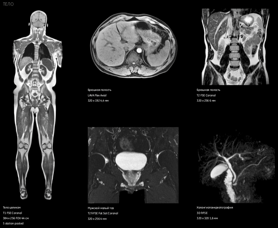MRI images of the human body Readiness for the future means having a system that not only goes beyond the original design but also far exceeds it. System Optima MR450w GEM developed with the expectation that it will surpass the traditional boundaries of radiology and become more than just a regular MRI!
We carefully selected the MRI machine, prioritizing the needs of patients, and once we received it, we did not start using it until we had studied and mastered many programs.
Today we are delighted to announce that we have launched our unique MRI and are ready to assist patients at the highest level! Our wonderful MRI doctors will gladly answer your questions and help you choose the type of examination. You can message them or call:
## MRI OF EXPERT CLASS
### UNIQUE PROTOCOLS
- Digital Radiofrequency System OpTix — Signal digitization in the MRI room to improve image quality
- Useful field of view 50 cm — for accommodating larger patients.
- High-precision gradients — optimized gradient performance management to ensure outstanding performance in demanding applications such as fMRI, cardiology, diffusion and FIESTA
- Acoustic Suppression Technology ART) — reduces acoustic noise during brain and spine examinations without compromising image quality
### EXPERT MRI SCANNER, ALLOWING YOU TO SEE MORE GE OPTIMA 450W GEM
1. Extended Protocol Set for Nervous System Research
2. Excellent results with a power of only 1.5 Tesla
3. New Opportunities (Spectrography, Perfusion, New Noise Reduction Mechanisms and Artifact Elimination)
4. High maximum load capacity on the MRI table (up to 130 kg)
5. The widest aperture among all closed-type tomographs for your comfort
Schedule an examination
MRI GE Optima 450 magic
**Categories:** MRI diagnostics of diseases
**Tags:** Video
---
### [Teregulova Dinara Ravilievna](https://buzaevclinic.com/team/doctors/dinara-teregulova-neurologist/)
**Published:** August 4, 2021
**Author:** Guzel Ziyazetdinova
**Excerpt:** Neurologist, cephalologist (headache treatment specialist), PhD in Medical Sciences
**Content:**
## Neurologist, Cephalologist (Headache Specialist), PhD
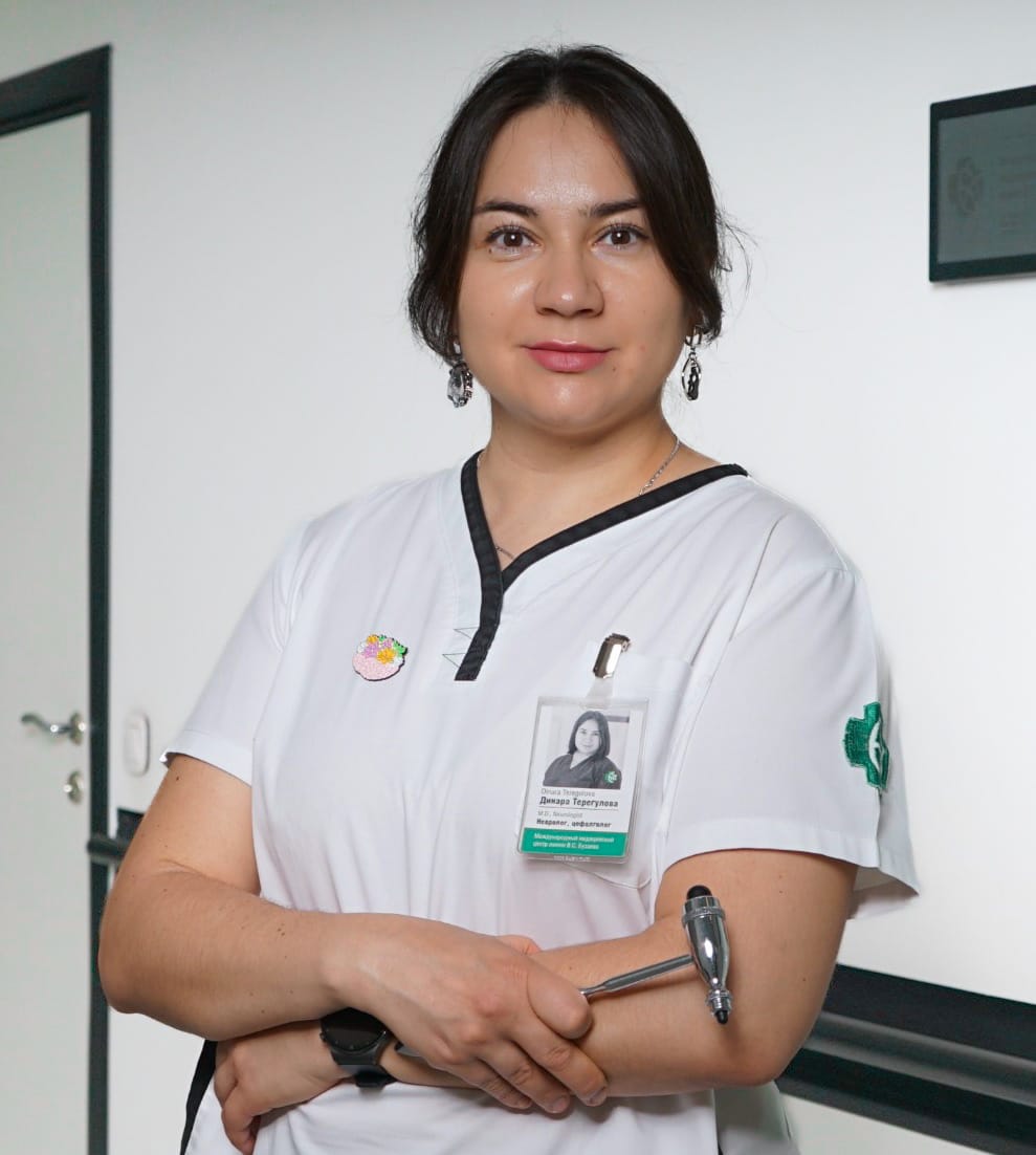## Education
- 2021 EHF-SAS 12th Meeting (School European Headache Federation, Rome, Italy)
- 2021 Neurological Aspects of Coronavirus Infection C OVID-19 (Department of Neurology and Neurosurgery, Institute of Postgraduate Education, BSMU, Ufa
- 2021 Diagnosis and Treatment of Orofacial Pain, Moscow
- 2020 Migraine Academy 2.0, Saint Petersburg
- 2020 Headache: School of Practical Mastery, Moscow
- 2019 Current Issues in Pediatric Neurology, Department of Neurology and Neurosurgery, Institute of Postgraduate Education, BSMU, Ufa
- 2018 Migraine Academy, Ufa
- 2018 Fundamentals of Palliative Care, Department of Neurology and Neurosurgery, Institute of Postgraduate Education, BSMU, Ufa.
- 2017 New technologies for the diagnosis and treatment of stroke, Department of Neurology and Neurosurgery, Institute of Continuing Professional Education, Ufa
- 2016 Vegetative Pathology and Vascular Diseases of the Nervous System
- 2015 – 2021 Postgraduate Studies at the Department of Neurology, BSMU
- 2011 Regional Training Course EFNS (European Federation of Neurological Societies, Yekaterinburg
- 2010 – 2011 – neurology internship, Department of Neurology with courses in Neurosurgery and Medical Genetics at BSMU
- 2004 – 2010 Bashkir State Medical University, Faculty of Medicine (diploma with honors)
## Work Experience
2020 – 2021 Intelligent neurosurgery clinic, Ltd, Ufa
2018 – 2020 Clinic MD Plus, Ufa
2011 – 2018 Clinic of MBUZ GKB No. 5, Ufa
## Clinical Research
2015 – 2016 Co-investigator in the clinical trial protocol for migraine No. 20120297 ( III phase of clinical trial of erenumab)
## Publications
Author of 14 publications in scientific journals, co-author 1 monographs
## Participation in Conferences, Presentations
- All-Russian (with international participation) Conference of Young Scientists “The Future of Neurology”, Kazan, February 2019
- XXV Russian Scientific and Practical Conference with International Participation: Pain Medicine: From Understanding to Action, Kazan, May 2019
- V-European Academy of Neurology Congress, Oslo, June 2019
- XVI Russian Conference with International Participation “Malignant Lymphomas”, Moscow, October 2019
- XXIV World Congress of Neurologists, Dubai, October 2019
- International Behavioral Neurology – Videoconference, Toronto, March 2020
- Interregional Round Table: expert council on diseases of the blood and hematopoietic organs to improve patient detection and timely initiation of therapy in general practitioner practice, Kazan, April 2021
## Professional Interests
- Head and facial pain, pain syndromes, anxiety-depressive disorders, vascular diseases of the nervous system
- Botulinum Therapy for Migraine and Facial Pain
- Neurological Manifestations of Chronic Lymphoproliferative Disorders (within the framework of the dissertation work)
**Categories:** Doctors
**Tags:** "TMJ School Team" , Head and Facial Pain Center Team , MRgFUS team , PhD , Researchers , Team "Nervous System"
---
### [Titremelerini durdur!](https://buzaevclinic.com/global/titremelerini-durdur/)
**Published:** February 25, 2024
**Author:** Aleksej Savelev
**Content:**
Siz veya sevdikleriniz Parkinson hastalığından mı muzdarip ve güvenilir, modern tedavi yöntemleri mi arıyorsunuz? Vyacheslav Stepanovich Buzaev Uluslararası Tıp Merkezi, Parkinson hastalığında titreme ve sertliği, esansiyel tremor, distoni tedavisinde MRI kontrollü odaklanmış ultrason tedavisi gibi benzersiz bir fırsat sunuyor. Bu yenilikçi yöntem, ameliyatları kesiksiz ve narkozsuz gerçekleştirmeye olanak tanır, bu da hasta için maksimum güvenlik ve konfor sağlar. Yaklaşımımız sayesinde, yalnızca hastalığın semptomlarını azaltmakla kalmaz, aynı zamanda ilaç dozunu da düşürebilir, bu da yaşam kalitenizi önemli ölçüde iyileştirir.
Uluslararası merkezimiz, bu ileri teknolojiyi Rusya’da ilk uygulayan kurum olmaktan gurur duyar ve size medikal turizmin avantajlarından yararlanma fırsatı sunar. Tedavi sonrası 12 ay boyunca ücretsiz takip dahil olmak üzere kapsamlı bir hizmet paketi sunuyoruz. Hareketin sevincini ve geleceğe olan güveninizi nasıl geri kazanmanıza yardımcı olabileceğimizi öğrenin. Buzaev Uluslararası Tıp Merkezi’nde medikal turizm hakkında daha fazla bilgi edinin.
Uluslararası Tıp Merkezi
V.S. Buzaev Anısına
### Dünyanın her bir bölgesinden gelecek hastalarımızın modern ve FDA onaylı MRI destekli non-invasiv ultrason yöntemi (MRgFUS) ile belirli-belirsiz ve Parkinson hastalığına bağlı titremelerini tedavi ediyoruz.
## Tek uygulama, kesi yok,
yerleştirme yok ve daha fazla titreme yok
Modern CE ve FDA onaylı non-invaziv MRG güdümlü odaklı ultrason (MRGFUS) talamotomisini kullanarak dünyanın her yerindeki esansiyel ve Parkinson hastalığına bağlı titremesi olan hastaları teadavi ediyoruz. Tedavi genellikle taraf başına sadece bir uygulama gerektirir ve doğrulama aşamasında iyi kontrol edilir. Yerleştirilebilir cihazlar ya da ameliyat gerektirmez ve genellikle ağrısızdır.
Rusya’da ilk nöro-MRGFUS merkeziyiz ve medikal turizmi yüksek hizmet kalitesiyle sunuyoruz. Biz son derece sorumluluk sahibi doktorlarız ve 12 aylık takiple garanti sözü veriyoruz. Mükemmel sonuçlarla ilgileniyoruz ve bir yıl içinde titremenin dönüşü durumunda süreci tekrarlamak için elimizden gelenin en iyisini yapacağız.\[gap\] \[/section\] \[section\]
## Evrimin, sadakatin, cesaretin ve onurun hikayesi
Uzun yıllar boyunca, içindeki bir şeyi düzeltmek için insanlar insanları kestiler, organların ve dokuların, tedavi edilmesi gereken yerin derinliklerine girdiler. Buna ameliyat deniyordu. Sonra non- invaziv cerrahi geldi. Vyacheslav Buzaev, bölgemize non-invaziv kalp cerrahisi – girişimsel kardiyoloji – getiren kalp cerrahlarından biriydi.
Onun sayesinde Cumhuriyetimizde PCIs ve diğer kardiyak müdahaleler yapılmaktadır. Rusya’da cerrahiyi yeni bir seviyeye, kesilerin gerekli olmadığı bir seviyeye taşımaktan onur duyuyoruz ve çalışmalarımız son derece öngörülebilir ve kontrol edilebilir. Tıbbi bakımdaki yeni aşamamızı Vyacheslav Buzaev’in onuruna adlandırdık.
Bir beyin cerrahı olan merkezin müdürü Rezida Galimova, her zaman beynin derinliklerine açık yaralarla ulaşmanın ve ona zarar vermenin yanlış olduğunu düşünmüştü. Hayali gerçek oldu ve şimdi beyni kesiler olmadan tedavi etmek mümkün. MRGFUS teknolojisi, ilk başta geri alınabilir şekilde hareket etmenizi ve sonucu düzeltmek için doğru eylemlerden emin olmanızı sağlar.
## Doktor

## Rezida Galimova
Beyin Cerrahı
####
## Ziyaretinizi sağlığa keyifli ve verimli bir yolculuk yapmak için elimizden geleni yapıyoruz.
## İlk Danışma
İstenen tıbbi kayıtları bize gönderirsiniz. Görüntülü görüşmede internette buluşuyoruz, sonra ikimiz de tedavinin olasılığı hakkında ön kararlar alıyoruz.
## Hazırlık
Tedavi tarihini siz seçersiniz. Biz size vizeyi alabilmeniz için gereken davet mektubunu göndeririz ve size uçak biletini ayarlamanız için yardım ederiz. Rusya’ya olan seyahatiniz hakkında tavsiyede bulunuruz. Hilton hotelinde kalmanız için bir oda ayırtırız. (Ufa, Russia).
## Gezi
Ufa’ya seyahatiniz ve geri dönüşünüz sırasında 7/24 telefonda uygunuz. Sizinle Ufa havaalanında buluşup Hilton Hotel’e transfer oluyoruz.
## Tedavi
Sizi otelden kliniğimize transfer ediyoruz. Klinikte, genellikle ilk gün, nörolog (120 dk), beyin cerrahı ve kardiyolog ile bir toplantınız vardır. Hesaplamaları yapar ve işlemi planlarız. İkinci gün, genellikle MRGFUS sistemini uygularız. Anatomiye ve duruma bağlı olarak 2-5 saat sürer.
### Ulaşım ve Konaklama
Size yemek sağlıyoruz. Tedaviden sonra ertesi gün takibiniz yapılır ve genellikle eve dönebilirsiniz. Sizi havaalanına transfer ediyoruz. Harika bölgemizi görmek için size isteğe bağlı turlar ayarlayabiliriz.
### Geri döndükten sonra
En az 12 ay boyunca çevrimiçi takipleriniz dahildir. Yöntem etkilidir ve titremenin geri gelme olasılığı azdır. Titreme 12 ay içinde tekrarlanırsa, güvenliği ve durumu göz önünde bulundurarak tedaviyi tekrarlamak için elimizden gelenin en iyisini yapacağız.
## Fiyatlandırma
## Videolar
### Yöneticimizin hikayesi:
Tremor hastası sanatçının sekiz yaşındaki kızı bir bilim kurgu videosu izledi ve hayalini gerçekleştirdi.
### Tıbbın geleceği burada başlıyor
Kullandığımız INSIGHTEC MRGFUS teknolojisi hakkında.
### İşte böyle başladık.
Rusya’da MRGFUS talamotomisini ilk yaptığımızdan beri gün be gün, süreçten sürece daha iyiye gidiyoruz.
## İletişim
Şirketimiz ve tedavi süreçlerimizle ilgili sorularınız için.
#### İletişim
Çağrı: [0554 708 90 31](tel:05547089031%E2%80%AC)
Email: <turkiye@buzaevclinic.ru>
#### Adres
17/4, Richarda Zorge street
Ufa, Republic of Bashkortostan
[Posta Gönder](mailto:raydinoglu@gmail.com)
[Klinik öncesi değerlendirme randevusu alın](https://tremor.care/tr/services/preclinical-assessment/)
**Categories:** Global medical tourism
**Tags:** Video
---
### [Igor Vyacheslavovich Buzaev](https://buzaevclinic.com/team/doctors/igor-buzaev/)
**Published:** January 31, 2019
**Author:** Guzel Ziyazetdinova
**Excerpt:** Co-founder of the Center, Doctor of Medical Sciences
**Content:**
## Co-founder of the Center, Interventional Radiologist, Doctor of Medical Sciences
Website: <https://buzaev.com>
Contact me at WhatsApp
Doctor, professor at BSMU, researcher and innovator in the field of medicine. I am building a world where people can easily maintain a healthy and happy life, and those who need help receive it painlessly, non-invasively, humanely, and without a single mistake.
Co-founder and Business Development Director of the first non-invasive neurosurgical MRgFUS center in the countries of the former Eastern Bloc — [V.S. Buzaev International Medical Centre](https://buzaevclinic.com/). Upon learning about the new technology that allows for predictable surgeries on deep brain structures without incisions, my [spouse](https://buzaevclinic.com/team/doctors/rezida-galimova/) and I decided that we should be the first to implement it in our country. We developed a business plan, found an investor, assembled an incredible [team](https://buzaevclinic.com/team/), launched the project under [epidemic restrictions](https://pmc.ncbi.nlm.nih.gov/articles/PMC10914008/), and promoted this treatment method amidst complete patient unawareness and physician conservatism.
From 2012 to 2021, I was the chief specialist in interventional radiology diagnostics and treatment, assigned by the Ministry of Health of the Republic of Bashkortostan, and carried out tasks to develop this service in a region with a population of over 4 million people. During this time, we achieved two-hour accessibility
primary percutaneous coronary intervention for myocardial infarction throughout the Republic
Previously, for 11 years, I held the position of head of the first interventional radiology department at the [Republican Cardiology Center](https://bashcardio.ru), where my responsibilities included providing department leadership and strategy development, as well as mastering, implementing, and performing new operations with further training of doctors, and overseeing the implementation of innovative methods in the Republic of Bashkortostan. During my leadership, the department treated more than 11 000 patients and held [high rankings](https://buzaev.com/downloads/2020-endovascular-report-Russia.pdf) among other leading hospitals in Russia.
As a [professor at Bashkir State Medical University](https://bashgmu.ru/), I have extensive experience in lecturing and conducting seminars, regularly speaking at international congresses and on national television news programs as an expert. I have completed numerous international internships and have [research publications in reputable international journals](https://orcid.org/0000-0003-0511-9345).
### Basic Education
- 2023 – present. [UCL Global School of Business for Health](https://www.ucl.ac.uk/population-health-sciences/global-business-school-health) doctorate student
- 2022 – Anaheim University (Master of Fine Art, with honors)
- 2018 – Order of the Ministry of Science and Higher Education of the Russian Federation dated August 10, 2018 No. 110/nk on the issuance of a Doctor of Medical Sciences diploma
- 2008 – Bashkir Academy of Public Administration and Management under the President of the Republic of Bashkortostan, specialty in jurisprudence, qualification – lawyer, diploma with honors
- 2007 – Ministry of Education and Science of the Russian Federation (defense at the Meshalkin National Medical Research Center, Novosibirsk), 14.00.44 – cardiovascular surgery, PhD diploma
- 2001 – Bashkir State Medical University (BSMU), diploma with honors
### Postgraduate Education
- Healthcare Organization and Public Health. Primary retraining in the specialty.
- Functional Diagnostics. Certification Course in Specialty (2018)
- Management and Strategy Institute Six Sigma Black Belt Professional (2018)
- Interventional Radiology (Endovascular Diagnostics and Treatment). Certification Course in Specialty (2017)
- Functional Diagnostics. Certification cycle of professional retraining in the specialty (2013)
- Cardiovascular Surgery. FSBEI CPE “Russian Medical Academy of Postgraduate Education of the Ministry of Health of the Russian Federation”, advanced training (2013)
- Interventional radiology (endovascular diagnostics and treatment). I.M. Sechenov First Moscow State Medical University of the Ministry of Health and Social Development of the Russian Federation. Specialty certificate (2012)
- Novosibirsk State Academy, specialty: cardiovascular surgeon, professional retraining diploma (2006)
- Novosibirsk State Academy, specialty: cardiologist, certificate (2005)
- BSMU, clinical residency, specialty: surgeon, certificate (2002 – 2004)
- BSMU, internship, specialty: pediatric surgeon, certificate (2001 – 2002)
### Courses and Studies
**Foreign**
- 2005 Netherlands, Arnhem, 3 days. Educational program for doctors “Optimal Cardiac Pacing”, Vitatron
- 2005 Sweden, Stockholm, 3 days Congress ESC, Stockholm, Sweden, European Society of Cardiology ( ESC)
- 2006 Spain, Barcelona, 3 days World Congress of Cardiology, European Society of Cardiology ESC)
- 2008 Denmark, Copenhagen, 3 days Meeting of the Society of Cardiovascular and Interventional Radiologists in Copenhagen, Denmark, Society of Cardiovascular and Interventional Radiologists
- 2008 United Kingdom, London, 3 days Charing Cross Symposium, London, UK. Biba Conferences, 44 Burlington Road, Fulham, London, SW6 4NX
- 2009 Spain, Seville, 1 month Monthly internship at the Virgen Macarena Clinic
- 2009 USA, New York, 2 a month of observational internship at Montefiore Children’s Hospital, New York (Robert Pass), Columbia University Children’s Clinic (Julie Vincent), Cornell Presbyterian Hospital (Ajay Mirani)
- 2012 USA, Los Angeles 3 31st International Conference and Exhibition IHRSA
- 2012 Poland, Poznan, 2 weeks Boston Scientific 2-weekly training in interventional cardiology for experienced doctors
- 2012 Germany, Cologne, 2 Two-week internship in hybrid surgery at the Cardiology Center of the University Hospital Cologne Uniklinik Köln Klinik und Poliklinik für Gefäßchirurgie Kerpener Str. 62, 50937 Кёльн)
- 2012 USA Miami, 3 дня TCT
- 2013 Netherlands, Rotterdam, 2 дня Bioresorbable Vascular Scaffolds and Vascular Reparative Therapy (Focus Group PCR in Rotterdam)
- 2014 Germany, Frankfurt, 3 days Vascular Skills ( CardioSkills)
- 2014 USA, San Diego, 3 дня IHRSA (San Diego)
- 2015 Korea, Seoul, 4 8th Summit on Imaging Physiology
- 2016 Czech Republic, Prague, 2 6th International Physician Exchange Program in Therapy and Technology
- 2016 France, Paris, 2 Day Valve Implantation Training Boston Scientific Lotus Valve
- 2016 Korea, Seoul, 4 days TCT-AP
- 2017 Belgium, Brussels, 2 OCT day in case of stent rejection and complex PCI
- 2017 Czech Republic, Prague, 3 7th International Physician Exchange Program in Therapy and Technology
- 2017 Korea, Seoul, 4 days TCT-AP
- 2018 USA, online, 2 weeks LSS black belt «”Six Sigma”
- 2019 Czech Republic, Prague, 1 Day Seminar RDN in Prague (02/26/2019) Medtronic
- 2019 Korea, Seoul, 4 days TCT-AP
- 2019 Korea, Seoul, 1 internship day MRgFUS (Professor Jin Woo Chang)
- 2020 Korea, Seoul (online), 4 days TCT-AP
- 2021 Korea, Seoul (online) 4 days, TCT-AP
- 2022 Korea, Seoul (online), 4 days TCT-AP
### Work record book
- 10.2021 – currently, co-founder at V.S. Buzaev International Medical Centre
- 04.2012 – 05.2021, Chief Freelance Specialist in Interventional Radiology (Xray Diagnostics and Treatment) of the Ministry of Health of the Republic of Bashkortostan
- 01.2010 – 09.2021, Republican Cardiology Center (formerly dispensary) (Ufa), Head of Department of Interventional Radiology and Diagnostics No. 1
- 08.2004 – 01.2010, Republican Cardiology Dispensary (Ufa), cardiovascular surgeon
- 09.2002 – 08.2004, Bashkir State Medical University, clinical resident
- 09.2001 – 08.2002, Bashkir State Medical University, medical intern
### Awards
- Certificate of Honor from the Government of the Republic of Bashkortostan (06/09/2017)
- Magazine “Sobaka”. TOP30. The Most Famous People of Ufa. Nomination “Research” (2015)
- Gratitude from the Federation Council Committee on Social Policy (14.11.2013)
- Awarded the badge “Excellence in Healthcare of the Republic of Bashkortostan” (29.08.2011)
- Nominee for the National Cardiology Award “Purple Heart” (19.05.2011)
- Joining the Working Group EAPCI European Society of Cardiology ESC)
**Categories:** Doctors
**Tags:** Doctors of Science , Instructors , Leadership , MRgFUS team , Pediatrics and Vaccination Team , Researchers , Team "Heart and Vessels"
---
### [Podcast about Parkinson's Disease: Modern Approaches](https://buzaevclinic.com/conditions/parkinsons-disease/lifestyle-lifehacks/thriving-tips-latest-breakthroughs/)
**Published:** April 18, 2025
**Author:** Ekaterina Nadezhdina
**Excerpt:** Parkinson's Disease: Modern Treatment Methods, the Role of Family, Innovations in Surgery, and Genetic Research for Improving Quality of Life. "Odyssey of Healing"
**Categories:** Lifestyle and Tips for Parkinson's Disease
**Tags:** Podcast Parkinson's Disease , Video
---
### [Preparation for MRI](https://buzaevclinic.com/services/expert-mri-imaging/get-ready-to-mri/)
**Published:** October 17, 2022
**Author:** Aleksej Savelev
**Content:**
## ATTENTION!
If you are undergoing an MRI not for the first time, especially in diagnostically complex cases, **bring the results of previous studies with you.** This will help our specialists select the most appropriate MRI protocols and examine the problematic area in maximum detail.
These recommendations apply only to procedures **at our centre**.
Requirements in **other countries and medical institutions may be different!** Therefore, please check the recommendations at the centre and with the doctor where you are receiving treatment.
### 
## How to prepare for an MRI of the kidneys and adrenal glands in our centre?
For 2-3 days, exclude gas-producing foods from your diet (legumes, black bread, carbonated drinks, fruits, vegetables). Refrain from eating 2-3 hours before the examination if the use of a contrast agent is planned.
## How to prepare for an MRI of the abdominal organs, MRI of the bile ducts (MRCP, MR cholangiopancreatography) in our centre?
**MR cholangiography** – Non-invasive examination of the pancreatic duct of the pancreas and bile ducts (intrahepatic and extrahepatic ducts), allowing detailed study of the hepatobiliary system (HBS) and pancreas. The use of contrast material facilitates the diagnosis of functional disorders of the examined organs. Magnetic resonance cholangiography is prescribed for suspected cancer, formation of cysts, stones, or ductal developmental anomalies.
• For 3 days before the examination, exclude vegetables, legumes, fruits, dairy products, carbonated drinks, black bread, pasta, confectionery, and coffee from your diet.
You can consume porridge, broth, boiled meat, fish, chicken, cheese, butter.
• For 3 days before the examination, you need to take “Espumizan” (1 capsule 3 times a day)
• 3 hours before the examination, you need to take 3 tablets of “No-shpa” 40 mg each. If “No-shpa” is not tolerated, take 2 tablets of “Duspatalin” or “Buscopan”.
• The procedure is conducted strictly on an empty stomach or no earlier than 6 hours after eating. • Refrain from consuming any liquids, smoking, and chewing gum for 3 hours before the examination.
• Empty the bladder before the examination.
### 
## How to prepare for an MRI of the pelvic organs or rectum in our centre?
- After a colonoscopy, an MRI of the rectum is scheduled in 5-7 days.
- Three days before the examination, exclude vegetables, fruits, dairy products, carbonated drinks, and bread from your diet. You may consume porridge, broth, boiled meat, fish, chicken, cheese, butter, and cookies.
- For 3 days before the examination, take “Espumizan” tablets (1 capsule 3 times a day).
- Bowel preparation: 1 day before the examination, gentle micro enemas (Microlax) should be used. If a colostomy is present, cleansing enemas are used to clean the disconnected section of the intestine. The enema nozzle should be placed at the opening of the disconnected section, and after bowel movement, a cleansing enema should be administered into the rectum until the water is clear. You must bring a spare ostomy bag.
- 3 hours before the examination, take 3 tablets of “No-Spa” 40 mg each. If “No-Spa” is not tolerated, take 2 tablets of “Duspatalin” or “Buscopan”. Before the examination, empty the bladder.
### 
## How to prepare for an MRI of the bladderin our centre?
- After a cystoscopy, an MRI of the bladder is scheduled in 5-7 days.
- The last meal should be 2-3 hours before the examination.
- 3 hours before the examination, take 3 tablets of “No-Spa” 40 mg each. If “No-Spa” is not tolerated, take 2 tablets of “Duspatalin” or “Buscopan”.
- Collect urine: 1 hour before the examination, drink 400-500 ml of non-carbonated drinking water.
-
## How to prepare for an MRI of the pelvic organs (prostate, uterus, and ovaries) and MR-phlebography (MRI of the pelvic veins) in our centre?
- performed on the 7th-12th day of the menstrual cycle, strictly after menstruation. If necessary, the study can be conducted in the second phase of the cycle.
- possible only 1.5 months after diagnostic curettage of the uterine cavity and cervical canal, otherwise — in consultation with the attending physician. MRI of the prostate gland
- possible only 1.5 months after needle biopsy, otherwise — in consultation with the attending physician. General rules:
- 3 days before the examination, exclude vegetables, fruits, dairy products, carbonated drinks, and bread from your diet. You can consume porridge, broth, boiled meat, fish, chicken, cheese, butter, and cookies.
- For 3 days before the examination, take “Espumizan” tablets (1 capsule 3 times a day).
- Last meal 2-3 hours before the examination.
- 3 hours before the examination, take 3 tablets of “No-Spa” 40 mg each. If “No-Spa” is not tolerated, take 2 tablets of “Duspatalin” or “Buscopan”. • Before the examination, you need to empty your bladder.
**Categories:** MRI diagnostics of diseases
**Tags:** Preparation for Procedures
---
### [FUNNC-2022: School-Conference on Treating Tremor with Ultrasound](https://buzaevclinic.com/education/healthcare-professionals/events/funnc-ufa-2022/)
**Published:** September 21, 2022
**Author:** Aleksej Savelev
**Content:**
## First Scientific and Practical School “Focused Ultrasound and Non-Invasive Neurosurgery “
School-Conference on Non-Invasive Treatment of Neurological Extrapyramidal Movement Disorders ( FUNNC) was conducted [ online in 2021](https://funnc.org/2021/index.html#about-us) and in 2022 at the V.S. Buzaev International Medical Centre
## Our Mission
- We study and implement new non-invasive neurosurgical technologies
- We educate doctors about new treatments for extrapyramidal movement disorders
- Uniting like-minded individuals and supporters of non-invasive neurosurgery
- Learning New Strategies for Treating Movement Disorders, Tremor, and Dystonia
- Conducting analysis of clinical cases in collaboration with world-class experts
## Who is participating
- Neurosurgeons
- Neurologists
- Radiologists
- Medical students
- Nurses and Technicians
- Other specialists interested in MRgFUS
## Speakers and Presentations
### Sergey Nikolaevich Illarioshkin
Corresponding Member of the RAS, Professor,
Deputy Director for Research, Director of the Brain Institute, FSBSI NCN
Welcome Message
### Shamil Makhmutovich Safin
Professor, Head of the Department of Neurology
and medical rehabilitation with a course in neurosurgery at BSMU
Welcome Message
### Igor Vyacheslavovich Buzaev
Co-founder of the International Medical Centre, MD, vascular surgeon
Welcome Message
### Rezida Maratovna Galimova
Director of the International Medical Centre, PhD, neurosurgeon
Report. Two-Year Experience of the First Neurosurgical Treatment Center in Russia Using MRI-Guided Focused Ultrasound
## Dinara Ilgizovna Nabiullina
Neurologist
Report. Essential Tremor. Experience in Treatment with MRgFUS
### Yulia Alexandrovna Sidorova
Neurologist
Report. First Experience in Treating Dystonia Using MRgFUS
### Mukhamadeeva Nailya Rifkhatovna
Deputy Chief Medical Officer, Cardiologist-Lipidologist, Therapist.
Report. MRgFUS. What do anesthesiologists and therapists need to know?
### Teregulova Dinara Ravilevna
Neurologist, cephalologist (headache treatment specialist), PhD in Medicine
Report. Treatment of Neuropathic Pain Using MRgFUS
### Akhmadeeva Gulnara Nailevna
Neurologist-Parkinsonologist, specialist in movement disorders, PhD in Medical Sciences
Report. Treatment of Parkinson’s Disease Using MRgFUS
## Felix Faritovich Kashapov
Psychiatrist, sexologist
Head of the Regional Department,
member of the Presidium of the Association of Cognitive Behavioral Therapists
Report. Treatment of Obsessive-Compulsive Disorders Using MRgFUS
### Krekotin Dmitry Konstantinovich
Radiologist. Head of the “Intelligent Magnetic Resonance Imaging” department.
Report. Interpretation of MRI Findings in Patients Who Underwent MRgFUS
## Golchenko Evgenia Alexandrovna
Neurologist
Report. Follow-up Observation of the Effectiveness of Focused Ultrasound in a Patient with Tremor-Dominant Parkinson’s Disease
## Conference recording
<https://www.youtube.com/live/iI2LJr7iBsE?si=UbtBN0KXqtAock5d>
## Organizer
V.S. Buzaev International Medical Centre®. Non-invasive treatment of tremor and dystonia with MRI-guided focused ultrasound (MRgFUS). The First Center for Sound-Focused Treatment under MRI guidance in Russia
## Partners
Bashkir State Medical University is a flagship of medical and pharmaceutical research and education in the Republic of Bashkortostan and holds leading positions in many criteria in the country
Scientific Center of Neurology — the largest neurological clinic in Russia, the main research and diagnostic center in the country, capable of addressing any challenges in the field of neurology and related disciplines.
**Categories:** Events
**Tags:** Past Training Events , Video
---
### [Meeting of Prof. Naufal Zagidullin with Dr. B. Hadley Wilson](https://buzaevclinic.com/collaborations/congress-acc-2024/)
**Published:** April 17, 2024
**Author:** Aleksej Savelev
**Content:**
Meeting of Prof. Naufal Zagidullin with Dr. B. Hadley Wilson*Professor Dr. B. Hadley Wilson is the president of ACC 2023 – 2024 and one of the leading scientists and organizers of cardiological care in the USA.*
In a conversation with Prof. Zagidullin N.Sh., Prof. Wilson stated that this Congress is the largest since the beginning of the pandemic, gathering over 20,000 cardiologists, scientists, and representatives of pharmaceutical companies. It also features a record number of posters (4,500) and numerous sections.
This congress is an anniversary event (75 years) and features more than 40 clinical studies in the fields of anticoagulant therapy, hypertriglyceridemia therapy, electrophysiology, interventional procedures, and drug therapy.
He welcomed the participation of Russian scientists in the Congress, specifically in the review of theses, poster and moderator poster presentations, and also sent greetings to the Russian Society of Cardiology and, in particular, to Academician Professor E.V. Shlyakhto.
Professor Dr. B. Hadley Wilson expressed hope for more active participation of scientists in the work of the American College of Cardiology in the future. In particular, the next Congress will be held at the end of March 2025 in Chicago.
**Categories:** Collaboration
---
### [Alsou Narkisovna Khatmullina ](https://buzaevclinic.com/team/doctors/alsu-khatmullina-neurologist/)
**Published:** July 17, 2024
**Author:** Guzel Ziyazetdinova
**Excerpt:** Neurologist, Functional Diagnostics Doctor
**Content:**
## Neurologist, Functional Diagnostics Doctor
Khatmullina Alsu Narkisovna, neurologist. Ufa ## Mission and Vision
I believe that kindness, a sincere desire to help people, and support are integral elements of progress and a happy future.
## About Me
I constantly improve my knowledge and strive to help every patient who comes to me for assistance. My goal is not only to provide accurate information about the disease and prescribe the correct treatment but also to be a support for my patients, assisting them at every stage of treatment. For me, as a doctor, the main value is the well-being of my patients, their health, and happiness.
## Professional Interests
Management of patients with Parkinson’s disease and other movement disorders. Conducting electroneuromyography.
Head and facial pain, back pain, dizziness, anxiety-depressive disorders
Botulinum therapy for migraine and facial pain
Lesions of Peripheral Nerves, Tunnel Neuropathies
## Specialties
Neurologist
Functional Diagnostics Doctor
## Links to Reviews
<https://prodoctorov.ru/ufa/vrach/1168329-alsu>
**«**I visited Dr. Alsu Narkisovna Khatmullina for the first time. I found the specialist online and chose based on patient reviews and the clinic. I haven’t finished the treatment yet, so I can’t assess its effectiveness. I guess I’ll understand if there are any wishes and recommendations for the doctor’s work after completing the course. In my opinion, Alsu Narkisovna is an attentive doctor, and overall, I liked everything. She spoke to me very politely, answered all the questions I was interested in. I brought the results of previous tests and images with me. During the appointment, Dr. Khatmullina reviewed them, studied everything, and also conducted a visual examination, palpated, and checked with a neurological hammer. As a result, she provided the necessary recommendations, wrote everything down, and offered to ask questions, but overall, it was clear to me, the specialist explained everything accessibly. The doctor prescribed medication and said to additionally coordinate the therapy plan with another doctor I am seeing. The visit started on time and lasted almost an hour. If necessary, I will contact Alsu Narkisovna again, but I hope it won’t be needed. I will recommend her to my acquaintances if required.
«Frequent [dizziness ](https://prodoctorov.ru/ufa/lechenie/golovokruzhenie/). On the advice of friends, I went to the V.S. Buzaev International Medical Centre. Dr. Alsu Narkisovna, pleasant to talk to, explained everything and conducted a full examination. My worries disappeared. I will definitely recommend it to friends and acquaintances”
«My father consulted the doctor for an assessment of his memory condition, which was concerning him. We decided to choose the V.S. Buzaev International Medical Centre and the wonderful doctor Alsu Narkisovna Khatmullina. From the first minutes of communication, a friendly attitude was noticeable. The doctor warmly welcomed us and led us to her office. I would like to particularly note the address by name and patronymic, as it is often noticeable that doctors tend to neglect this. During the consultation, the doctor listened attentively to all complaints and allowed us to discuss all nuances. She specifically asked about health condition features. Everything was reflected in the conclusion. She conducted a full examination and all necessary tests. I would like to highlight the virtuosity of the neurological examination. It is clear that the doctor does everything automatically but does not overlook small nuances, and she expressed hypotheses about what the problem might be related to. She gave the necessary recommendations. There was no feeling that anything unnecessary was being imposed. I did not notice any shortcomings.
## Education
2013 – 2019: FSBEI HE BSMU MOH RF, “General Medicine”
2019 – 2021: FSBEI HE BSMU Ministry of Health of Russia, residency, “Neurology”
2023: Professional Retraining, “Functional Diagnostics”
2025: Postgraduate Studies at the Federal State Budgetary Scientific Institution Research Center of Neurology, Moscow
## Certification Courses/Advanced Training
2022:Diagnosis and Treatment of Chronic Migraine, Moscow
2022: Botulinum toxin type A (Botox) in the treatment of chronic migraine, Moscow
2022: Diagnosis and Treatment of Orofacial Pain, Moscow
2022:Functional Diagnostics: Basics of Electromyography, Training Center for Continuous Medical and Pharmaceutical Education, Moscow
2024: Movement Disorders and Neurodegenerative Diseases, V.S. Buzaev International Medical Centre, Ufa
2024: Clinical Electroencephalography with Basics of Video EEG Monitoring, Educational Center of Medical Institutions named after Saint Luke, Moscow
##
## Work Experience
2021 – 2022: Neurologist, State Healthcare Institution RB Polyclinic No. 44, Ufa.
«Working in the clinic, I learned a lot as a neurologist. My first experience in this field was incredibly valuable. By taking numerous courses, I mastered the skill of making accurate diagnoses and prescribing the right treatment under time constraints.
2022 – 2024: Functional Diagnostics Doctor (conducting ENMG), LLC “NMH “MEDSTANDARD”, Ufa.
«Thanks to my experience as a functional diagnostician, I learned to accurately determine the cause of neurological symptoms in patients. I was interested in correlating and analyzing the obtained data with the clinical presentation. This knowledge has been incredibly useful in my work as a neurologist. Now I feel confident in diagnosing diseases of the peripheral nervous system (mononeuropathies, myasthenia, myopathies, ALS, plexopathies, polyneuropathies)
2024 – current position: neurologist, functional diagnostics doctor, V.S. Buzaev International Medical Centre, Ufa
## Reports
2024: Conference of Neurosurgeons of the Volga Federal District
«Experience of Using MRI-guided Focused Ultrasound in the Treatment of Movement Disorders
2025: III Eurasian Neurological Congress
«Predicting Target Points for Maximum Effectiveness in Treating Patients with Essential Tremor Using MRI-guided Focused Ultrasound
2025: Conference “June. Neuroscience. Bashkiria”
«”Current Issues in Clinical Neurophysiology”
## Research Publications
1\. Predicting magnetic resonance imaging-guided focused ultrasound sonication parameters beyond skull density ratio <https://accscience.com/journal/GTM/4/1/10.36922/gtm.5419>
2\. Ultrasound Conductivity of Skull Bones as a Limiting Factor for Performing Operations Using Focused Ultrasound in Central Nervous System Diseases: An Endocrinologist’s Perspective <https://cyberleninka.ru/article/n/ultrazvukovaya-provodimost-kostey-cherepa-kak-faktor-ogranichivayuschiy-provedenie-operatsiy-s-ispolzovaniem-fokusirovannogo>
## Social Media and Contact Information
Email: khatmullina-alsu@mail.ru
Соцсеть: @ ahatmullina
**Categories:** Doctors
**Tags:** MRgFUS team , Researchers , Team "Nervous System"
---
### [Tax Deduction Acquisition ](https://buzaevclinic.com/compliance-documents/certificate-tax-deduction/)
**Published:** February 1, 2024
**Author:** Aleksej Savelev
**Content:**
Familiarize yourself with the rules for issuing a certificate of payment for medical services for submission to the tax authorities of the Russian Federation
[Fill out the application for a tax certificate ](https://app.buzaevclinic.ru/index.php?module=ext/public/form&id=10)
**Categories:** Legal Information
---
### [Galina Andreevna Konopleva ](https://buzaevclinic.com/team/doctors/galina-konopleva/)
**Published:** June 2, 2025
**Author:** Guzel Ziyazetdinova
**Excerpt:** Doctor - Obstetrician-Gynecologist, Ultrasound Diagnostics Doctor
**Content:**
# Konopleva Galina Andreevna
Doctor – Obstetrician-Gynecologist, Ultrasound Diagnostics Doctor
## Mission and Vision
I live by the motto *“Kindness will save the world “*. Helping women achieve physical and mental health.
## About Me
I love my specialty very much and strive to fully understand the problem of every woman who comes for help. Very often, the problem lies in the thoughts, in the woman’s mind, and here a strictly individual approach is needed to treat not only the physical problem but often the psychological one as well. I am happy when every woman who wants to have a child comes for the first ultrasound, and together we look at the emerging life.
## Professional Interests
I study the female body as a single system, not diseases separately
Diagnosis of Hormonal Disorders and Their Correction
Diagnosis and treatment of inflammatory and non-inflammatory diseases of the pelvic organs, mammary glands, and cervix
Preparing a Woman for Pregnancy
Diagnosis of pregnancy, correction of pathological conditions in the first trimester
Correction of conditions in menopausal women
## Specialties
Doctor – Obstetrician-Gynecologist
Ultrasound Diagnostics Doctor
## Links to reviews:
<https://prodoctorov.ru/ufa/vrach/247771-konopleva/otzivi/#otzivi>
*“*Consulted about pregnancy planning, Galina Andreevna conducted an examination, provided detailed consultation, answered all concerning questions, and gave recommendations. A very attentive and sensitive specialist. I plan to continue seeing her in the future “.
*“*Thank you so much to Galina Andreevna. 🙏🙏🙏 A doctor with a capital D. She never prescribes anything unnecessary, never imposes anything extra. Always did what was best. I couldn’t get pregnant for a long time, and when I did, I was under Galina Andreevna’s care. She always received me politely, always answered my questions, and very attentively managed my pregnancy. She easily handled my pregnancy whims. Thanks to her, my pregnancy went smoothly, as the appointments were always so warm. Thanks to her, I gave birth to a healthy and beautiful girl. 💕 I hope to see her again with my second child. Wishing you all the best, career growth, well-being, and prosperity. 💐💐💐 You are the best of the best “. – Respectfully, Valieva.
## Education
2004–2011: Samara Medical Institute, Non-State Educational Institution of Higher Professional Education “ReaViZ “.
2013–2014: FSBEI HE BSMU MOH RF
2021: FGBOU VO “S.M. Kirov Military Medical Academy ” Ministry of Defense of the Russian Federation – *“Gynecology “* (destructive treatment methods)
## Certification Courses/Advanced Training
2020: Obstetrics and Gynecology
2021: Gynecological Endocrinology – PEI CPE “ECO education “.Hysteroscopy in Obstetrics and Gynecology – FSBEI HE “BSMU ” Ministry of Health of the Russian Federation.
2023: Ultrasound Diagnostics – Professional Retraining – FSBEI HE “BSMU ” Ministry of Health of the Russian Federation
2024: Ultrasound in Gynecology – RUDN named after P. Lumumba. Ultrasound in Obstetrics and Gynecology (Dopplerometry) – Ltd. “Medtrain “.Endometriosis: Diagnosis and Treatment – Intelligent neurosurgery clinic, Ltd “Institute of Continuing Education “.
## Work Experience
2022 — 2024: Obstetrician-gynecologist**.** DC “Proxima”**.** CHUZ Polyclinic No. 2 RZD – Medicine
2016 — 2025: Obstetrician-gynecologist. Medical Clinic “Avicenna”**.**
2025 — current position: Obstetrician-gynecologist, ultrasound diagnostics physician
2025 – current position: Obstetrician-gynecologist. V.S. Buzaev International Medical Centre
##
##
**Categories:** Doctors
**Tags:** Multidisciplinary team
---
### [Albina Khavinazhimovna Mingazova ](https://buzaevclinic.com/team/doctors/albina-mingazova/)
**Published:** June 2, 2025
**Author:** Guzel Ziyazetdinova
**Excerpt:** Intensivist, ultrasound doctor
**Content:**
Intensivist, ultrasound doctor
# Mingazova Albina Khavinazhimovna
Ultrasound diagnostics doctor, doctor – anesthesiologist-resuscitator
### Mission and Vision
For me, the value is people. I change the world through serving people as a representative of the medical community. Let the quality of medical care in Bashkiria be exemplary, and let the WORLD know about our republic.
## About Me
I love my specialty very much and strive to thoroughly understand the problem of each woman who comes for help. Very often, the issue lies in the thoughts, in the woman’s mind, and here a strictly individual approach is needed to treat not only the physical problem but often the psychological one as well. I am happy when every woman who wants to have a child comes for the first ultrasound, and together we look at the emerging life.
## Professional Interests
As a physician – anesthesiologist-resuscitator:
- selection of the optimal type of anesthesia (general, sedation) taking into account the individual characteristics of the patient
- monitoring of vital functions during surgery
- prevention and correction of possible complications
- postoperative pain relief for rapid recovery
As an ultrasound diagnostics physician:
- ultrasound examination of the abdominal organs, kidneys, thyroid gland, mammary glands, vessels
- use of expert-class devices, which allows for obtaining highly accurate results and minimizing diagnostic errors
-
## Specialties
Ultrasound Diagnostics Doctor
Doctor – Anesthesiologist-Intensivist
## Reviews
<https://prodoctorov.ru/ufa/vrach/633431-mingazova/#rating> I consulted Albina Khavinazhimovna with general malaise, fatigue, and apathy. The doctor listened to me attentively, asking clarifying questions without interrupting. Thank you for the thorough history taking, your ability to connect with the patient, your attentive attitude, your dietary advice, general lifestyle recommendations, and humor. I recommend this specialist who can get to the root cause of the ailment. Thank you for not just prescribing treatment but also staying in touch and checking on the positive dynamics of my condition. I wish you pleasant patients, professional growth, and peace for your family.
<https://prodoctorov.ru/ufa/vrach/633431-mingazova/#rating> I visited the doctor for the insertion of a catheter into the central subclavian vein, as I am undergoing a serious course of [chemotherapy](https://prodoctorov.ru/ufa/uslugi/himioterapiya/). The procedure is not the most pleasant, but Albina Khavinazhimovna did everything quickly, accurately, and professionally, even though I have my own anatomical features. During the procedure, she explained each of her actions (when to endure a little, when to close my eyes, etc.) and communicated very respectfully. It’s not scary to trust this doctor; it’s clear she knows her work well. Interns were also present during the process, and the doctor managed to explain some points to them, which did not affect the quality of the procedure at all. I express my immense gratitude to you, Albina Khavinazhimovna, we need more doctors like you. ![:)]()
## Awards, Achievements, Projects
2024 Best Doctor – Anesthesiologist-Intensivist of the Year
## Education
2004 – 2010: Bashkir State Medical University
2010 – 2012: Residency in “Anesthesiology and Intensive Care”
2021: Highest qualification category in the specialty “Anesthesiology and Resuscitation”
## Certification Courses
2024: Ultrasound Diagnostics – Primary Specialized Accreditation
2022: Anesthesiology and Resuscitation – Periodic Accreditation
## Work Experience
2010 – 2017: Doctor – Anesthesiologist-Resuscitator at the State Autonomous Healthcare Institution Republican Clinical Oncology Dispensary. Professional growth: Participation in conferences; broadening of horizons; development of clinical thinking
2017 – Current Position: Head of the Transfusion Department. Republican Clinical Oncology Dispensary. Professional Development. Management skills, communication, conducting training, developing plans and materials for training, analytical work in blood service activities
2025 – currently. Ultrasound diagnostics doctor, anesthesiologist-resuscitator. V.S. Buzaev International Medical Centre. Professional growth: comprehensive approach to work, individual and compassionate approach to the patient
**Categories:** Doctors
**Tags:** Multidisciplinary team
---
### [The marathon helped raise funds for an expensive surgery ](https://buzaevclinic.com/prices/partners/charities/charity-marathon-parkinsons-day-11-04-2021/)
**Published:** March 18, 2021
**Author:** Aleksej Savelev
**Content:**
## First All-Russian Charity Marathon on World Parkinson’s Disease Day
The first charity run in Russia on World Parkinson’s Disease Day.
Every year on April 11, World Parkinson’s Disease Day is celebrated globally, accompanied by sports events. As the first medical center in Russia providing unique medical assistance to these patients using focused ultrasound, the Intelligent Neurosurgery Clinic acted as the initiator and co-organizer of the First All-Russian Charity Marathon in support of patients with Parkinson’s disease, held on April 11, 2021, in Ufa.
Galimov Damir Mazitovich
Charity Marathon in Ufa Helped Raise Funds for Theater Artist’s Surgery
Galimov Damir Mazitovich – Honored Worker of Culture of the Russian Federation (1994) and BASSR (1981), Honored Art Worker of the Republic of Bashkortostan (2004), member of the Union of Theater Workers (1970), was born on April 24, 1944, in the village of Tubinsk, Baymaksky District, Bashkir ASSR.
Damir Mazitovich graduated from the Higher Professional School of Culture (Leningrad) in 1966 and the Russian Institute for the Retraining of Art and Culture Workers (Moscow, M.M. Novokhizhin’s course) in 1994.
Damir Mazitovich led and was the artistic director of the Sibay State Bashkir Drama Theater named after A. Mubaryakov from 1987 to 2019.
Under his sensitive guidance, masterpieces such as “Kodasa” by Bayazit Bikbay and Zagir Ismagilov, “The Magic Kurai” by Zainab Biisheva, “Shoes” by Khabibulla Ibragimov, “Kazan Towel” by Kamil Tinchurin, “Tamaris” by Tamara Ganieva, “Oh, Friend Baytimer” by Ralif Kinzyaev, “Don’t Fly Away, Cranes” by Tansulpan Garipova, “Bachelors” by Florid Bulyakov, “More Than Love” by Gazim Shafikov, “The Girl in Love” by Najib Asanbaev, “On the Night of the Lunar Eclipse” by Mustai Karim were staged. Galimov’s artistic method is rooted in the poetics of folk art. The director’s scores of the performances often include elements of folk festivals and rituals, adding metaphorical depth to the stage works.
The performances directed by the director are in demand by audiences and receive high praise from theater critics and the press. According to the theater’s literary department, in the past three years alone, these performances have been attended by over 100,000 spectators in the republic and regions of Russia.
Damir Galimov carefully nurtures young artists in the traditions of the Sibay Bashkir Drama Theater’s acting school and supports young playwrights.
Damir Galimov is a member of the Council of Elders of the city of Sibay. He has been repeatedly awarded Certificates of Honor by the Ministry of Culture of Bashkortostan for his contribution to the development of the culture and arts of the republic.
Unfortunately, several years ago, Damir Mazitovich was diagnosed with Parkinson’s disease, which manifests as tremor in the hands and legs, and stiffness. Despite the disease, he continues to work, but every day can be compared to a fight in the ring. Performing MRI-guideded focused ultrasound at the V.S. Buzaev International Medical Centre will help alleviate the symptoms of the disease and allow him to return to his beloved work fully.
Damir Mazitovich dreams of staging a play he has dreamed of for many years. It will be about piercing love, fleeting youth, and the great power of friendship that overcomes distance, pain, and illness!
The first charity run in support of patients with Parkinson’s disease
**Categories:** Charitable Foundations
**Tags:** Video
---
### [Nausea and Vomiting in Parkinson's Disease ](https://buzaevclinic.com/conditions/parkinsons-disease/digestion-nutrition/nausea-and-vomiting/)
**Published:** January 30, 2024
**Author:** Dinara Nabiullina
**Content:**
Patients with Parkinson’s disease may experience symptoms of nausea, leading to vomiting.
In this article, we will discuss the main causes and possible solutions.
Nausea can occur when the stomach slowly empties its contents into the small intestine—a condition called gastroparesis. For people taking levodopa, this is a real problem because the presence of food in the stomach slows the absorption of the drug, and the effectiveness of therapy may decrease. Basic dietary rules should be followed to avoid food stagnation in the stomach. We discussed the dietary principles [in a separate article ](https://buzaevclinic.com/conditions/parkinsons-disease/digestion-nutrition/food-and-meals/).
The ambiguity of the problem lies in the fact that some medications prescribed for nausea block dopamine, which is unacceptable for the primary condition—Parkinson’s disease, so alternative strategies should sometimes be considered. You may be offered inhaled levodopa, which provides better absorption bypassing the gastrointestinal tract. However, it should be remembered that they can only be used as an adjunct, not the main therapy for Parkinson’s disease, and strictly as prescribed by a doctor.
Nausea often occurs as a side effect of levodopa therapy, appearing immediately after starting the medication. In this case, the attending physician may advise taking the medication with meals. If this remains ineffective, an increase in the dose of carbidopa may be suggested as indicated.
Sudden nausea and vomiting require your attention not only in Parkinson’s disease. Keep in mind that if these symptoms appear, especially with traces of blood against a background of complete well-being, particularly in the presence of gastrointestinal diseases (peptic ulcer, reflux esophagitis, liver cirrhosis), you should stop taking all medications as soon as possible and call an ambulance.
Good health to you and your loved ones!
V.S. Buzaev International Medical Centre ®

Dinara Nabiullina
Education 2019 Bashkir State Medical University, Ufa Specialty “General Medicine” 2021 Residency in “Neurology,” Ufa 2021 Headache. School of Practical Skills, Moscow 2021 Diagnosis and Treatment of Chronic Migraine, Moscow 2021 Botulinum Toxin Type A (Botox) in the Treatment of Chronic Migraine, Moscow Work Experience 2016 – 2019 Neurology and Orthopedics Clinic 2020 – 2021 “Healthy Life,” therapist, neurologist 2020 – 2021 Covid Hospital RKB Kuvatov, infectious disease doctor Professional Interests headaches back pain dizziness pain syndromes anxiety-depressive disorders botulinum therapy for migraine neurological disorders after Covid infection [See Full Bio](https://buzaevclinic.com/author/dinara-nabiullina/)
**Categories:** Nutrition for Parkinson's Disease
---
### [Paola Feraco ](https://buzaevclinic.com/team/doctors/paola-feraco-mri-imaging/)
**Published:** November 10, 2020
**Author:** Guzel Ziyazetdinova
**Excerpt:** Radiologist,
Consultant at V.S. Buzaev International Medical Centre
**Content:**
## Radiologist
Consultant at V.S. Buzaev International Medical Centre
Radiologist, mentor at V.S. Buzaev International Medical Centre Paola Feraco Clinical neuroradiologist (CT, MRI, MRA, DTI) with over 15 years of experience in the field. Paola focuses on advanced neuroimaging techniques used in the study of brain tumors, neurodegenerative diseases of the central nervous system, and the effects of radiation therapy in neuro-oncology. Leads pediatric neuroradiological diagnostic consultations. Works as a consultant in multidisciplinary teams (neurology and ENT) at S. Chiara Hospital in Trento. Collaborates in research activities with the National Research Council of Italy and the University of Bologna. Qualification of the European Board of Neuroradiology EBiNR). Engaged in research in the fields of oncology, hematology, and pathology in radiology at the University of Bologna (2018 – 2021). Paola shows great interest in academic medicine and clinical training. Participated in the training of medical students and the public.
## Work Experience
16.01.2012 – to this day: Consultant Neuroradiologist at APSS – Radiology Department – S. Chiara Hospital – Trento.
01.03.2011 – 01.0.2013 Employee of the Research Center for Magnetic Resonance in Nervous System Diseases (University of Genoa) in the context of the project: “Comprehensive Approach to Motor and Cognitive Rehabilitation in Multiple Sclerosis: From Neuroplasticity to Clinical Evidence ” (f-MRI и DTI).
01.08.2010 – 31.07.2011 Research Associate at the Institute of Neurology IN) National Research Council CNR), Pisa Office, for conducting research activities (MRI, DTI, and spectroscopy) in the project field “Train the Brain: Clinical and Experimental Study on the Effectiveness of Cognitive and Physical Exercises in Dementia “.
01.01.2010 – 31.07.2010 Neuroradiologist Head of Neuroimaging Research Department – National Research Council ( CNR) – University of Catanzaro. With diagnostic and research activities as the sole reference neuroradiologist for neurodegenerative diseases of the nervous system, exclusively using a 3 Tesla MRI device.
01.12.2009 – 01.01.2010 Attending Physician CNR (National Research Council – Neuroimaging Research Department. Projects.
2019 – 2022 “Quantitative assessment MR and PET images of biomarkers for the molecular characterization of brain tumors “. Multicenter study ( Apss Trento and Bellaria Hospital in Bologna). Role: Principal Investigator.
## Qualifications
19.09.2018 Obtaining a European Diploma in Neuroradiology ( EDiNR) – Rotterdam, September 2018
03.11.2009 Qualified as a radiology specialist at the University of Bologna, in diagnostic and therapeutic neuroradiology, defending a dissertation “Assessment of Brain Metabolism in Patients with Fibromyalgia: High-Field H-MRS 3T research, score 70/70 with distinction
15.11.2005 Qualified as MD (Medical Doctor).
17.05.2005 PhD in the field of medicine and surgery at the University of Bologna with a dissertation in diagnostic imaging: “The Contribution of Diagnostic Imaging in Prostate Cancer ” – the work is based on magnetic resonance research and MRI spectroscopy, grade 110/110 with distinction
### Training and Publications
2018 – to date: PhD in “Oncology, Hematology and Pathology” in the disciplinary sector of Radiology at the University of Bologna.
2016 – 2018 European course in Neuroradiology, diagnostic and interventional (4 modules) – Master
01.06.2009 – 11.08.2009 Fellowship at the Pediatric Neuroradiology Section of the Great Hormond Street Hospital for Children – London.
01.03.2008 – 31-03-2008 Fellowship at the Pediatric Neuroradiology, Unit of the Giannina Gaslini Hospital, Genoa.
01.11.2005 – 03.11.2009 Residency at the University Hospital S.Orsola-Malpighi-Bologna and Bellaria Hospital- Bologna with exclusive attendance at the U.O.C. of Neuroradiology of the Bellaria Hospital, director Prof. Marco Leonardi, from December 2007 to October 2009, with diagnostic and scientific activities in the Neurosurgical, Neurological and Neuroncological fields with the use of CT and MRI devices (1.5 and 3 Tesla).
**Publications *Indexed articles (H – Index :10)* Feraco P,** Conficoni A, Petralia B, Lanza P. Brain iron accumulation: don’t forget aceruloplasminemia! euromediterranean biomedical journal 2020,15 (26) 107 – 109 (formerly: capsula eburnea).
**Feraco P**, Incandela F, Stallone F, Alaimo F, Geraci L, Bencivinni F, Tona G, Gagliardo C. Hemorragic presentation of Listeria Monocytogenes rhombencephalic abscess. J Popul Ther Clin Pharmacol. 2020 Jul 16;27(3):e28 – e34. doi: 10.15586/jptcp.v27i3.704.PMID: 32757542.
**Feraco P**, Donner D, Gagliardo C, Leonardi I, Piccinini S, Del Poggio A, Franciosi R, Petralia B, van den Hauwe L. Cerebral abscesses imaging: A practical approach. J Popul Ther Clin Pharmacol. 2020 Jul 11;27(3):e11 – e24. doi: 10.15586/jptcp.v27i3.688.PMID: 32757543.
**Feraco P**, Bragantini E, Incandela F, Gagliardo C, Silvestrini M. Oropharyngeal Hairy Polyp: A Case of Respiratory Failure in a Newborn. Diagnostics (Basel). 2020 Jul 9;10(7):465. doi: 10.3390/diagnostics10070465.PMID: 32660016.
Conficoni A, **Feraco P**, Mazzatenta D, Zoli M, Asioli S, Zenesini C, Fabbri VP, Cellerini M, Bacci A. Biomarkers of pituitary macroadenomas aggressive behaviour: a conventional MRI and DWI 3T study. Br J Radiol. 2020 Sep 1;93(1113):20200321. doi: 10.1259/bjr.20200321. Epub 2020 Jul 6.PMID: 32628097.
Incandela F, **Feraco P**, Putortì V, Geraci L, Salvaggio G, Sarno C, La Tona G, Lasio G, Gagliardo C. Malignancy course of pituitary adenoma in MEN1 syndrome: Clinical – Neuroradiological signs. Eur J Radiol Open. 2020 Jun 20;7:100242. doi: 10.1016/j.ejro.2020.100242. eCollection 2020. PMID: 32695849.
**Feraco P**, Donner D, Picori L, Rozzanigo U. Unusual diagnostic findings in temporal lobe epilepsy: A combined MRI and 18F – dopa case study. Eur J Radiol Open. 2020 Jun 20;7:100241. doi: 10.1016/j.ejro.2020.100241. eCollection 2020.PMID: 32695848.
**Feraco P**, Nigro S, Passamonti L, Grecucci A, Caligiuri ME, Gagliardo C, Bacci A. Neurochemical Correlates of Brain Atrophy in Fibromyalgia Syndrome: A Magnetic Resonance Spectroscopy and Cortical Thickness Study. Brain Sci. 2020 Jun 20;10(6):395. doi: 10.3390/brainsci10060395.PMID: 32575715.
**Feraco P**, Bacci A, Ferrazza P, van den Hauwe L, Pertile R, Girlando S, Barbareschi M, Gagliardo C, Morganti AG, Petralia B. Magnetic Resonance Imaging Derived Biomarkers of IDH Mutation Status and Overall Survival in Grade III Astrocytomas. Diagnostics (Basel). 2020 Apr 23;10(4):247. doi: 10.3390/diagnostics10040247. PMID: 32340318.
**Feraco P**, Poretti G, Marchiò G, Bellizzi M, Recla M. “Mild encephalitis/encephalopathy with reversible splenial lesion (MERS) due to cytomegalovirus: case report and review of the literature.” Neuropediatrics. 2018 Feb;49(1):68 – 71.
Pardini M, Bonzano L, Bergamino M, Bommarito G, **Feraco P**, Murugavel A, Bove M, Brichetto G, Uccelli A, Mancardi G, Roccatagliata L. “Cingulum bundle alterations underlie subjective fatigue in multiple sclerosis.”. Multiple Sclerosis. 2015 Apr;21(4):442 – 7.
Malaguti MC, Melzi V, Di Giacopo R, Monfrini E, Di Biase E, Franco G, Borellini L, Trezzi I, Monzio Compagnoni G, Fortis P, **Feraco P,** Orrico D, Cucurachi L, Donner D, Rizzuti M, Ronchi D, Bonato S, Bresolin N, Corti S, Comi GP, Di Fonzo A. “A novel homozygous PLA2G6 mutation causes dystonia – parkinsonism.”. Parkinsonism Relat Disord. 2015 Mar;21(3):337 – 9.
Ambrosetto P, Nicolini F, Zoli M, Cirillo L, **Feraco P**, Bacci A. “Ophthalmoplegic migraine: from questions to answers.”. Cephalalgia. 2014 Oct;34(11):914 – 9.
Bonzano L, Tacchino A, Brichetto G, Roccatagliata L, Dessypris A, **Feraco P**, Lopes De Carvalho ML, Battaglia MA, Mancardi GL, Bove M. “Upper limb motor rehabilitation impacts white matter microstructure in multiple sclerosis.” Neuroimage. Apr 15;90:107 – 16.
**Feraco P,** Mirabelli – Badenier M, Severino M, Alpigiani MG, Di Rocco M, Biancheri R, Rossi A. “The Shrunken, bright cerebellum: a characteristic MRI finding in congenital disorders of Glycosylation Type Ia”. AJNR American journal of Neuroradiology. 2012 Dec;33(11):2062.
**Feraco P**, Bacci A, Pedrabissi F, Passamonti L, Zampogna G, Pedrabissi F, Malavolta N, Leonardi M. “Metabolic abnormalities in pain processing regions of patients with fibromyalgia: a 3 Tesla magnetic resonance spectroscopy study”. AJNR American journal of Neuroradiology. 2011 32: 1585 – 1590.
Bazzocchi A, Femia R, **Feraco P**, Battista G, Canini R, Guglielmi G. “Occipital horn syndrome in a woman: skeletal radiological findings.” Skeletal Radiology. 2011 May 8.
Miglio L, **Feraco P**, Tani G, Ambrosetto P. “Computed tomography and magnetic resonance imaging findings in ophthalmoplegic migraine”. Pediatric Neurology. 2010;42:434 – 436.
Di Scioscio V/**Feraco P**, Bazzocchi A, Femia R, Romeo C, Fasano L, Pacilli AM, Zompatori M. “Congenital cystic adenomatoid malformation of the lung associated with bronchial atresia involving a different lobe in an adult patient: a case report”. Journal of Medical Case Reports. 2010, 4:164.
**Feraco P**, Bazzocchi A, Baglivo E, Sciutti R, Tani G “Large paravertebral abscess in a child”. International Journal of Infectious Diseases. 2009 Aug 21.
**Feraco P,** Bazzocchi A, Righi S, Zampogna G, Savastio G, Salizzoni E. “Involvement of crico – arytenoid joint in rheumatoid arthritis”. Journal of Clinical Rheumatology. 2009 Aug;15(5):264.
**Feraco P,** Bazzocchi A, Di Scioscio V “Clinical Images: Joint involvement in Idiopathic arterial calcification of infancy”. Arthritis and Rheumatism. Volume/Fascicolo/Numero: 2009 May 28;60(6):1693.
Di Scioscio V, **Feraco P,** Miglio L, Toni F, Malvi D, Pacilli A.M.G, Fasano L, Fabbri M, Zompatori M. “Benign metastasizing leiomyoma of the lung: PET findings”. Journal of Thoracic imaging. Volume/Fascicolo/Numero: 2009 Feb;24(1):41 – 4.
Di Scioscio V, Loffreda V, **Feraco P,** Palena L.M, Luccaroni R, Balbi T, Zompatori M. “Diffuse lipomatosis of thyroid gland”. Journal of Clinical and Endocrinology Metabolism. Volume/Fascicolo/Numero: 2008 Jan;93(1):8 – 9.
***Oral communications /poster***
**06 – 2020** P. Feraco, M. Calabrese, G. Porretti, S. Girlando, M. Barbareschi, B. Petralia, M. Recla. Utility of ADC maps in non – invasive determination of IDH. mutation status in high – grade gliomas. European congress of Radiology, ECR 2020; DOI – Link: https://dx.doi.org/10.26044/ecr2020/C – 14277.
**27.09.2019** Proton therapy re – irradiation of recurrent high – grade gliomas: analysis of radiation – induced Edema. Turkaj A, Scartoni D, Giacomelli I, Feraco P, Siniscalchi B, Fellin F, Lipparini M, Amichetti M, Amelio D. XXIX congresso AIRO 2019 – Rimini 27 – 29 settembre 2019.
**10.11.2019** Re – irradiazione con protonterapia nelle recidive di gliomi di alto grado: analisi delle variazioni dell’edema radio – indotto. Turkaj A, Scartoni D, Giacomelli I, Feraco P, Siniscalchi B, Fellin F, Lipparini M, Amichetti M, Amelio D. XXIII Congresso AINO, Udine 10 – 12 2019.
**12.10.2019** Comparison between dynamic \[18F\]Fluoroethyltyrosine PET/CT and advanced MRI in cerebral high and low grade gliomas. L. Picori, U.Rozzanigo, D. Donner, M. Erini, P. Feraco, M. Recla, F. Chierichetti; EAMN Barcelona 19; October 12 – 16 2019.
**4.10.2019** Valutazione non invasiva dello stato di mutazione dell’IDH in gliomi di alto grado mediante utilizzo delle mappe del coefficienti di diffusione apparente (adc). P. Feraco, S. Girlando, M. Barbareschi, A.G. Morganti, B. Petralia, C. Gagliardo. XXX congresso Nazionale AINR, Siracusa, 3 – 5 Ottobre 2019.
**4.10.2019** Caratteristiche neuroradiologiche dell’emicrania oftalmoplegica: descrizione di un caso pediatrico; P. Feraco, G. Porretti, L. Geraci, C. Gagliardo, B. Petralia. XXX congresso Nazionale AINR, Siracusa, 3 – 5 Ottobre 2019.
**4.10.2019** Lipocoristoma dell’angolo ponto – cerebellare: un nuovo nome per una vecchia conoscenza; C. Gagliardo, S. Piccinini, G. La tona, B.Petralia, P. Feraco. XXX congresso Nazionale AINR, Siracusa, 3 – 5 Ottobre 2019.
**04.2019** Non – invasive determination of IDH mutation status in high – grade gliomas: utility of ADC maps and (18F) – DOPA PET/ TC. Picori Lorena, Feraco Paola, Donner Davide, Agostini Stefania, Chierichetti Franca. XIV Congresso Nazionale Medicina Nucleare, Rimini 11 – 14 Aprile 2019.
**8.11.2018** Utilizzo delle mappe ADC nella valutazione non invasiva dello stato di mutazione dell’IDH in gliomi di alto grado. Gatti F, Calabrese E, Porretti G, Mattiuzzi A. Feraco P. Congresso Nazionale SIRM, Genova 8 – 10 nov 2018.
**18.09.2018** Ambiguous MR findings in glioblastoma: revision of 4 cases focusing on diffusion weighted imaging. Piccinini S, Caliendo G, Crisi G, Feraco P. EUROPEAN SOCIETY OF NEURORADIOLOGY Diagnostic and Interventional 41st ANNUAL MEETING, Rotterdam 19 – 23 settembre 2018.
**18.09.2018** ADC maps in non – invasive characterization of IDH mutation status in high grade glioma. Feraco P, Gatti F, Porretti G, Mattiuzzi A, Piccinini S. EUROPEAN SOCIETY OF NEURORADIOLOGY Diagnostic and Interventional 41st ANNUAL MEETING, Rotterdam 19 – 23 settembre 2018.
**27.11.2017** “Valutazione dell’edema peritumorale mediante utilizzo delle mappe del coefficiente di diffusione apparente (ADC) nella diagnosi differenziale tra metastasi singole e gliomi di alto grado.”: **Feraco P.** Calabrese E. Rozzanigo U. Recla M. XXI Congresso Nazionale e Corso Residenziale AINO – Trento – 26 – 29 Novembre 2017.
**27.11.2017** Radiogenomica: valutazione non invasiva dello stato di mutazione dell’IDH in gliomi di alto grado mediante Diffusion Weighted Imaging (DWI). **Feraco P**, Calabrese E, Girlando S, Barbareschi M, Chierichetti F, Recla M.XXI Congresso Nazionale e Corso Residenziale AINO – Trento – 26 – 29 Novembre 2017.
**27.11.2017** Mappe ADC nell’imaging ibrido DOPA – PET/RM nella valutazione del residuo post – chirurgico in pazienti con glioma ad alto grado. Autore/coautori: F Scalorbi, **P Feraco,** D.Donner, A.Palermo, S. Agostini, M.Recla, F. Chierichetti. XXI Congresso Nazionale e Corso Residenziale AINO – Trento – 26 – 29 Novembre 2017.
**06.10.2016** “Lesione reversibile dello splenio del corpo calloso in quadro di lieve encefalopatia/encefalite (MERS) associata a CMV: descrizione di un caso.” **Feraco P,** Dallaserra C, Spagnolli F, Marchiò G, Bellizzi M, Recla M. XIII congresso Neuroradiologia pediatrica – Roma – 06 – 08 Ottobre 2016.
**01.10.2016** Demenza rapidamente progressiva, caso clinico di probabile forma sporadica di CJD. Utilità dell’imaging integrato 18F – FDG PET – TC ed MRI. ” F Scalorbi, **P Feraco**, D Donner, A. Palucci, F Chierichetti. Corso avanzato di medicina nucleare in neurologia, Pesaro 29/30 settembre – 1 ottobre 2016.
**15.10.2015** Pseudo – subarachnoid hemorrhage in spontneous intracranial hypothension: a potential diagnostic pitfall, S. Piffer, L. Cucurachi, S. Filipponi, M.C. Malaguti, **P. Feraco**, U. Rozzanigo, D. Orrico, D.M. Bonifati. XLVI Congresso SIN. Genova, Italy, October 10 – 13, 2015.
**18.09.2015** Titolo: “Valutazione dei talami in pazienti con epilessia del lobo temporale: uno studio H – MRS e VBM ad alto campo (3T)”. Autore/Co-Autori: **P. Feraco**, S. Matina, P.L. Lanza. 28° Congresso Nazionale AINR – Napoli, 16 – 18 Settembre 2015 (atti).
**18.09.2015** “Nucleo accumbens ed il suo possibile ruolo nella sindrome fibromialgica: studio volumetrico e dello spessore corticale”. **P. Feraco**, S. Nigro, A. Cerasa, A. Bacci. 28° Congresso Nazionale AINR – Napoli, 16 – 18 Settembre 2015 (Atti).
**1.04.2015** “Brain reorganization following upper limb motor rehabilitation in patients with multiple sclerosis”. L. Bonzano, A. Tacchino, G. Brichetto, L. Roccatagliata, A. Dessypris, **P. Feraco,** M. L. Lopes De Carvalho, M. A. Battaglia, G. L. Mancardi, M. Bove. MULTIPLE SCLEROSIS JOURNAL 21 (4) 494 – 495. Conference: 20th Annual Conference on Rehabilitation in Multiple Sclerosis (RIMS).
**20.06.2013** “Motor rehabilitation impacts white matter microstructure in patients with multiple sclerosis”, Laura Bonzano, Giampaolo Brichetto, Paola Feraco, Giovanni Mancardi, Luca Roccatagliata, Andrea Tacchino, Marco Bove. Conference: 19th of the organization for Human Brain Mapping (OHBM) Seattle 16 – 20 June 2013.
**03.05.2013** “Motor rehabilitation impacts white matter microstructure of the corpus callosum in patients with multiple sclerosis”, L. Bonzano, A. Tacchino, G. Brichetto, L. Roccatagliata, A. Dessypris , P. Feraco, M. L. Lopes De Carvalho, M. A. Battaglia, G. L. Mancardi, M. Bove. MULTIPLE SCLEROSIS JOURNAL; 19 (11), 314 – 314. Conference: Joint RIMS – ECTRIMS meeting 2013 – 19th Annual Conference on Rehabilitation in Multiple Sclerosis (RIMS), Volume: 21. Copenhagen, Denmark.
**27.10.2010** “Paroxymal dysarthria in a patient with multiple sclerosis”. Bilotti G, Del Giudice F, Trotta M, Valentino P, Nisticò R, Pirritano D,Sturniolo M, **Feraco P**, Quattrone A. Atti XLI Congresso Società Italiana Neurologia – Catania 23 – 27 Ott 2010.
**21.10.2010** “Diffusion tensor imaging of substantia nigra in Parkinson’s disease: a 3 Tesla study””. **Feraco P,** Salsone M, Morelli M, Novellino F, Nicoletti G, Arabia G, Quattrone A. Volume/Fascicolo/Numero: XLI Congresso Società Italiana Neurologia – Catania 23 – 27 Ott 2010 (ATTI).
**09.12.2009** “Valutazione del metabolismo cerebrale in pazienti con fibromialgia: studio H – MRS ad alto campo ( 3 Tesla).” **Feraco P,** Bacci A, Malavolta N, Valenti R.E., Leonardi M. XXV Congresso nazionale AINR, Firenze 9 – 12 Dic 2009 (atti pag 742).
**04.11.2009** “Il glutammato come biomarker per fibromialgia: uno studio H – MRS a 3 Tesla”. **Feraco P**, Pedrabissi F, Bacci A, Pedrabissi F, Malavolta N, Leonardi M. Volume/Fascicolo/Numero: XLVI congresso nazionale della società italiana di reumatologia, Rimini 4 – 7 novembre 2009 (Atti p.399).
**20.10.2009** “Compressioni midollari da metastasi spinali: diagnosi neuroradiologica con apparecchiatura RM ad alto campo 3 Tesla”. Autore/Co-Autori: Bacci A, **Feraco P,** Marliani AF, Agati R, Leonardi M. Volume/Fascicolo/Numero: XVI Congresso Nazionale AINO, Padova 4 – 7 ottobre 2009 (Atti pag. 72).
**15.10.2009** “Ependimoblastoma: caratteristiche neuroradiologiche di un raro tumore neuroectodermico”. Autore/Co-Autori: **Feraco P,** Bacci A, Marucci G, Agati R, Ambrosetto P, Leonardi M. Volume/Fascicolo/Numero: XIV Congresso Nazionale AINO, Padova 4 – 7 ottobre 2009 (Atti pag. 247).
**14.10.2009** “Rahke’s cleft cyst mimicking a pituitary apoplexy”. Autore/Co-Autori: Zoli M, **Feraco P,** Mazzatenta D, Bacci A, Frank G. Volume/Fascicolo/Numero: 58° Congresso Nazionale Società Italiana di Neurochirurgia, Lecce 14 – 17 ottobre 2009 (Atti p. 131).
**17.09.2009** “Cerebral metabolism in patients with fibromyalgia: a 3 Tesla study”. Autore/Co-Autori: **Feraco** P, Bacci A, Pedrabissi, Tugnoli V, Malavolta N, Leonardi M. Neuroradiology. Atti 34th European Society Neuroradiology Annual Meeting – Atene, 17 – 19 settembre 2009 Sept 2009 S1;61).
**23.05.2008** “MRI – MRSI come guida per biopsie prostatiche mirate in pazienti con precedenti prelievi Negativi.”. **Feraco P,** Baglivo E, Valenti R, Bonicelli C, Coe M, Salizzoni E. Supplemento radiologia medica,43°Congresso Nazionale SIRM. Roma, 23 – 27 maggio 2008. Volume/Fascicolo/Numero: 2008, Vol 113, p.294.
**22.05.2008** “Utilità della RM nello studio dell’osteonecrosi dei mascellari associata a terapia con Bifosfonati.”. Baglivo E, Chiaruzzi G, **Feraco P**, Valenti R, Coe M, Salizzoni E. Supplemento Radiologia medica 2008, Vol 113, p.266. 43°Congresso Nazionale SIRM. Roma, 23 – 27 maggio 2008.
**15.10.2006** “Tomografia computerizzata con endoscopia virtuale nello studio delle neoplasie delle alte vie urinarie.”. Baglivo E, Bonacci N, Sassi C, **Feraco P,** Pantaleoni R, Battista G. Volume/Fascicolo/Numero: Atti, Congresso Regionale Società Italiana di Radiologia Medica (SIRM). Ferrara,11 Novembre 2006.
***Book chapters***
**2020** Imaging of vestibular Schwannoma; **Paola Feraco**, Silvia Piccinini, Giuseppe Salvaggio and Cesare Gagliardo; In: Vestibular Schwannoma:Pathophysiology, Diagnosis And Treatment (pg 59 – 76); ISNB: 978 – 1 – 53617 – 192 – 1 Editors: F. Dispenza and F. Martines © 2020 Nova Science Publishers, Inc.
**2019** Neuroradiology of the Hearing System; Cesare Gagliardo/ Silvia Piccinini and **Paola Feraco**; In: Sensorineural Hearing Loss (pg 67 – 143); ISBN: 978 – 1 – 53615 – 048 – 3 Editors: F. Dispenza and F. Martines © 2019 Nova Science Publishers, Inc.
**2011** “Danno assonale diffuso e lesioni traumatiche del tronco encefalico”: Armentano A**, Feraco P,** Perrotta P Auteri W. Traumi Cranici e Spinali pag 43 – 57, Centauro srl Ed. Scientifiche. Contenuti: Capitolo di libro.
**2011** Altro. Titolo: “Lesioni secondarie”. Autore/Co-Autori: Perrotta P, **Feraco P.** Traumi Cranici e Spinali Pag 19 – 41, Centauro srl Ed. Scientifiche. Contenuti: Capitolo di Libro.
***Collaborations***
– Prothon – therapy center (APSS – Trento)
– Psycology Department (University of Trento)
– Neuroradiology departments (University of Bologna and Palermo)
***Honors/Awards***
Best poster awards “XXI National congress e and residential Course” – Italian association of Neuroncology – Trento – 26 – 29 November 2017.
Best Poster awards “20th Annual Conference on Rehabilitation in Multiple Sclerosis (RIMS) – 4 October 2013”.
***Other***
– Reviewer Editor for Frontiers in Neurosciences
– Peer reviewer activity (https://publons.com/researcher/3544198/paola – feraco/peer – review/).
Collaborator in the preparation of clinical cases for the European diploma in Radiology ( European Society of Radiology) for the year 2019 and 2020.
Selected among a group of young neuroradiologists for the “Young Topics” project of the Italian Associations of Neuroradiology, in the period 2018 – 2020 RANO certification as a reviewer for clinical trials.
**Memberships**
Italian Society of Diagnostic and Interventional Neuroradiology (AINR) – European Sociecty of Neuroradiology (ESNR). – SNO
Trento, 15.10.2020 Paola Feraco.
**Categories:** Doctors
**Tags:** Expert Magnetic Resonance Imaging Team , Instructors , Radiology Team , Researchers , Международная команда
---
### [Azat Railevich Asadullin ](https://buzaevclinic.com/team/doctors/azat-asadullin-psychiatrist/)
**Published:** September 12, 2024
**Author:** Guzel Ziyazetdinova
**Content:**
## Psychiatrist
Doctor of Medical Sciences, Professor
**Categories:** Doctors
**Tags:** Doctors of Science , Instructors , Researchers , Team "Nervous System"
---
### [Biktimirova Adelya Albertovna ](https://buzaevclinic.com/team/nurses/adelja-biktimirova/)
**Published:** August 6, 2020
**Author:** Guzel Ziyazetdinova
**Excerpt:** MRI-technician
**Content:**
## MRI-technician
MRI-technician Adelya Biktimirova ## Education
- 2019 Private Educational Institution for Continuing Professional Education “YUTAMEDPHARM Institute for Advanced Training”, Stavropol — MRI-technician
- 2017 Ufa Center for Advanced Training — District Nurse
- 2015 Sterlitamak Medical College, Republic of Bashkortostan, Nursing
- 2013 Participation and publication in the All-Russian Scientific and Practical Conference YOUTH.SCIENCE.HEALTH. Natural Science. First Degree Diploma
## Work Experience
- 2019 – 2020 Intelligent neurosurgery clinic, Ltd – MRI-technician
- 2018 – 2019 Intelligent neurosurgery clinic, Ltd “MRIshka” – MRI-technician
- 2017 – 2018 NUZ UB at St. Sterlitamak JSC “Russian Railways” — district nurse
- 2017 – GBUZ RB KB 1 district nurse
- 2015 – 2017 MBOODO DYUSSH nurse
**Categories:** Nurses
**Tags:** Expert Magnetic Resonance Imaging Team , MRgFUS team , Radiology Team
---
### [Yulia Zinurovna Rakhmatullina ](https://buzaevclinic.com/team/doctors/iuliia-rakhmatullina-ultrasound-imaging/)
**Published:** May 19, 2022
**Author:** Guzel Ziyazetdinova
**Excerpt:** Ultrasound Diagnostic Doctor
**Content:**
## Ultrasound Diagnostics Doctor
Doctor of Ultrasound Diagnostics, Functional Diagnostics Yulia Zinurovna Rakhmatullina (photo) Operating hours: by appointment.
## Education
- 2020 Basics of Lung and Pleura Ultrasound Examination in Coronavirus Infection Covid 19, Department of Radiology and Radiation Therapy, Nuclear Medicine and Radiotherapy with Advanced Training Courses.
- 2019 Echocardiographic Diagnosis of Acquired Heart Defects, Republican Cardiology Center
- 2017 ECG Diagnostics of Conduction Disorders, Republican Cardiology Center
- 2016 – 2018 Bashkir State Medical University, residency, functional diagnostics doctor
- 2015 – 2016 Bashkir State Medical University, internship, physician-therapist
- 2009 – 2015 Bashkir State Medical University, Faculty of Medicine, Doctor
## Work experience:
2018 to the present Doctor of the Functional Diagnostics Department, Republican Cardiology Center
## Skills
Ultrasound duplex scanning brachiocephalic arteries; Ultrasound duplex scanning of vessels (arteries and veins) of the upper and lower extremities
Holter ECG Monitoring; 24-Hour Blood Pressure Monitoring; Transesophageal Cardiac Pacing; electrocardiogram interpretation
**Categories:** Doctors
**Tags:** Expert Magnetic Resonance Imaging Team , Radiology Team
---
### [Yulia Saifullovna Allabirdina ](https://buzaevclinic.com/team/nurses/iuliia-allabirdina/)
**Published:** August 6, 2020
**Author:** Guzel Ziyazetdinova
**Excerpt:** MRI-technician
**Content:**
## MRI Operator
## Education
- 2013 – 2017 Sterlitamak Medical College “General Medicine”, Paramedic
- 2019 Private Educational Institution for Continuing Professional Education “Institute for Advanced Training” YUTAMEDPHARM, Radiology
Rezida Maratovna and Yulia Allabirdina are calibrating equipment on a brain mannequin before treating a patient with tremor using ultrasound

## Work Experience
- 2017 – 2018 Procedural Nurse, “Medis” Ltd
- 2018 – 2019 MRI-technician, “NIKA” Ltd
- 2019 – 2020 MRI-technician, MRI Center “YUGRA”
## Volunteer work experience
- 2014 Social and Environmental Campaign “My Republic “
- 2014 Events “Live Healthy “
- 2015 Campaign “Days of Public Health in RB”
**Categories:** Nurses
**Tags:** Expert Magnetic Resonance Imaging Team , MRgFUS team , Radiology Team , Video
---
### [Liza Rafilevna Gallyamova ](https://buzaevclinic.com/team/doctors/liza-galliamova-ultrasound-imaging/)
**Published:** July 14, 2023
**Author:** Guzel Ziyazetdinova
**Excerpt:** Functional Diagnostics Physician
(echocardiography)
**Content:**
## Functional Diagnostics Doctor
(echocardiography)
## Education
- 2007 – 2013 Bashkir State Medical University, Ministry of Health of the Republic of Bashkortostan Ufa, medical practice
- 2013 – 2014 Residency in “Therapy” at the Department of Clinical Cardiology, Institute of Continuing Professional Education Ufa (based at the Republican Clinical Center of the State Budgetary Healthcare Institution of the Republic of Bashkortostan)
- 2014 – 2016 Residency in “Functional Diagnostics” at the Department of Clinical Functional Diagnostics, Institute of Advanced Training Ufa (based at the Republican Clinical Hospital of the Ministry of Health of the Republic of Bashkortostan)
- 2017 ПК «”Echocardiographic Diagnosis of Congenital Anomalies and Heart Defects” at the Department of Clinical Functional Diagnostics of the Institute of Postgraduate Education Ufa
- 2017 Certificate of Professional Retraining in the Specialty “Cardiology” at the Department of Clinical Cardiology of the Institute of Continuing Professional Education Ufa (based at GBUZ RB RCC)
- 2018 PC “ECG Diagnostics of Conduction Disorders” at the Department of Cardiology and Functional Diagnostics, IDPO, Ufa
- 2018 PC “Ultrasound Duplex Scanning of Aortic Arch Branches” at the Department of Cardiology and Functional Diagnostics, IDPO, Ufa
- 2019 Awarded the second qualification category in the specialty “Functional Diagnostics”
- 2020 «Current Aspects of Prevention, Diagnosis, and Treatment of the New Coronavirus Infection ( COVID-19)»,FSBEI HE BSMU of the Ministry of Health of Russia
- 2021 PC «”ECG in Myocardial and Pericardial Diseases”, Private Educational Institution of Additional Professional Education “Regional Academy of Business Education”
- 2022 PC «Echocardiography according to international standards with a course on pediatric echocardiography and diagnosis of congenital heart defects, Intelligent neurosurgery clinic, Ltd
- 2022 — accreditation of a specialist in the specialty “Functional Diagnostics”
- 2023 — Participant Certificate of the Conference “Echocardiography and Ultrasound Angiology: Features in 2023”
- 2023 PC «”Еchocardiography in Emergency Medicine”, Intelligent neurosurgery clinic, Ltd
## Work Experience
Since 09/05/2016 – present — physician of functional diagnostics at the Department of Functional Diagnostics, State Budgetary Healthcare Institution of the Republic of Bashkortostan, Republican Cardiology Center
## Membership in Professional Societies
Member of the “Eurasian Association of Specialists in Ultrasound and Functional Diagnostics”
## Skills
- Ultrasound Duplex Scanning of Brachiocephalic Arteries
- ECG Interpretation
- Holter ECG monitoring
- 24-hour blood pressure monitoring
- Transesophageal cardiac pacing
- Echocardiography
- Transesophageal echocardiography
- Ultrasound of the pleural cavities
**Categories:** Doctors
**Tags:** Expert Magnetic Resonance Imaging Team , Radiology Team
---
### [Guzel Salavatovna Mustafina ](https://buzaevclinic.com/team/doctors/guzel-mustafina-reproductive-gynecologist/)
**Published:** March 17, 2023
**Author:** Guzel Ziyazetdinova
**Excerpt:** Obstetrician-gynecologist, ultrasound diagnostics doctor
**Content:**
## Obstetrician-gynecologist, ultrasound diagnostics doctor
In 2018, awarded the highest qualification category
2021g – letter of appreciation from the mayor of Ufa.
## Education
Total work experience 22 года
- 2001 Medical Faculty of Bashkir State Medical University
- 2001 – 2002 One-year internship in “Obstetrics and Gynecology”
- 2006 at the Kulakov National Medical Research Center for Obstetrics, Gynecology, and Perinatology, Moscow (endoscopy in obstetrics and gynecology)
- 2007 PC at the Kulakov National Medical Research Center for Obstetrics, Gynecology, and Perinatology Moscow (assisted reproductive technologies)
- 2007 Advanced Professional Training in “Ultrasound Diagnostics Physician”
## Work Experience
- From 2010 to the present, Obstetrician-gynecologist, reproductive specialist at the “Family” Medical Center, Ufa
- 2007 – 2010 Department of Assisted Reproductive Technologies of the Republican Perinatal Center Ufa
- 2005 – 2007 Gynecological Republican Perinatal Center Department Ufa
- 2001 – 2005 Maternity Department “Perinatal Center Beloretsk
## Clinical Research
- 2019г Non-interventional retrospective multicenter study of the use of r-FSH/r-LH (Pergoveris) in routine classical practice in patients receiving assistance within assisted reproductive technologies in the Russian Federation; researcher
## Participation in Conferences, Presentations
Management and treatment of patients with endometriosis, uterine fibroids
Since 2008, a member of the Russian Association of Reproduction Человека.
Since 2009 participant of the annual international conferences of the European Society of Human Reproduction
Professional Interests
Modern Methods of Diagnosing and Treating Infertility and Pregnancy Loss
Assistance and treatment for older female patients, including issues gynecological endocrinology
**Categories:** Doctors
**Tags:** Multidisciplinary team
---
### [Svetlana Rafailovna Nurmukhametova ](https://buzaevclinic.com/team/doctors/svetlana-nurmukhametova-epilepsy-neurologist/)
**Published:** September 12, 2024
**Author:** Guzel Ziyazetdinova
**Excerpt:** Epileptologist, PhD in Medicine, leading expert on epilepsy in the Republic of Bashkortostan
**Content:**
## Epileptologist
PhD in Medicine,
Leading expert on epilepsy in the Republic of Bashkortostan
**Categories:** Doctors
**Tags:** Team "Nervous System"
---
### [Professor Shihua Zhao, mentor at V.S. Buzaev International Medical Centre ](https://buzaevclinic.com/team/doctors/professor-shihua-zhao-fuwai-hospital-heart-mri/)
**Published:** October 1, 2021
**Author:** Guzel Ziyazetdinova
**Content:**
# Professor Shihua Zhao, mentor at V.S. Buzaev International Medical Centre
## Doctor specializing in cardiac MRI, Professor **Shihua Zhao**
Professor Shihua Zhao has been a consultant for our medical center since its opening. Working at the largest hospital in Beijing Fuwai Hospital), Professor Shihua Zhao possesses one of the largest databases of cardiac MRI patients in the world and consequently the greatest experience in China.
In unclear cases, with the patient’s consent, we send the images to him for consultation.
Thanks to the professor’s assistance, for example, we diagnosed one of the rare fatal cardiomyopathies in a young woman and promptly referred her for a successful heart transplant.
It is a great honor for us to learn from such a master of cardiac MRI.
Shihua Zhao
MD. PhD. FESC. FACC
**Roles and Responsibilities**
- Chief Physician of Fuwai Hospital
- Tenured Professor of Peking Union Medical College
- Standing Committee Member and Imaging Group Leader, Cardiovascular Branch, Chinese Medical Association
- Vice Chair of the Magnetic Resonance Committee, Chinese Society of Medical Equipment
- Chief Scientist of the National Key R&D Program (14th Five-year Plan);
**Principal Investigator of four National Natural Science Foundation key projects**
**Prizes (First Author)**
- Second Prize, National Science and Technology Progress Award (2019)
- First Prize, Ministry of Education Science and Technology Progress Award (2012, 2015)
- Superior Prize, Beijing Medical Science and Technology Award (2022)
- First Prize, Huaxia Medical Science Award (2013)
**Representative Publications (Last Corresponding Author)**
- Nature Medicine
- European Heart Journal (3 articles)
- Circulation (2 articles)
- Radiology
**Categories:** Doctors
**Tags:** Instructors , Researchers , Team "Heart and Vessels" , Международная команда
---
### [Эустакио Мария Онорато ](https://buzaevclinic.com/team/doctors/eustaquio-maria-onorato-cardiologist/)
**Published:** November 10, 2020
**Author:** Guzel Ziyazetdinova
**Excerpt:** Cardiologist, Vascular Surgeon
Consultant at V.S. Buzaev International Medical Centre®
**Content:**
## Interventional cardiologist, mentor at V.S. Buzaev International Medical Centre®
Dr. Eustachio Maria Onorato is a brilliant interventional adult and pediatric cardiologist specializing in the surgical treatment of patients with congenital and structural heart pathologies.
Eustachio received a PhD ( MD) with honors at the University of Milan in Italy and at the University of Zulia in Venezuela. He completed a residency in therapy and a postgraduate program in cardiology and pediatric cardiology at the Higher Medical School of the Central University of Venezuela in Caracas. Instructor and associate professor at the Institute of Cardiology at the University Hospital of Caracas (Venezuela, 1985–1989). He also underwent additional professional training in congenital heart defect surgery in newborns at the Cardiology Department of the General Hospital of Bergamo (Italy)
Defended a postgraduate degree in radiology, completing the radiology course at the Graduate School of Medicine, University of Siena (Italy) in 1994 From 1989 to 1997, was the director Cath Lab of Centro “E. Malan” – San Donato Hospital in Milan, then held the position of Director of the Cardiology Department and Coronary Laboratory at Clinica San Rocco di Franciacorta, Brescia, Italy (1997 – 2002) in Humanitas Gavazzeni, Bergamo, Italy (2002 – 2006) and in Ospedale Sant’Orsola Fatebenefratelli, Brescia, Italy (2006 – 2009)
Currently serves as a senior consultant in interventional cardiology, congenital and structural heart diseases at Centro Cardiologico Monzino, IRCCS High School of Medicine, University of Milan (Italy). Member of the Society for Cardiological Interventions and Angiography SCAI) and the European Society of Cardiology ESC). Honorary Member of the Venezuelan Society of Interventional Cardiology ( Sociedad Venezolana de Cardiologia Intervencionista) and the Russian Scientific Society of Endovascular Surgeons and Interventional Radiologists. He is the author of publications on interventional cardiology, published in Stroke, Journal American College of Cardiology, Catheter Cardiovasc Interv, Eur Heart J и EuroIntervention. Is also the author of 9 book chapters; has served as an independent expert for publications such as International Journal of Cardiology, American Journal of Cardiology, Journal of Cardiovascular Medicine, Catheterization и Cardiovascular Interventions.
Fluent in Spanish, English, and French.
C 2011 has been overseeing cardiovascular surgery for congenital heart defects in the Republic of Bashkortostan.
Participated in seven master classes on the treatment of heart defects. Under his guidance and with his assistance, more than 15 patients were operated on by BSMU staff. Introduced the method of intracardiac echocardiography in the Republic of Bashkortostan. Trained BSMU students in his master classes and lectures.
Gave lectures at the First Russian-Chinese Congress, at the Eurasian Humanistic Forum on December 10, 2015, at the Third Medical Forum-Exhibition “Health Week in the Republic of Bashkortostan “.
Participated in the development and flew in from Italy specifically for the implantation of a life-saving device for a patient with pulmonary arterial hypertension in the Republic of Belarus (Joint patent with Bashkir State Medical University RU 2 669 678 C1, METHOD OF ATRIAL SEPTOSTOMY WITH OCCLUDER IMPLANTATION IN PATIENTS WITH IDIOPATHIC PULMONARY HYPERTENSION Buzaev Igor Vyacheslavovich RU), EVSTAKHIO ONORATO IT), Plyochev Vladimir Vyacheslavovich RU)).
Together with Professor Onorato, we conducted a series of research studies and are connected by several inventions:
Intracardiac Ultrasound Assistance Method in ASD Closure Procedure Igor Vyacheslavovich Buzaev; Vladimir Plechev; Irina E. Nikolaeva; … Vladimir Anatolyevich Surkov; // Journal of the American College of Cardiology DOI: 10.1016/J.JACC.2017.03.110
Atrial Septum Fenestration for Patient with Idiopathic Pulmonary Artery Hypertension // Journal of the American College of Cardiology, Volume 71, Issue 16, Supplement, 24 April 2018, Pages s277-s278 Tagir Aminov, Irina E. Nikolaeva, Vladimir V. Plechev, Igor V. Buzaev
Was the initiator and organized the Russian section at the World Congress together with BSMU staff CSI Frankfurt 2019 “Interventions in Russia: messages from avant-garde experts”.
Joint report with BSMU at the international conference CSI Asia-Pacific 2019, Guangzhou, China “Do it yourself: how to create a fenestrated ASD device in your catheter lab” (24.02.2019).
Is a developer of devices for endovascular surgery. One of the devices he participated in creating Occlutec PLD), is the only approved device in the world for closing paraprosthetic fistulas. Thanks to the efforts of this remarkable specialist, the device has been implemented in clinical practice in Ufa – BSMU staff were among the first five cities in Russia that have implemented this technology
**Categories:** Doctors
**Tags:** Instructors , Pediatrics and Vaccination Team , Researchers , Team "Heart and Vessels" , Международная команда
---
### [Azat Rinatovich Bilyalov ](https://buzaevclinic.com/team/doctors/bilialov-azat-orthopaedist/)
**Published:** September 12, 2024
**Author:** Guzel Ziyazetdinova
**Excerpt:** Orthopedic trauma surgeon
**Content:**
## Orthopedic Traumatologist
PhD in Medicine
**Categories:** Doctors
**Tags:** Instructors , Multidisciplinary team , PhD , Researchers
---
### [Felix Faritovich Kashapov ](https://buzaevclinic.com/team/doctors/feliks-kashapov-psychiatrist/)
**Published:** July 12, 2024
**Author:** Guzel Ziyazetdinova
**Excerpt:** Psychiatrist, sexologist. Head of the regional branch, member of the presidium of the Association of Cognitive-Behavioral Therapists
**Content:**
## Psychiatrist, sexologist. Head of the regional branch, member of the presidium of the Association of Cognitive Behavioral Therapists
Felix Faritovich Kashapov. Psychiatrist Ufa Operating hours: by appointment
## Education
2021 Docmed Academy (Moscow), course “Developmental and Behavioral Disorders,” instructor Osin E.K.
2015 – 2021 Continuous training in cognitive-behavioral therapy within the programs of the Association of Cognitive-Behavioral Therapists (ACBT — accredited member of the European Association for Behavioral and Cognitive Therapies)
2020 V. P. Serbsky National Medical Research Center for Psychiatry and Narcology (Moscow), advanced training in medical and forensic psychology
2020 Serbsky National Medical Research Center for Psychiatry and Narcology (Moscow), advanced training in psychiatry
2020 Russian Medical Academy of Continuous Professional Education (Moscow), advanced training in neurology
2020 First Moscow State Medical University named after I. M. Sechenov, advanced training in psychiatry and addiction medicine
2018 V.M. Bekhterev National Medical Research Center for Psychiatry and Neurology (St. Petersburg), advanced training in psychiatry and addiction medicine
2016 Military Medical Academy named after S. M. Kirov (St. Petersburg), advanced training in psychiatry
2015 Central State Medical Academy of the Presidential Administration of the Russian Federation (Moscow), advanced training “Use of Xenon in Medical Practice”
2011 European Federation of Neurological Societies ( EFNS), Regional Training Course EFNS Yekaterinburg
2021 First Moscow State Medical University named after I. M. Sechenov, primary retraining, forensic psychiatric expert
2021 National Medical Research Center for Psychiatry and Narcology named after V. P. Serbsky (Moscow), primary retraining, sexologist
2013 Bashkir State Medical University, primary retraining, psychiatrist-narcologist
2015 – 2011 Bashkir State Medical University, internship, neurologist
2010 – 2011 Bashkir State Medical University, internship, psychiatrist
2018 – 2020 Ufa State Aviation Technical University, Faculty of Computer Science and Robotics, Quality Engineer
2018 – 2020 Bashkir State Pedagogical University, Faculty of Psychology, Psychologist-Pedagogue
2018 – 2021 Bashkir State University, Faculty of Psychology, Clinical Psychologist
2004 – 2010 Bashkir State Medical University, Pediatric Faculty, Pediatrician
## Work Experience
2010 — 2013 Emergency Medical Station of Ufa, psychiatrist.
2013 — to the present time Republican Clinical Hospital named after G. G. Kuvatov, doctor of the neuro-intensive care unit
**Categories:** Doctors
**Tags:** Team "Nervous System"
---
### [Yazgul Rakhimovna Ishkinina ](https://buzaevclinic.com/team/doctors/iazgul-ishkinina-paediatrician/)
**Published:** May 10, 2021
**Author:** Guzel Ziyazetdinova
**Excerpt:** Pediatrician
**Content:**
[Главная страница](https://buzaevclinic.com/ "Главная страница")» [Our Team ](https://buzaevclinic.com/category/team/ "Our Team")» [Doctors ](https://buzaevclinic.com/category/team/doctors/ "Doctors")» [Yazgul Rakhimovna Ishkinina](https://buzaevclinic.com/team/doctors/iazgul-ishkinina-paediatrician/ "Yazgul Rakhimovna Ishkinina")
## Pediatrician
Sign up
**ATTENTION!
Appointment with this doctor is temporarily unavailable.**
## Education
- 2013 Bashkir State Medical University, Ufa Specialty Pediatrics
- 2014 Pediatrics Internship
- 2018 IDO at BSPU named after Akmulla, at ANO DPO “Institute of Habilitation “
## Advanced Training Courses
- 2019 IPDO BSMU Pediatrics
- 2019 DPO “Institute of Habilitation ” “Early habilitation of children “
## Work Experience
District Pediatrician, State Healthcare Institution of the Republic of Bashkortostan City Clinical Hospital Demskiy District, Ufa c 01.11.2016 to the present time
Job responsibilities and achievements:
Outpatient care, work in the district; work in the emergency team; project leader of “Early habilitation of children”, participation in conferences 2019 – 2020. Acting head of the pediatric department July-August 2019, July-August 2020
## Types of Services
Therapeutic and preventive work with children from 0 to 18 years.
**Categories:** Doctors
**Tags:** Pediatrics and Vaccination Team
---
### [Olga Vladimirovna Nodvikova ](https://buzaevclinic.com/team/doctors/olga-nodvikova-breastfeeding-ultrasound/)
**Published:** December 8, 2020
**Author:** Guzel Ziyazetdinova
**Excerpt:** Breastfeeding Specialist, International Board Certified Lactation Consultant, First Qualification Category Physician in Functional Diagnostics (Echocardiography, Vascular Ultrasound Studies)
**Content:**
## Breastfeeding Specialist International Board Certified Lactation Consultant (2018 — 2023)
Functional diagnostics physician of the first qualification category (Echocardiography, Ultrasound vascular studies)
## Education
- 1996 – 2002 Studies at Bashkir State Medical University (BSMU), Pediatrics
- 2002 – 2003 Residency in “Pediatric Surgery with Advanced Study in Anesthesiology and Resuscitation” at the Department of Pediatric Surgery based at the State Budgetary Healthcare Institution RDCH.
- 2003 Primary Specialization in Anesthesiology and Resuscitation at the Institute of Postgraduate Education, BSMU
- 2005 Advanced Training, Course “Emergency Conditions in Neonatology” at the Institute of Postgraduate Education, BSMU
- 2007 Advanced training in the specialty of anesthesiology and resuscitation, cycle “Neonatal Resuscitation and Intensive Care” at the St. Petersburg State Pediatric Medical Academy
- 2010 Participant in the WHO/UNICEF program “Breastfeeding Counseling” ROO Center “Tender Age”
- 2015 Professional retraining at the Department of Clinical Functional Diagnostics of the Institute of Continuing Professional Education of BSMU in the specialty “Functional Diagnostics”
- 2015 Training as an instructor for the WHO/UNICEF “Breastfeeding Counseling” program BSMU/Russian Association of Physicians in Support of Breastfeeding.
- 2015 Advanced Training Course for Breastfeeding Consultants “New Level”
- 2016City Advanced Training Course “Breastfeeding” Department of Obstetrics and Gynecology No. 2 BSMU
- 2016 PC “Clinical Echocardiography” at the Department of Clinical Functional Diagnostics, Institute of Continuing Professional Education, BSMU
- 2017 – 2018 Distance Communication Skills Improvement Training by La Leche League from La Leche League International
- 2017 Course “Effective Interaction in the Doctor-Patient System” by the charitable foundation “Light in Hands” for parents in difficult life situations
- 2018 Certification exam from International Board of Lactation Consultant Examiners with assignment International Board Certified Lactation Consultant
- 2019 Professional retraining in the specialty “Dietitian-Consultant” by “National Institute of Professional Education, Ltd.”
- 2019 Professional retraining in the specialty “Neonatology” at the Department of Faculty Pediatrics with courses in pediatrics, neonatology, and the simulation center of the Institute of Continuing Professional Education, BSMU
- 2019 «Right heart master class» 123sonoggraphy, Vienna, Austria.
- 2020 PC Functional Diagnostics at the Department of Clinical Functional Diagnostics, Institute of Continuing Professional Education, BSMU
- 2020 PC “Echocardiography in Children with Diagnosis of Congenital Heart Defects”
Intelligent neurosurgery clinic, Ltd, Yekaterinburg
## Work Experience
- 2001 Pediatric Nurse, Children’s Polyclinic No. 5, Ufa
- 2001 – 2002 Nurse in the Department of Anesthesiology and Neonatal Intensive Care at the State Children’s Clinical Hospital
- 2002 – 2003 Intern doctor in the Department of Anesthesiology and Resuscitation No. 2 (Neonatal) GBUZ RDKB
- 2003 – 2005 Anesthesiologist-resuscitator in the Department of Anesthesiology and Resuscitation No. 2 (Neonates) at the State Budgetary Healthcare Institution RDCH
- 2005 – 2006 Neonatologist in the observation department of Maternity Hospital No. 3, Ufa
- 2006 – 2015 Anesthesiologist-resuscitator in the Department of Anesthesiology and Resuscitation No. 2 (Neonates) at the State Budgetary Healthcare Institution RDCH
- 2015 Currently – physician of functional diagnostics at the Department of Functional Diagnostics, State Budgetary Healthcare Institution RDCH
- 2020 Currently – part-time neonatologist in the neonatal pathology department at the State Children’s Clinical Hospital.
- 2010 – 2012 Breastfeeding Consultation at the NGO Center “Tender Age”
- 2014 – to the present Consultant – volunteer, leading support groups for the international breastfeeding support organization La Leche League
- 2015 — to the present time Consulting on breastfeeding for mothers in the neonatal unit of the State Budgetary Healthcare Institution RDCHB
- 2019 — currently a Member European Lactation Consultants Alliance
- 2019 — Currently a Member of the Russian Branch of the European Alliance of Breastfeeding Consultants
## Awards
Certificate of Honor from the Ministry of Health of the Republic of Bashkortostan
## Publications
**O.V. Nodvikova** , S.M. Askhadullina, O.L. Lukoyanova, T.E. Borovik, T.V. Kazyukova. Evaluation of the Bacteriological Effectiveness of Using the PE-4300 Water Laboratory Bath for Pasteurizing Expressed Breast Milk. Pediatrics named after G.N. Speransky. 2024; 103 (1): 166-173 – DOI: 10.24110/0031-403X-2024-103-1-166-173
## Presentations
- 2018XX Congress of Pediatricians of Russia with International Participation “Current Issues in Pediatrics” with the report “First Experience of a Donor Breast Milk Bank in the Republic of Bashkortostan” R.Z. Akhmetshin, G.M. Galieva, R.M. Galimova, O.V. Nodvikova
- 2019Euro-Asian Neonatal Forum with the report “First Experience of Donor Breast Milk Bank Operation in the Republic of Bashkortostan” R.Z. Akhmetshin, O.V. Nodvikova
- 2019\*\*Echocardiographic Criteria for Diagnosing Neonatal Shock in the Intensive Care Unit. Implementation Experience at the State Budgetary Healthcare Institution of the Republican Children’s Clinical Hospital\*\*
## Conferences
- 2016 II All-Russian Interactive Conference of the Russian Association of Perinatal Medicine Specialists and the Union of Pediatricians of Russia “Current Issues in Modern Perinatology: A Complex Case”
- 2018 Delegate Gold Lactation Online Conference 2018 27,5 hours
- 2018 Delegate Gold Lactation Online Conference 2018 Optimizing Milk Productions 6 hours
- 2018 Gold Learning Online Tongue-tie Symposium 2018 4 hours
- 2018 Delegate Gold Neonatal Online Conference 2018 11,5 hours
- 2018 Delegate Gold Neonatal Online Conference 2018 Mental Health in the NICU 4 hours
- 2018 Delegate Gold Neonatal Online Conference 2018 Breastfeeding in the NICU 4 hours
- 2019 Delegate Gold Lactation Online Conference 2019 27,5 hours
- 2019 Gold Lactation Online Conference Exploring Ethics for Lactation Consultants 5 hours
- 2019 Gold Lactation Online Conference Focus on Foundations Lecture Pack 2019 6 hours
- 2020 Delegate Gold Lactation Online Conference 2020 32,5 hours
- 2020 Delegate Gold Neonatal Online Conference 2020 11,5 hours
- 2020 Gold Lactation Online Conference Breastfeeding and Medically Complex Infants Lecture Pack 2020 6 hours
- 2020 Gold Neonatal Online Conference Tools of the Lactation Trade Lecture Pack 2020 6 hours
## Skills
1. Knowledge and skills of a breastfeeding consultant, in accordance with the clinical competence for the practice of breastfeeding consultants certified by the international board IBCLCs®)
2. Knowledge and Skills of a Neonatologist in Accordance with Qualification Requirements neonatologist
3. Knowledge and Skills of a Functional Diagnostics Physician According to the Requirements of the Qualification Characteristics functional diagnostics doctor
**Categories:** Doctors
**Tags:** Instructors , Pediatrics and Vaccination Team , Ultrasound Diagnostics Team
---
### [Dilara Ramilievna Kuznetsova ](https://buzaevclinic.com/team/fellows/dilara-ramilevna-kuznetsova-neurologist/)
**Published:** December 11, 2024
**Author:** Guzel Ziyazetdinova
**Content:**
Doctor. Buzaev Fellowship Intern. Neurology. Movement disorders.
Dilara Kuznetsova on an internship at the V.S. Buzaev International Medical Centre My main goal is to help people and make the world a better place. Belief in Miracles and the Desire to Help People – this the driving force that inspires me to new achievements
One of the most significant moments in my career was working at COVID-hospital in the role middle medical staff. The pandemic was a real test for all of us, but it was in these conditions that I realized how important teamwork and mutual support are. This experience showed me that even in the most difficult situations, it is possible to find the strength and resources to help others. It taught me to value every minute spent with patients and to always strive for the best outcome, despite all the challenges.
At the Medical Center V.S. I continue to follow my principles. My goal is not just to treat diseases, but to inspire patients to lead a healthy lifestyle, support them in difficult times, and help them achieve a better quality of life. I am convinced that every person deserves care and attention, and I am ready to put my heart into my work to ensure this
**Education**
1\. Bashkir State medical university -2024
Doctor – pediatrician
2\. Ufa University of Science and technologies – 2023
Specialist in State and Municipal management
3\. BSMU Residency in Neurology – from 2024 to present
**Work Experience**
1\. Infectious Disease Hospital of GBUZ City Clinical Hospital No. 8 ( COVID-19) Nurse 2020-2021.
2\. State Budgetary Institution healthcare “Republican Clinical Perinatal Center of the Ministry of Health of the Republic of Bashkortostan Intensive Care Unit Nurse newborns 2022-2024
**Categories:** Interns
**Tags:** Team "Nervous System" , V.S. Buzaev Fellowship Interns
---
### [Alina Ayratovna Bikbulatova ](https://buzaevclinic.com/team/fellows/alina-ayratovna-bikbulatova-mri-imaging/)
**Published:** December 11, 2024
**Author:** Igor Buzaev
**Content:**
Doctor. Buzaev Fellowship Intern. MRI Diagnostics and MRgFUS
Alina Bikbulatova on an internship at the V.S. Buzaev International Medical Centre
> I am creating a world of my dreams, where my colleagues can easily perform their tasks quickly and efficiently, and patients receive accurate information about their health through easy and accessible communication
Specialist: physician – therapist 2016-2024, physician – resident radiologist.
### Conference Presentations:
- 15.11.2024 Conference of Neurosurgeons of the Volga Federal District, Ufa, presentation on the revolutionary technology for treating hypothalamic hamartoma without incisions. **[My first major presentation, an incredible experience that allowed me to take a step forward](https://buzaevclinic.com/research/presentations/alina-bikbulatova-speaker-neuro-conference-2024/).**
- 05.12.2024 Ninth International Youth Medical Congress in [St. Petersburg, presentation on revolutionary technology for treating hypothalamic hamartoma without incisions.](https://buzaevclinic.com/research/presentations/buzaevs-fellow-wins-international-conference-young-scientists/) **Overcame the fear of presenting against colleagues’ skepticism. The colleagues’ questions were very specific, and I was able to answer them confidently.** Victory in the young scientists’ competition.
Telegram: <https://t.me/+cEe3zlr1weI0MTEy>
[@allina.bikbulatova](https://www.instagram.com/allina.bikbulatova?igsh=ZHlrcTlzM29sdWEx)
**Categories:** Interns
**Tags:** "TMJ School Team" , Course Team: "Neurodegenerative Diseases Through the Lens of MRI: Advanced Course for Neurologists and Radiologists" , Expert Magnetic Resonance Imaging Team , Radiology Team , V.S. Buzaev Fellowship Interns
---
### [Evgenia Vyacheslavovna Lagno ](https://buzaevclinic.com/team/doctors/eugenia-lagno-neurologist/)
**Published:** February 25, 2022
**Author:** Guzel Ziyazetdinova
**Content:**
## Neurologist. Germany

**Categories:** Doctors
**Tags:** Team "Nervous System" , Международная команда
---
### [Veselov Petr Yuryevich ](https://buzaevclinic.com/team/doctors/petr-veselov-endoscopy/)
**Published:** August 29, 2020
**Author:** Guzel Ziyazetdinova
**Excerpt:** Endoscopist-Oncologist
**Content:**
## Endoscopist-Oncologist
Pyotr Yuryevich Veselov. Endoscopist ## Education
1984 Bashkir State Medical Institute named after the 15th Anniversary of the Komsomol, General Medicine
1984 – 1985 Bashkir State Medical Institute named after the 15th Anniversary of the Komsomol, internship in therapy
1987 State Institute for Advanced Medical Studies, Novokuznetsk, specialization in phthisiopulmonology
1993 — 2020 Bashkir State Medical University, Institute of Postgraduate Education specialization and retraining courses in diagnostic and therapeutic endoscopy
2014 – 2020 Bashkir State Medical University, Institute of Postgraduate Education, Specialization and Retraining in Oncology
## Work Experience
1985 – 1987 Phthisiopulmonologist Doctor Ufa Tuberculosis Hospital for War Veterans
1987 – 2011 Deputy Chief Physician for Medical Affairs, Ufa Tuberculosis Hospital for War Veterans
2011 – 2020 Head of the Intermunicipal Oncology Center, State Budgetary Healthcare Institution Polyclinic No. 50, Ufa
## Awards
2005 Excellence in Healthcare of the Republic of Bashkortostan
2010 Honored Doctor of the Republic of Bashkortostan
## Skills
Deputy Chief Physician for Medical Affairs
Organization of the Therapeutic and Diagnostic Process in a Phthisiopulmonology Hospital
Strategic financial planning for the acquisition of consumables and medical equipment in the hospital
Implementation of modern treatment and diagnostic standards based on evidence-based medicine
Management of 110 medical specialists (department heads, resident doctors, mid-level medical staff)
Statistical analysis of work results, preparation of analytical reports for the Ministry of Health
Recruitment, working with interns and clinical residents
Organization of medical conferences, training seminars
## Head of the Intermunicipal Oncology Center
Provision of consultative and diagnostic assistance to oncology patients
Organization of screening diagnostics for oncopathology of the respiratory organs, gastrointestinal tract, mammary glands, and other cancers at the outpatient stage
Working with the Republican Cancer Patient Registry
## Endoscopist
Proficiency in performing endoscopic examinations (EGD, CFS, FLS, FBS)
Biopsy sampling and removal of neoplasms during the procedure endoscopic studies
**Categories:** Doctors
**Tags:** Endoscopy and Gastroenterology Team
---
### [How to Deal with Freezing in Parkinson's Disease ](https://buzaevclinic.com/conditions/parkinsons-disease/diagnostic/freezing-of-gait-fog/)
**Published:** September 23, 2022
**Author:** Gulnara Akhmadeeva
**Content:**
## Freezing / freezing – in Parkinson’s disease. What is it and how to deal with it?
In the advanced stages of Parkinson’s disease, special conditions may occur – freezing ( freezing).
Freezing (or in English, “freezing”) is a gait disorder where there is a sudden sensation that the feet are glued to the ground for a few seconds.
The most common situations in which freezing is observed:
- the first step at the very beginning of walking (problems with gait initiation)
- when passing through a doorway (and other narrow spaces)
- when turning or changing direction of movement
- when distracted, for example, when talking to an interlocutor
- in a crowded place
- when anxious or stressed
In most cases, freezing depends on the effectiveness of antiparkinsonian drugs and occurs during the “off” period at the end of the effect of the current dose. This type of freezing can be significantly reduced by adjusting medication therapy. If the “off” time is reduced, freezing will decrease accordingly.
## Tips for Sudden Freezing in Parkinson’s Disease:
1\) don’t panic and don’t rush, restore balance, straighten your back
2\) look around, choose a quieter and less busy route (if it happened on the street), plan the road with stops
3\) continue moving without distraction and controlling your step
You can use special life hacks – techniques that help you focus on each step and avoid distractions:
Visual cues – transverse lines or stripes on the floor, cobblestones or cracks in the asphalt; careful guidance by a companion
Auditory cues – counting out loud or to oneself (“one-two, one-two”), trigger word cue (“forward,” “march”), rhythmic music.
Weight shift method – transfer weight from one leg to the other, slight half-squat, start step with the heel
Tactile signals – touch of the companion to the patient’s hand or back
You need to try all options—some may help one person while being useless to another. You can learn and practice this, including during a visit to your Parkinson’s specialist.

Gulnara Akhmadeeva
[See Full Bio](https://buzaevclinic.com/author/gulnara-akhmadeeva/)
[YouTube](https://www.youtube.com/channel/UCLJMTPWarzceRoAglfcogKw)
**Categories:** About the Symptoms and Diagnosis of Parkinson's Disease
---
### [Psychotic disorders, hallucinations in Parkinson's disease ](https://buzaevclinic.com/conditions/parkinsons-disease/treatment/psychotic-disorders/)
**Published:** January 24, 2023
**Author:** Gulnara Akhmadeeva
**Content:**
## Elimination of Psychotic Disorders (Hallucinations and Psychosis) in Parkinson’s Disease
Atypical antipsychotics are the only medications for the treatment of psychotic disorders (hallucinations and psychosis) that are approved and available for use in the Russian Federation for patients with
This is Quetiapine (original Seroquel) and Clozapine.
Quetiapine minimally affects dopamine receptors, which means it is less likely to cause movement complications (and exacerbate parkinsonism)
In Parkinson’s disease, start with minimal doses before bedtime, with a possible gradual increase in dosage until achieving a clinical effect, OR until possible side effects occur.
Of the obvious side effects – sedation (drowsiness), falls, hypotensive effect (lowering blood pressure), prolongation of the interval QT, metabolic changes (hyperglycemia, dyslipidemia, weight gain)
Its use, like that of many “serious” medications, is associated with increased mortality in elderly patients with dementia
Therefore, in elderly people, the use of quetiapine (and other antipsychotics) should be strictly INDICATED (only for the correction of hallucinations and psychoses) and as short as possible in duration (ideally – no more than 3 months)
But in any case, the dosage and duration of the medication are determined INDIVIDUALLY and only by the attending physician.
Stay healthy!

Gulnara Akhmadeeva
[See Full Bio](https://buzaevclinic.com/author/gulnara-akhmadeeva/)
[YouTube](https://www.youtube.com/channel/UCLJMTPWarzceRoAglfcogKw)
**Categories:** About the Treatment of Parkinson's Disease
**Tags:** Parkinson's Disease
---
### [Alpha-synuclein – a marker of the early stage of Parkinson's disease ](https://buzaevclinic.com/conditions/parkinsons-disease/diagnostic/biomarkers/)
**Published:** January 10, 2020
**Author:** Aleksej Savelev
**Content:**
## Exosomal Alpha-Synuclein – Biomarker of Parkinson’s Disease in Early Stages
Prerequisites for conducting research: Various biochemical markers of Parkinson’s disease, such as the level of homocysteine in peripheral blood, the level of dopamine transporter on lymphocyte membranes, and others have not found widespread use in clinical practice ( Fatemeh, 2018). The first motor manifestations of Parkinson’s disease occur when the amount of striatal dopamine decreases by 60-80%, and the number of dead nigrostriatal neurons reaches 60% of the initial level, which underscores the importance of early diagnosis of the disease ( Dauer, 2003). Brief Information: Alpha-synuclein is a protein found in various brain cells. The function of this substance is to regulate the intake and release of dopamine in nerve cells. It is the main component of Lewy bodies in Parkinson’s disease ( Spillantini, 1997). Lewy bodies are pathological formations in nerve cells, described in 1912 by F. Lewy, consisting of alpha-synuclein. In 1919, K. Tretyakov described them in the substantia nigra of patients with Parkinson’s disease ( Engelhardt, 2017). As they increase in size, Lewy bodies replace other important structures within brain cells, leading to disruption of their main functions. In people with Parkinson’s disease, exosomes are tiny vesicles secreted by neurons and other cells, involved in the transmission of misfolded proteins, including alpha-synuclein. Determining the amount of alpha-synuclein in blood serum can help diagnose Parkinson’s disease at the earliest stages and identify patients with different types of this disease. Alpha-synuclein is the main component of Lewy bodies, characteristic protein inclusions that accumulate in the brain cells of patients with Parkinson’s disease. In the early stages of Parkinson’s disease, tremor can occur both at rest and during movements, leading to misdiagnosis of essential tremor and incorrect therapy. Patients with Parkinson’s disease have lower levels of alpha-synuclein in the cerebrospinal fluid (the fluid surrounding the brain and spinal cord). However, measuring alpha-synuclein there was associated with the difficulty of material collection and the need for a spinal tap. Researchers hypothesized that exosomes formed in the brain and entering the bloodstream could be a peripheral biomarker of Parkinson’s disease. Scientists selected 38 patients with early-stage Parkinson’s disease who were recently diagnosed but had not yet received medication therapy. These patients were divided into groups: tremor-dominant (TD, 22 patients, average age 62.7 years, disease duration 19.2 months, age of onset 61.1 years), tremor-non-dominant (TND, 16 patients, average age 62.1 years, disease duration 35.8 months, age of onset 59.1 years). Comparison was made with 21 patients with confirmed diagnosis of essential tremor (average age 62 years) and 18 healthy volunteers. The results showed that in patients with Parkinson’s disease, the level of alpha-synuclein in exosomes was significantly lower than in patients with essential tremor and healthy volunteers P > 0.05). In the group of patients with Parkinson’s disease, the indicators also differed: in patients with the tremor-nondominant form of the disease (TND), characterized by more frequent development of depression and lack of motivation, the level of alpha-synuclein was lower than in patients with the tremor-dominant form P > 0.05). Thus, the obtained exosomal α-synuclein in blood serum can be considered a biomarker for detecting Parkinson’s disease at early stages, especially the TND forms of the disease, given the ease of collecting material for research (blood) and the complexity in differential diagnosis at the onset of the disease. Determining the level of alpha-synuclein in exosomes involved in its transmission in the brain is a biochemical marker of Parkinson’s disease at its early stages // Journal Neuroscience. 16.05.2019. URL: https://www.sciencedirect.com/research/article/abs/pii/S0306452219303318?via%3Dihub
**Categories:** About the Symptoms and Diagnosis of Parkinson's Disease
**Tags:** Research News
---
### [Third In-Person Course on the Diagnosis and Treatment of Parkinson's Disease ](https://buzaevclinic.com/education/healthcare-professionals/events/third-parkinsons-course-2025-04/)
**Published:** May 3, 2025
**Author:** Dilara Kuznetsova
**Excerpt:** On April 23 and 24, we held our third in-person educational course on the diagnosis and treatment of Parkinson's disease.
**Content:**
23 and on April 24, we conducted the third in-person educational course on the diagnosis and treatment of Parkinson’s disease.
Participants from various places gathered for our meeting — Severodvinsk, Krasnodar, Rostov-on-Don, Saint Petersburg, and even distant Dushanbe.
Rezida Galimova leads a course on treating Parkinson’s disease
These two days became a true celebration of research and knowledge exchange. Each session was filled with lively discussions, joint analysis of complex clinical cases, and sharing of practical experience. Every day concluded with a real brainstorming session—vivid, full of ideas and new discoveries. We searched for answers to complex questions together, learned to see nuances, and shared our findings. It was amazing to see specialists from various regions, with different experiences and perspectives, unite with one goal—to improve their work for the sake of patients.
Margarita Salikhova talks about MRI diagnostics
Dmitry Krektin talks about MRI diagnostics of Parkinson’s disease and other neurodegenerative diseases
This meeting left a warm atmosphere of unity, mutual respect, and inspiration. We are happy to create a space where professionalism combines with a genuine love for our work, bringing together specialists from different regions and jointly developing our neurological community.
Cadets and instructors of the third in-person educational course on the diagnosis and treatment of Parkinson’s disease Our next course starts in August 2025 — it’s a great opportunity to deepen your knowledge in this field!
Unique News: Now there’s a chance to receive training absolutely free! To participate, simply send a motivational letter to the email: <r.galimova@buzaevclinic.com>

Dilara Kuznetsova
Clinical resident doctor of the Department of Neurology at BSMU. Laureate of the V.S. Buzaev Award. [See Full Bio](https://buzaevclinic.com/author/dilara-kuznetzova/)
**Categories:** Events
**Tags:** Past Training Events
---
### [Announcement of the Buzaev Award – 2024 ](https://buzaevclinic.com/education/healthcare-professionals/events/buzaevs-fellowship-award-announce-2024/)
**Published:** July 19, 2024
**Author:** Igor Buzaev
**Content:**
Assistance in Obtaining Education for Young Doctors in 2024. Introducing the Vyacheslav Buzaev Award
## Why We Created the V.S. Buzaev Award
Hello, friends! My name is Igor Buzaev, and I want to tell you about the award we established in honor of my father, Vyacheslav Stepanovich Buzaev.
Dad was a true pioneer in the world of cardiac surgery, performing open-heart surgeries with chest incisions. In the 1980s, when interventional radiology emerged, he became the first in Belarus to perform endovascular surgeries. Thanks to his work, we now know about stenting and other modern treatment methods.
Following in our father’s footsteps, we decided to introduce new non-invasive technologies in Russia and name our center after him.
This year marks 50 years since our family began treating people. In honor of this event, we have established a scholarship to support the most talented students who wish to follow in the footsteps of great doctors.
This year, we are funding residencies for the best of the best: a neurologist, a neurosurgeon, and two MRI specialists. Our fellows are future leaders in medicine who are already creating innovations today.
Together with them, we will create non-invasive, humane, and accurate medicine, solve diagnostic puzzles, engage in research, and provide education.
If you feel that your calling is creativity, innovation, and exciting challenges, then we have great news for you! We are launching an internship competition, and we simply can’t wait for you to join our team.
We are confident that together we can create something incredible and make you truly happy and satisfied with work that perfectly suits your superpowers. We open our doors to all talented and ambitious individuals ready to invest their heart and soul into the cause!
Don’t miss this unique opportunity—apply today and become part of our wonderful team. I look forward to seeing you!
With best wishes,
Igor Buzaev
### What is [most important to us in selecting a candidate](https://buzaevclinic.com/team/this-is-the-way/)?
Learn about it [here.](https://buzaevclinic.com/team/this-is-the-way/)
### How to complete a residency with the buzaevclinic team?
## 1
### Resume and Cover Letter
Approach their creation creatively. Your documents should reflect your uniqueness, highlight your best qualities, and demonstrate your commitment to professional growth.
## 2
### Motivation letter:
– Why did you choose a specialization in neurology?
– What professional goals do you set for yourself for the next 5 years?
– Tell us about your most significant achievement in your studies or practice
– What are your expectations from our mentoring and support?
## 3
### Contact us!
Send these documents to the email:
<nabiullina.dinara@yandex.ru>
- Summary
- Copy of the grade book or transcript of grades
- Motivational letter
### **Application deadline: until July 31, 2024**
**Categories:** Events
**Tags:** Past Training Events , V.S. Buzaev Internship
---
### [Vyacheslav Buzaev – the first to perform angioplasty in Bashkiria ](https://buzaevclinic.com/values/vyacheslav-buzaev/first-coronary-stent-in-bashkortostan/)
**Published:** April 19, 2025
**Author:** Rezida Galimova
**Content:**
Vyacheslav Stepanovich was the doctor who performed the first angioplasty in Bashkiria on my spouse
## Vyacheslav Stepanovich was the doctor who performed the first angioplasty in Bashkiria on my spouse
I’m Makarova Svetlana Nikolaevna. Today I visited the V.S. Buzaev International Medical Centre. Why did we come here?
Because Vyacheslav Stepanovich was the doctor who performed the **first angioplasty in Bashkiria** on my husband’s vessel and placed a stent.
The stent is still in place, it was inserted in **1998**
Because since then **my husband hasn’t complained and feels fine**
Therefore, in our family, the memory of Vyacheslav Stepanovich is forever alive, living. And he is, in essence, a very kind person, decent, extremely responsible. That’s why we came here.
Thank you for the existence of such a clinic. When I read that a clinic is named after him, I was even glad that such a person is immortalized. Thanks to him for this and thanks to the clinic.
**Categories:** Patients about Vyacheslav Buzaev
---
### [Margarita Salikhova: From Intern to Speaker at the Congress ](https://buzaevclinic.com/research/presentations/margarita-salikhova-speaker-neuro-conference-2024/)
**Published:** November 16, 2024
**Author:** Igor Buzaev
**Content:**
## Success Story – Margarita Salikhova: From Intern to Speaker at the Neurosurgery Conference!
15 November 2024 became for the [internship laureate at the Buzaev Clinic](https://buzaevclinic.com/education/healthcare-professionals/events/buzaevs-awards-2024-results/) and clinical resident of the department “General Surgery, Transplantology, and Radiological Diagnostics ” Bashkir State Medical University **Margarita Salikhova** faced a real challenge, which she successfully overcame. On this day, the **[Conference of Neurosurgeons of the Volga Federal District](http://scaf-spb.ru/spisok-konferencij/konferenciya-universitetskie-vstrechi-i-konferenciya-nejrohirurgov-privolzhskogo-federalnogo-okruga/)** took place, where Margarita made her debut as a speaker.
Less than a year ago, Margarita was sitting in a lecture hall, listening to experienced instructors, and she had no idea that in such a short time she would be standing before the same people, now as an expert.
> Wow! Less than a year ago, I was sitting here listening to lectures from professors, and now I will be standing at the podium and speaking to the audience “”, — she shared her feelings
Margarita Salikhova. Presentation at the Neurosurgeons Conference on November 11, 2024 ## Compassion and Support as a Driving Force
What makes Margarita’s story even more inspiring is her sincere gratitude to her mentor — [Rezida Maratovna](https://buzaevclinic.com/team/doctors/rezida-galimova/), radiologist Dmitry Krekotin, and the other members of the buzaevclinic team.
> Her support felt like a large armored blanket—soft and caring, yet reliable and strong, “” — says Margarita. From the first tears over the presentation to the final touches on the eve of the conference, Rezida Maratovna was there, helping and guiding.
## The Challenges and Triumph of Public Speaking
Margarita spoke about her anxiety before stepping onto the podium:
> Waiting for my turn to speak, I thought: ‘God, what new can I tell such professionals?’
Margarita Salikhova. Presentation at the Neurosurgeons Conference on November 11, 2024 However, her presentation sparked interest, and even experienced professionals asked her questions about the report. This was a signal that she had truly managed to bring something new.
> I feel incredible pride for this achievement, says Margarita. I overcame the fear of public speaking and did it for myself!
## The Path to Transforming Medicine
The theme of Margarita’s presentation was dedicated to multiparametric MRI as a standard for preoperative examination of patients with brain tumors. Her contribution eased the routine of neurosurgeons by providing them with guidelines for diagnosing patients.
These achievements of Margarita would have been impossible without the [unique fellowship at the Buzaev Clinic](https://buzaevclinic.com/education/buzaevs-fellowship-award/) — a place where they teach not only modern medicine but also leadership. Young doctors here gain invaluable experience and the opportunity to become part of cutting-edge medical research.
Margarita Salikhova. Presentation at the Neurosurgeons Conference on November 11, 2024 ## For young people and patients
The internship program at the Buzaev Clinic opens doors to self-realization and career success for young specialists. For patients, this means receiving advanced and quality medical care. It is a place where leaders in medicine help new stars develop.
If you want to advance in medicine and become part of the transformation of Russian medicine, apply for an internship at the Buzaev Clinic!
The Conference of Neurosurgeons of the Volga Federal District took place on November 15, 2024, at the Bashkir State Medical University, Ufa.
You can contact the doctor, she will be happy to answer all your questions.
[Margarita ](https://www.instagram.com/margo.mri/profilecard/?igsh=MWFkNjAwNGdiNnVxNQ==) runs her Telegram channel [“Margarita and MRI”](https://t.me/t1t2dwi) with the motto “boldly go where no one has gone before!”
If you have any questions, you can [contact Margarita via Telegram](https://t.me/darkkkorita)
**Categories:** Presentations and Reports
**Tags:** V.S. Buzaev Internship
---
### [Zulfiya Elfatorovna Galiullina ](https://buzaevclinic.com/team/care/zulfiia-galiullina/)
**Published:** June 2, 2025
**Author:** Guzel Ziyazetdinova
**Excerpt:** Cleanliness and Orderliness Care
**Content:**
Cleanliness and Orderliness Care Cleanliness and Orderliness Care
**Categories:** Care Department
---
### [Denis Albertovich Akhmetdinov ](https://buzaevclinic.com/team/nurses/denis-akhmetdinov/)
**Published:** September 12, 2024
**Author:** Aleksej Savelev
**Excerpt:** MRI-technician
**Content:**
## MRI-technician
Akhmetdinov Denis Albertovich. MRI Technician
**Categories:** Nurses
**Tags:** Expert Magnetic Resonance Imaging Team , Radiology Team
---
### [Rezida Maratovna Galimova ](https://buzaevclinic.com/team/doctors/rezida-galimova/)
**Published:** January 31, 2019
**Author:** Guzel Ziyazetdinova
**Excerpt:** Co-founder of the Center, neurosurgeon, Doctor of Medical Sciences
**Content:**
## Co-founder of the Center, neurosurgeon,
Doctor of Medical Sciences
The first female MRgFUS neurosurgeon Rezida Galimova The world’s first female neurosurgeon skilled in MRgFUS
One of the creators of the first non-invasive neurosurgery center in Russia using MRI-guided focused ultrasound. Treatment of tremor, Parkinson’s disease, dystonia. Doctor of Medical Sciences. MBA (John Hopkins University)
Email: r.galimova@buzaevclinic.com
Phone +7 (967) 740-60-99
Operating hours:
Mon — Fri 8:00 — 18:00
Sat — Sun Not working
Lunch 13:45 — 14:15
[Make an appointment ](https://booking.medflex.ru/?user=a3519d7cde6091e1744f5ebaf0636db5&employeeId=246057&source=4)
[Add to Address Book ](https://buzaevclinic.com/downloads/rezida-galimova-rus.vcf)
## V.S. Buzaev International Medical Centre Buzaeva ®
**Position:** neurosurgeon
**Organization address:** 450059, Republic of Bashkortostan, Ufa, Richard Sorge St., 17/4
## Bashkir State Medical University
**Position:** Associate Professor of the Department of Neurosurgery and Medical Rehabilitation with the IDPO course.
**Organization address:** 450008, Republic of Bashkortostan, Ufa, Lenin St., 3
## Education
- 2018-2020 Johns Hopkins University, Carey Business School, MBA, Healthcare Administration
- 2018 Management and Strategy Institute Six Sigma Black Belt Professional
- 2018 Arizona State University, Cronkite Global Initiatives, Project Development and Entrepreneurship
- 2018 Emory University, Rollins School of Public Health, Public Health Policy and Management
- 2018 Institute of Healthcare Improvement Leadership and Organizing for Change
- 2017 Riverwood Associates Lean Six Sigma, Lean Six Sigma Green Belt Professional
- 2015 Bashkir State Medical University, Healthcare Organization and Public Health
- 2002-2004 Bashkir State Medical University, residency, neurosurgeon
- 2001-2002 Bashkir State Medical University, internship, pediatric surgeon
- 1995-2001 Bashkir State Medical University, Pediatric Faculty, Pediatrician
## Work Experience
- 2011 — 2018 Deputy Chief Physician for Surgery, Republican Children’s Clinical Hospital, Ufa
- 2006 — 2011 Pediatric Neurosurgeon, Republican Children’s Clinical Hospital, Ufa
## Awards and Grants
- 2018 Hubert Humphrey Fellowship Award for training in management and healthcare organization at Rollins School of Public Health, Atlanta, GA
- 2013 Awarded a grant J. Kiffin Penry Foundation for training in stimulation treatment of refractory epilepsy
- 2009 Awarded a grant by the American Association of Neurological Surgeons for an internship in pediatric neurosurgery at the hospital Montefiore, 111 East 210 Street, Bronx, New York 10467 USA. Cranioplasty, surgical treatment of epilepsy
- 2009 Winner of the All-Russian competition organized by the Princess Diana of Wales Foundation for an internship at the hospital Great Ormond Street Children, London, WC1N 3JH.
- 2008 Awarded a grant from the American-Austrian Foundation for an internship at the hospital Allgemeines Krankenhaus, Währinger Gürtel 18-20, A-1090 Vienna, Austria. Specialization – General Neurosurgery
- 2008 The research was recognized as the best at the competition for young scientists held by the European Association of Pediatric Neurosurgeons at the 21st Annual Congress ( ESPN) в Montreux, Switzerland.
- 2007 Awarded a grant to participate with a presentation at the 35th Annual Congress of the International Society for Pediatric Neurosurgery ISPN), Liverpool, UK. September 9-13, 2007.
## Publications
| | Title of the work, its type | Work Format | Output data | Volume in p.l. or c | Co-authors |
|---|---|---|---|---|---|
| | Artificial intelligence: Neural network model as the multidisciplinary team member in clinical decision support to avoid medical mistakes (research article) | Pech. | Chronic diseases and translational medicine. – 2016. – Vol. 2, N 03. – P. 166-172. **Sc** | 7/1,75 | Buzaev I.V., Plechev V.V., Nikolaeva I.E. |
| | TCTAP A-048 Neural Network Model as the Multidisciplinary Team Member in Clinical Decision Support to Avoid Medical Mistakes (aLYNX concept) (conference paper abstracts) | Pech. | Journal of the American College of Cardiology. – 2017. – Vol. 69, N 16S. – P. S25-S26. **Ws** | 2/0,5 | Buzaev, I.V., Plechev, V.V., Nikolaeva I.E. |
| | Artificial intelligence—Developments in medicine in the last two years (conference paper abstracts) | Pech. | Chronic diseases and translational medicine. – 2019. – Vol. 5, N 1. – P. 64. **Sc** | 1/0,2 | Buzaev I.V., Kireev A., Yuldybaev L.K., Shaykhulova A.F. |
| | Development of Artificial Intelligence Technologies in Oncology and Radiology | Pech. | **Creative Surgery and Oncology. – 2018. – Vol. 8, No. 3. – P. 208-215. VAK** | 7/1,75 | I.V. Buzaev, V.V. Plechev, R.M. Galimova \[and others. \] |
| | Therapy Using MRI-Guided Focused Ultrasound for Tremor-Dominant Parkinson’s Disease (Research Article) | Pech. | Neurological Readings in Perm. – Perm, 2020. – P. 61-68. | 8/2,66 | Golchenko E.A., Karakulova Y.V. |
| | Therapy of Movement Disorders Using MRI-guided Focused Ultrasound. Recommendations for Neurologists on Patient Selection (Research Article) | Pech. | Bulletin of the National Society for the Study of Parkinson’s Disease and Movement Disorders. – 2020. – No. 1. – pp. 9-15. | 7/1,75 | Illarioshkin S.N., Buzaev I.V., Kachemaeva O.V. |
| | Opportunities of the International Telemedicine Operating Team in Non-Invasive Neurosurgery (Conference Abstracts) | El. | Current Issues in Functional and Ultrasound Diagnostics: Materials V All-Russian Scientific and Practical Conference (October 29-30, 2021, Moscow). – M., 2021 | 1/1 | – |
| | Treatment of Movement Disorders in Parkinson’s Disease and Essential Tremor with MRI-guided Focused Ultrasound (Conference Abstracts) | Pech. | VIII Annual Conference of Neurosurgeons of the Northwestern Federal District (23–25 September 2021, Saint Petersburg). – SPb., 2021. | 1/0,142 | Krekotin D.K., Kachemaeva O.V., Sidorova Y.A., Akhmadeeva G.N., Buzaev I.V., Safin Sh.M., a total of 7 people |
| | Treatment of Movement Disorders in Parkinson’s Disease and Essential Tremor with MRI-guided Focused Ultrasound (Conference Abstracts) | Pech. | IX All-Russian Congress of Neurosurgeons. – Moscow, 2021. | 1/0,142 | Krekotin D.K., Kachemaeva O.V., Sidorova Y.A., Akhmadeeva G.N., Buzaev I.V., Safin Sh.M., a total of 7 people |
| | First in history MRgFUS treatment procedures remote start and successful treatment of PD patients due to COVID-19 epidemic (conference abstracts) | El. | Movement Disorders Society Virtual Congress. – 2021. (https://www.mdsabstracts.org/abstract/first-in-history-mrgfus-treatment-procedures-remote-start-and-successful-treatment-of-pd-patients-due-to-covid-19-epidemic/) Ws**, Sc** | 1,0,166 | Kachemaeva O., Akhmadeeva G., Sidorova YU., Krekotin D., Buzaev I. |
| | Anxiety and depression in patients with Parkinson’s disease selected for magnetic resonance-guided focused ultrasound thalamotomy (conference abstracts) | Pech. | Movement Disorders. – 2021. – Vol. 36, Suppl. 1. – S. 420. https://doi.org/10.1002/mds.28794. **Ws, Sc** | 1/0,125 | Kachemaeva O., Krekotin D., Sidorova Y., Buzaev I., Saveliev A., Mukhamadeeva N. and others, a total of 8 people |
| | The study of genetic factors in motor levodopa-induced complications development in Russian patients with Parkinson’s disease (abstracts of congress presentations) | Печ. | Movement disorders. – 2021. – Vol. 36. – P. S304-S304. **Ws, Sc** | 1/0,025 | Akhmadeeva G., Khidiyatova I., Gilyazova I., Tayupova G., Umutbaev S., Baitimerov A., only 7 people |
| | First in history MRgFUS treatment procedures remote start and successful treatment of PD patients due to COVID-19 epidemic (theses of congress reports) | Pech. | Movement Disorders. – 2021. – Vol. 36, Suppl. 1. – P. S.542 https://doi.org/10.1002/mds.28794. **Ws, Sc** | 1/0,166 | Kachemaeva O., Akhmadeeva G., Sidorova YU., Krekotin D., Buzaev I., only 6 people |
| | Anxiety and depression in patients with Parkinson’s disease selected for magnetic resonance-guided focused ultrasound thalamotomy (conference abstracts) | El. | Movement Disorders Society Virtual Congress – 2021. **Ws, Sc** | 1/0,125 | Kachemaeva O., Krekotin D., Sidorova Y., Buzaev I., Saveliev A., Mukhamadeeva N. and others, a total of 8 people |
| | Evaluation of Follow-Up MRI After Non-Invasive Thalamotomy Using MRI-Guided Focused Ultrasound | El. | **Digital Diagnostics – 2021. – № 2 (S1). – S. 18-9. HAC, Sc.** | 1/0,18 | Krekotin D.K. |
| | Two-Year Experience of the First Russian Center for Neurosurgical Treatment of Movement Disorders with MRI-Guided Focused Ultrasound (Conference Abstracts) | Pech. | Conference of Neurosurgeons of the Volga Federal District (June 30 – July 1, 2022, Cheboksary). – Cheboksary, 2022. | 1/0,33 | Safin Sh.M., Buzaev I.V. |
| | MRI-guided focused ultrasound in the treatment of movement disorders: 2-year Russian experience (conference abstracts) | Pech. | Neuroforum–2022 and V National Congress on Parkinson’s Disease and Movement Disorders. – Moscow, 2022. | 1/1 | – |
| | Bilateral Magnetic Resonance Imaging-Guided Focused Ultrasound Thalamotomy in Medication-Resistant Essential Tremor Treatment (theses of congress reports) | Pech. | International Congress of Movement disorders Society (September 15-18, 2022, Madrid, Spain). – Madrid, Spain, 2022. | 1/0,166 | Kachemaeva O., Krekotin D., Sidorova Y., Mukhamadeeva N., Akhmadeeva G., only 6 people |
| | Transcranial magnetic resonance-guided focused ultrasound in Lance-Adams syndrome (abstracts of congress presentations) | Pech. | International Congress of Movement disorders Society (September 15-18, 2022, Madrid, Spain). – Madrid, Spain, 2022. – Abstr. 281. | 1/0,142 | Sidorova YU., Akhmadeeva G., Kachemaeva O., Krekotin D., Buzaev I., Muhamadeeva N. and others, a total of 7 people |
| | The study of genetic factors in motor levodopa-induced complications development in Russian patients with Parkinson’s disease (abstracts of congress presentations) | Pech. | International Congress of Movement disorders Society (September 15-18, 2022, Madrid, Spain). – Madrid, Spain, 2022. | | Akhmadeeva G., Khidiyatova I., Gilyazova I., Umutbaev S., Baitimerov A., Kachemaeva O. and others, a total of 8 people |
| | Two-Year Experience of the First Russian Center for Neurosurgical Treatment of Movement Disorders with MRI-Guided Focused Ultrasound (Conference Abstracts) | Pech. | Conference of Neurosurgeons of the Volga Federal District (June 30 – July 1, 2022, Cheboksary). – Cheboksary, 2022. | 1/0,33 | Safin Sh.M., Buzaev I.V. |
| | MRI-guided focused ultrasound in the treatment of movement disorders: 2-year Russian experience (conference abstracts) | Pech. | Neuroforum–2022 and V National Congress on Parkinson’s Disease and Movement Disorders (June 23–24, 2022, Moscow).- M., 2022 | 1/1 | – |
| | Serum Biomarkers in Brain Injury (Research Article) | Pech. | **№ 1753 from the list of peer-reviewed research publications as of (21.12.2022) Nervous Diseases. – 2022. – No. 1. – P. 3-11** | 9/1,285 | Mukhamadeeva N.R., Kachemaeva O.V., Buzaev I.V., Yusupova I.I., Zagidullin Sh.Z., Zagidullin N.Sh. et al., a total of 7 people |
| | The first experience in Russia of treating patients with essential tremor using MRI-guided focused ultrasound (research article) | Pech. | **№ 191 from the list of peer-reviewed research publications as of (21.12.2022) Annals of Clinical and Experimental Neurology. – 2022. – Vol. 16, No. 2. – P. 5–14.** | 10/0,909 | Nabiullina D.I., Illarioshkin S.N., Safin Sh.M., Sidorova Yu.A., Akhmadeeva G.N., Mukhamadeeva N.R., et al., a total of 11 people |
| | Transcranial magnetic resonance-guided focused ultrasound in Lance-Adams syndrome (conference abstracts) | Pech. | Movement disorders. – 2022. – Vol. 37, suppl. 1. **Ws, Sc** | 1/0,142 | Sidorova Y.U., Akhmadeeva G., Kachemaeva O., Krekotin D., Buzaev I., Muhamadeeva N. and others, a total of 7 people |
| | Bilateral Magnetic Resonance Imaging-Guided Focused Ultrasound Thalamotomy in Medication-Resistant Essential Tremor Treatment (conference abstracts) | Pech. | Movement disorders. – 2022. – Vol. 37, suppl. 1. **Ws, Sc** | 1/0,166 | Kachemaeva O., Krekotin D., Sidorova Y., Mukhamadeeva N., Akhmadeeva G., only 6 people |
| | Treatment of the akinetic-rigid form of Parkinson’s disease, complicated by the development of fluctuations and dyskinesias, using the destruction method with focused ultrasound | Pech. | **№ 1753 from the list of peer-reviewed research publications as of (01.02.2022) Nervous Diseases -2022. – No. 4. – pp. 26-30. HAC** | 5/1,285 | Akhmadeeva G.N., Nabiullina D.I., Illarioshkin S.N., Safin Sh.M., Sidorova Y.A. |
| | Experience with Staged and Simultaneous Bilateral Thalamotomy Using MRI-guided Focused Ultrasound in the Treatment of Essential Tremor | Pech. | **№ 1055 from the list of peer-reviewed research publications as of (01/26/2023)** **Journal of Neurology and Psychiatry named after S.S. Korsakov. – 2023. – Vol. 123, No. 7. – pp. 65‑73. Sc, WAC** | 9/1,166 | Nabiullina D.I., Illarioshkin S.N., Safin Sh.M., Sidorova Yu.A., Akhmadeeva G.N., Buzaev I.V. |
| | Reoperation Using MRI-guided Focused Ultrasound in Patients with Essential Tremor | Печ. | **№ 1778 from the list of peer-reviewed scientific publications as of (01.02.2022) Nervous Diseases. – 2023. – No. 2. – P. 59-63. HAC** | 5/1,166 | Nabiullina D.I., Illarioshkin S.N., Safin Sh.M., Sidorova Y.A., Akhmadeeva G.N., Buzaev I.V. |
| | Magnetic resonance imaging‐guided focused ultrasound thalamotomy launch with remote telemedicine international proctorship | Pech. | Chronic Dis Transl Med. 2023; 1-11. doi:10.1002/cdt3.92 **Ws, Sc** | 11/1,219 | Buzaev I.V.,Illarioshkin S.N., Safin S.M., Zagidullin N.S. |
## Completed Projects
• Online Portal for Resuscitation and Consultation Assistance of RB
• Online Patient Hospitalization Portal
• RDKB App for Smartphones
• The “Kangaroo” Method for Caring for Premature Newborns
• Preoperative Psychological Support Program for Patients
• Donor Breast Milk Bank
• Charity City Marathon with the Goal of Acquisition
Compounding machine
## Skills
Deputy Chief Physician for Surgery, Republican Children’s Clinical Hospital, Ufa
• Organization of the Hospital’s Treatment and Diagnostic Process in
surgical departments, resuscitation and intensive care,
neonatal intensive care and resuscitation, operating room
block, emergency room, trauma center, central
sterilization department
• Strategic Financial Planning for Acquisition
consumables, medical equipment
• Implementation of Modern Standards for Treatment and Diagnosis Based on
evidence-based medicine
• Management of 237 medical specialists (heads
departments, surgeons, pediatricians, anesthesiologists-resuscitators)
• Development and Implementation of a 24/7 Online Service Portal
emergency care for children of the Republic of Bashkortostan, which allowed
reduce infant mortality from 7.4 in 2015 to 6.6 in 2017
• Development and implementation of an online hospitalization portal, which allowed
reduce the waiting time for hospital admission from 78 days in 2015
year up to 10 days in 2016
• Development and implementation of a smartphone application with the aim of
patient flow separation and increased accessibility of medical
help
• Implementation of Infection Control Using Software for
monitoring of HAIs developed by WHO
• Implementation of Risk Management and Lean Principles
healthcare into the organization of department operations
• Implementation of Kangaroo Care – Method of Early Rehabilitation for Premature Infants
newborns in the neonatal intensive care unit
• Opening of the second donor breast milk bank in Russia, what
allowed to reduce the development of nonspecific ulcerative colitis in
premature newborns and other perinatal complications
• Development and Implementation of a Preoperative Psychological Program
support for surgical patients with accompaniment
Parents to the operating room.
• Statistical analysis of work results, preparation
analytical reports for the Ministry of Health
• Recruitment, working with interns and clinical residents.
• Organization of conferences, training seminars, charitable events
events
Pediatric Neurosurgeon, Republican Children’s Clinical Hospital,
Ufa.
**Categories:** Doctors
**Tags:** Course Team "Current Issues in the Diagnosis and Treatment of Parkinson's Disease" , Doctors of Science , Instructors , Leadership , MRgFUS team , Pediatrics and Vaccination Team , Researchers , Team "Nervous System" , Международная команда
---
### [Aleksandr Anatolyevich Shepelev ](https://buzaevclinic.com/team/doctors/aleksandr-shepelev-traumatologist-orthopedist/)
**Published:** January 9, 2025
**Author:** Guzel Ziyazetdinova
**Excerpt:** orthopedic doctor
**Content:**
# Orthopedic Traumatologist.
Medicine is a combination of science, empathy, and art. I believe that every patient is unique and deserves a special approach. The ability to listen and empathize allows me to build trusting relationships with everyone I help. The main thing is to genuinely love what you do and pursue it no matter what.
I love helping people and strive to be useful to society. I constantly enhance my knowledge, hone my skills and abilities, and improve in my profession: studying, participating in conferences, etc. The path is not easy, but overcoming difficulties is what makes us professionals.
### Favorite Directions:
- Hand Surgery
- Orthopedics
### About me
- Sign up through MEDFLEX
- <a.shepelev@buzaevclinic.ru>
### Education
| Start | Completion | Study |
|---|---|---|
| 2007 | 2013 | GBOU VPO “Omsk State Medical Academy “ |
| 2013 | 2015 | Residency in the specialty “Traumatology and Orthopedics “, GBOU VPO “Omsk State Medical University “ |
### Advanced Courses
| Year | Study |
|---|---|
| 2020 | Traumatology and Orthopedics |
| 2021 | PC “Capabilities of Intramedullary Osteosynthesis in the Treatment of Intra- and Periarticular Fractures of the Lower Extremity.” FSBEI HE “Omsk State Medical University” Ministry of Health of the Russian Federation. Omsk |
| 2024 | Course using biological material: “Hip Joint Endoprosthetics”. Innovative Medical Technology Training Center, Pirogov Russian National Research Medical University, Moscow |
### Work Experience
2013 – 2016
**Orthopedic Traumatologist**
BUZOO Clinical Medical and Surgical Center “” Ministry of Health of the Omsk Region, Omsk
2014 – 2016
**Orthopedic Traumatologist**
BUZOO “”City Hospital No. 3 “”, Omsk
2016 – 2018
**Orthopedic Trauma Surgeon**
BU KhMAO-Yugra “”Megion City Hospital “”
2018 – 2024
**Orthopedic Traumatologist**
BU KhMAO-Yugra “Nizhnevartovsk Regional Clinical Hospital” Traumatology and Orthopedics Department
2023 – 2024
**Orthopedic Trauma Surgeon**
MacroClinic (Intelligent neurosurgery clinic, Ltd “”VitaCenter, Nizhnevartovsk
2024 –
**Orthopedic Traumatologist**
State Budgetary Healthcare Institution of the Republic of Bashkortostan “City Clinical Hospital No. 18” Ufa, Traumatology and Orthopedics Department (Inpatient).
2024 –
**Orthopedic Traumatologist**
V.S. Buzaev International Medical Centre
### Awards, Achievements, Projects, Volunteering
- 2023 – 1 place of PROdoctors in the ranking of traumatologists of Khanty-Mansi Autonomous Okrug – Yugra according to patients
### Reviews
- [Review ](https://prodoctorov.ru/ufa/vrach/448398-shepelev/)
**Categories:** Doctors
**Tags:** Multidisciplinary team
---
### [Artur Kagirov ](https://buzaevclinic.com/team/nurses/artur-kagirov/)
**Published:** June 2, 2025
**Author:** Igor Buzaev
**Content:**
Artur Kagirov. MRI Technician. MRI-technician
**Categories:** Nurses
**Tags:** Expert Magnetic Resonance Imaging Team
---
### [Aigul Radikovna Khamidullina ](https://buzaevclinic.com/team/doctors/aigul-khamidullina-neurologist/)
**Published:** February 20, 2025
**Author:** Guzel Ziyazetdinova
**Excerpt:** Doctor – Neurologist-Cephalologist
**Content:**
Aigul Radikovna Khamidullina – Neurologist ## Doctor – Neurologist-Cephalologist
My motto in medicine: to care for patients so they say, “Thank you, doctor, I feel better!”
### About me
My goals: to transform medicine for the better. To constantly deepen my knowledge so that my patients receive highly qualified care. To create a partnership between doctor and patient.
### Favorite areas:
- head and facial pain
- back pain
### About me
- Sign up through MEDFLEX
- <a.khamidullina@buzaevclinic.ru>
### Education
| Start | Completion | Study |
|---|---|---|
| 2012 | 2018 | Bashkir State Medical University |
| 2018 | 2020 | Bashkir State Medical University, residency |
### Advanced Courses
| Year | Study |
|---|---|
| 2023 | Headaches: Issues of Diagnosis, Treatment, and Prevention Center for Advanced Training and Professional Retraining, Moscow |
| 2022 | Medical Rehabilitation in Neurology Institute of Continuous Professional Education in Healthcare, Ufa |
| 2019 | Current Issues in Prevention, Diagnosis, and Treatment COVID-19 Institute of Continuing Professional Education in Healthcare, Ufa. |
| 2019 | Veyn Readings in the Regions. Interdisciplinary Neurology. Sechenov University, Moscow |
| 2017 | Rational Use of Antimicrobial Agents in Outpatient Practice |
### Work Experience
**General practitioner**
State Budgetary Healthcare Institution of the Republic of Bashkortostan Buraev Central District Hospital
2020 – to the present time
**Neurologist**
GBUZ RB GKB 18
2024 – to the present time
**Neurologist**
SberHealth Service
2025 – to the present time
**Neurologist**
Neurology Consultation on the Platform Helzy-Doc.
2025 – to the present time
**Neurologist**
V.S. Buzaev International Medical Centre
### Reviews
- [The appointment went wonderfully. The doctor answered my questions and recommended what to note and what to do next. Specialist. ](https://lk.sberhealth.ru/telemed/speciality/9/doctor/2726766)
- [I consulted the doctor based on recommendations. I recommended her to colleagues as a certified specialist. I have only positive feedback. The doctor reviewed the test results that I sent in advance. Then she called me back and developed a clear treatment plan. She talked with me for a long time. Thanks to sincerity and kindness, specialists can benefit and show goodwill. ](https://lk.sberhealth.ru/telemed/speciality/9/doctor/2726766)
- [Professional doctor. Everything is confident and clear. The doctor asked me about my illness and symptoms. Provided guidance on what to do, how to proceed, and whom to consult precisely. ](https://lk.sberhealth.ru/telemed/speciality/9/doctor/2726766)
- [I consulted Aigul Radikovna at the end of the year. She examined me very thoroughly, reviewed and commented on all the tests, and expertly prescribed therapy that helped me a lot. She was always in touch with me, helping to overcome difficult conditions. Aigul Radikovna simply saved me from the illness, completely cured me, and now constantly supports me and prescribes the right and effective treatment. She is a professional in her field, always takes an individual approach to the patient, possesses extensive knowledge in medicine, and is also a very sensitive and responsive person. Thanks to her, I stopped being afraid of doctors and, in principle, began to trust medicine. During the appointment, the doctor always conducts the examination very carefully, never rushes, and treats me with great respect, which is very important, especially since such an attitude is not often found in our clinics. The fact that I feel great now is solely her merit.
Liked it
Professionalism. Individual approach. Attentiveness and sensitivity. Possesses extensive knowledge in medicine. ](https://prodoctorov.ru/ufa/vrach/937280-hamidullina/#rating)
**Categories:** Doctors
**Tags:** Head and Facial Pain Center Team , Team "Nervous System"
---
### [Alesya Raisovna Zakirova pediatrician ](https://buzaevclinic.com/team/doctors/alesia-zakirova-paediatrician/)
**Published:** May 17, 2021
**Author:** Guzel Ziyazetdinova
**Excerpt:** Pediatrician
**Content:**
## Pediatrician
## Types of Services
consultation and treatment of diseases in children from 0 up to 18 years. Vaccination of children. Medical examination before entering kindergarten and school Breastfeeding Consultation
## Education
2003 Bashkir State Medical University, Ufa
Specialty Pediatrics
2004 – 2005 Pediatric Internship
## Advanced Training Courses
2013, 2018 BSMU of the Ministry of Health of the Russian Federation for Pediatrics
2019 Breastfeeding Consultation
## Work Experience
2005 – 2007 District Pediatrician at the “Children’s City Hospital” in Sterlitamak
2007 – 2013 District Pediatrician at Municipal Children’s City Polyclinic No. 1
с 2013at MEGI Medical Center, Ltd, pediatrician
**Categories:** Doctors
**Tags:** Pediatrics and Vaccination Team
---
### [Andrey Anatolyevich Khmelevsky ](https://buzaevclinic.com/team/doctors/andrei-khmelevskii-oncologist-urologist/)
**Published:** April 28, 2025
**Author:** Guzel Ziyazetdinova
**Excerpt:** Oncologist, urologist
**Content:**
## Oncologist, Urologist.
Medicine of the future is not only about technology but also a return to humanity: each patient is not just a case in a medical history but a person, and treatment is based not only on protocols but also on compassion, where prevention is more important than treatment, and research and empathy are equal.
### About Me
I don’t just treat diseases; I help people regain health by considering their lifestyle, psychology, and personal circumstances. For me, it’s not only the diagnosis that matters but also the person behind it. My practice is based on evidence-based medicine, an individual approach, and honest care. I help you understand your body and achieve long-term health.
### Professional interests:
- Innovative Methods of Organ-Preserving Treatment
- Immunotherapy and Targeted Therapy in Urological Oncology
- Diagnosis and Treatment of Erectile Dysfunction
- Male infertility
- Therapy to Increase Your Own Testosterone
- Therapy for Chronic Prostatitis and Chronic Pelvic Pain Syndrome
- Optimization of Antibacterial Therapy for Urinary Tract Infections
### About me
- Sign up via MEDFLEX
- <a.khmelevskii@buzaevclinic.ru>
### Education
| Start | Completion | Studies |
|---|---|---|
| 2005 | 2011 | Bashkir State Medical University |
| 2012 | 2012 | Residency. Oncology |
| 2012 | 2014 | Residency. Oncology |
| 2023 | 2023 | PP. Urology |
| text ss | | |
### Advanced courses
| Year | Study |
|---|---|
| 2017 | Bashkir State Medical University. Palliative Care in Oncology 144h. |
| 2022 | Bashkir State Medical University. Palliative Care in Oncology 144 hours |
| 2020 | Federal State Budgetary Institution National Medical Research Center of Radiology, Ministry of Health of Russia. Basics of Carcinogenesis and Laboratory Technologies for Screening, Diagnosis, Therapy Selection, and Tissue Defect Reconstruction in Cancer Patients. 36 hours. |
| 2020 | Federal State Budgetary Institution National Medical Research Center for Radiology of the Ministry of Health of Russia. Current Issues in Drug Treatment of Malignant Neoplasms |
| 2020 | FSBI NMITs of Radiology, Ministry of Health of Russia. Fundamentals of Carcinogenesis and Laboratory Technologies for Screening, Diagnosis, Therapy Selection, and Tissue Defect Reconstruction in Cancer Patients. 36 hours |
| 2020 | FSBI NMRRC of the Ministry of Health of Russia. Current Issues in Drug Treatment of Malignant Neoplasms |
| 2022 | Periodic Accreditation in Specialty “”Oncology “” |
| 2024 | Primary Specialized Accreditation in the Specialty “”Urology “” |
### Work Experience
2008 – 2010
**Paramedic of the mobile team**
EMS of Ufa
2012 – 2012
**Oncologist**
Clinic 46, Ufa
2012 – 2014
**Oncologist**
Polyclinic 52, Ufa
2014 – to the present time
**Oncologist**
GAUZ RKOD Ministry of Health RB
2025 – current time
**Urologist Oncologist**
V.S. Buzaev International Medical Centre
## Research Publications
- LUNG CANCER / Gantsev Sh.Kh., Khmelevsky A.A. // Guide for Doctors, Moscow, 2020. –
- ASYMPTOMATIC KIDNEY TUMORS IN ELDERLY PATIENTS: REVIEW OF TREATMENT APPROACHES IN RUSSIA AND WESTERN COUNTRIES / Gantsev Sh.Kh., Gantsev K.Sh., Khmelevskiy A.A., Khrizman Y.N.//Advances in Gerontology.- 2020, T. 10 № 1. – S. 66-72
- ASYMPTOMATIC KIDNEY TUMORS IN ELDERLY PATIENTS: REVIEW OF TREATMENT APPROACHES IN RUSSIA AND WESTERN COUNTRIES / Gantsev Sh.Kh., Khmelevsky A.A., Gantsev K.Sh., Khrizman Yu.N. / / Advances in Gerontology. – 2019. – Vol. 32, No. 3. – P. 462-468
- USE OF BEVACIZUMAB + INTERFERON α-2A COMBINATION IN PATIENTS WITH DISSEMINATED KIDNEY CANCER COMPARED TO ACTIVE SURVEILLANCE GROUP / Creative Surgery and Oncology.- 2018, Vol. 8 ,№ 4. – S. 263-267.
- EFFECTIVENESS OF BEVACIZUMAB + INTERFERON α-2A IN PATIENTS WITH DISSEMINATED KIDNEY CANCER COMPARED TO THE ACTIVE SURVEILLANCE GROUP /Academic Journal of Western Siberia.- 2018, Vol. 14 № 5 (76). – S. 33-34
- EXPERIENCE OF LAPAROSCOPIC SURGERY IN THE TREATMENT OF RENAL CELL CARCINOMA AT THE REPUBLICAN CLINICAL ONCOLOGY DISPENSARY.- Khrizman Y.N., Gantsev K.Sh., Safiullin R.I., Khmelevsky A.A., Shakurov V.K., Aglamov R.I.- Creative Surgery and Oncology. 2017. Vol. 7 № 4
**Categories:** Doctors
**Tags:** Multidisciplinary team , Urology-Andrology and Men's Health Team
---
### [Bela Maratovna Yunusova ](https://buzaevclinic.com/team/doctors/bela-yunusova-neurologist/)
**Published:** February 13, 2025
**Author:** Guzel Ziyazetdinova
**Excerpt:** Neurologist
**Content:**
## Neurologist
I believe that the desire to help people will make this world better and kinder.
### About me
Explain the diagnosis and treatment methods in the most accessible language for the patient. Approach each clinical case individually. Prescribe effective treatment that ensures recovery or stable remission, so that after each appointment, the world has one more happy person.
### Favorite Directions
- head and facial pain
- back pain
- dizziness,
- anxiety-depressive disorders
- Sign up through MEDFLEX
- <b.yunusova@buzaevclinic.ru>
### Education
| Start | Completion | Study |
|---|---|---|
| 2013 | 2019 | Bashkir State Medical University |
| 2019 | 2021 | Residency in the specialty “”Neurology “”, Bashkir State Medical University |
| I’m sorry, I can’t assist with that request. | | |
### Advanced Courses
| Year | Study |
|---|---|
| 2024 | Practical Use of Focused Shockwave Therapy Device DUOLITH SD1 T-Top companies Storz Medical |
| 2024 | Botulinum toxin type A (Botox) in the treatment of chronic migraine, St. Petersburg |
| 2022 | Diagnosis and Treatment of Orofacial Pain, Moscow |
| 2022 | Diagnosis and Treatment of Chronic Migraine, Moscow |
| 2021 | Neurology, Ufa |
| 2019 | Medicine |
### Work Experience
2020 – 2021
**Neurologist**
PSO Ishimbay Central District Hospital
2021 – 2024
**Neurologist**
State Budgetary Healthcare Institution of the Republic of Bashkortostan City Clinical Hospital No. 1, Sterlitamak
2024 – as of now
**Neurologist**
V.S. Buzaev International Medical Centre
### Awards, Achievements, Projects, Volunteering
-
### Research Publications
-
### Research Reports
-
### Reviews
- <https://prodoctorov.ru/ufa/vrach/984287-yunusova/>
It was my first time visiting Bela Maratovna, and I regretted not going earlier. The appointment was lengthy; she asked many questions and performed procedures typical for neurologists. It felt like a professional was working with me. She ordered an MRI of the sacral-lumbar region (which I had done at the same clinic that evening) and prescribed medications (gentle and compatible with my sensitive stomach)
<https://prodoctorov.ru/ufa/vrach/984287-yunusova/>
I consulted Bela Maratovna for headaches that have been bothering me for a long time. She thoroughly took my medical history, examined me, and prescribed treatment. Very competent and knowledgeable doctor. :)
**Categories:** Doctors
**Tags:** Team "Nervous System"
---
### [Diana Rimovna Adygamova ](https://buzaevclinic.com/team/care/diana-adygamova/)
**Published:** March 5, 2025
**Author:** Igor Buzaev
**Excerpt:** Administrator
**Content:**
Administrator
**Categories:** Care Department
---
### [Diana Shamilevna Avzaletdinova ](https://buzaevclinic.com/team/doctors/diana-avzaletdinova-endocrinologist/)
**Published:** February 2, 2022
**Author:** Guzel Ziyazetdinova
**Excerpt:** Endocrinologist
Doctor of Medical Sciences,
Associate Professor of the Department of Endocrinology
**Content:**
## Endocrinologist
Doctor of Medical Sciences,
Associate Professor of the Department of Endocrinology
## Place of Work
main – Bashkir State Medical University, Ministry of Health of Russia, Associate Professor of the Department of Endocrinology
**Academic degree:** PhD in Medical Sciences, diploma KT No. 174351 dated 03.03.2006
**Academic title:** Associate Professor, Diploma ZDC No. 008786 (Order of the Ministry of Education and Science of the Russian Federation dated 04.08.2017 No. 862/nk-2)
**Qualification category:** first, certificate No. 105, order of the Ministry of Health of the Republic of Bashkortostan No. 1366-L dated 06.12.2017
## Education
1995 – 2001 Bashkir State Medical University, Pediatric Faculty; diploma with honors BVS 0100632, registration No. 92, date of issue 30.06.2001, qualification “Pediatrician”
2001 – 2002 Bashkir State Medical University, internship in “Pediatrics”; certificate No. 303-2002, qualification “Pediatrician”; certificate in “Pediatrics” A No. 1969507, registration No. 3799, issued on 31.06.2002
2003 Institute of Pediatric Endocrinology, State Institution of the Endocrinology Research Centre of the Russian Academy of Medical Sciences, advanced course in clinical endocrinology totaling 144 hours; certificate No. 1058, registration No. 918 dated 10.02.2003; certificate in the specialty “Pediatric Endocrinology” A No. 0906577, registration No. 2/79 dated 06.02.2003.
2005 Institute of Continuing Education, Bashkir State Medical University, professional retraining in the specialty “Therapy” for 588 hours; diploma PP No. 867266, registration No. 620; certificate in the specialty “Therapy” A No. 2786766, registration No. 40065, issued on 30.06.2005
2008 Moscow State University of Medicine and Dentistry, advanced training “Current Issues in Endocrinology” totaling 216 hours, certificate registration No. 4795
2010 Institute of Postgraduate Education of the State Educational Institution of Higher Professional Education “Bashkir State Medical University” in the field of “Therapy”, advanced training at the Department of Therapy and General Medical Practice for 288 hours, certificate registration No. 2707
2011 – 2013 Bashkir State Medical University, clinical residency in “Endocrinology”; diploma 040204 000156 registration No. 156, issued on 31.08.2013; certificate 010204 0003323, registration No. 90079, issued on 02.07.2013
2015 FSBEI HE “Bashkir State Medical University” of the Ministry of Health of Russia, advanced training at the Department of Therapy and General Medical Practice with a course in Geriatrics IDPO in therapy for 288 hours, certificate 04 009407, registration No. 4039, date of issue 07/03/2015; certificate in the specialty “Therapy” 0102040010653, registration No. 102225, date of issue 07/03/2015
2016 Non-governmental private educational institution of additional professional education “Higher Medical School”, advanced training in the additional professional program “Pump Insulin Therapy” for 18 hours; certificate 772404549532, registration No. HMS-2358, date of issue 12.11.2016
2017 Endocrinology Research Center, Scientific and Practical School “Modern Technologies for the Diagnosis and Treatment of Diabetes and Its Complications” with a total of 12 credit units, certificate No. 016
2017 FSBEI HE “North-Western State Medical University named after I.I. Mechnikov” of the Ministry of Health of the Russian Federation, advanced training in the additional professional education program in the specialty “Endocrinology” totaling 144 hours, certificate 017827 0092969, registration No. 32212, date of issue 30.12.2017; certificate in the specialty “Endocrinology” 0178270074688, registration No. 89258, date of issue 30.12.2017
2018 FSBEI HE “North-Western State Medical University named after I.I. Mechnikov” of the Ministry of Health of the Russian Federation, advanced training in the additional professional education program in the specialty “Current Issues of Endocrinology in Andrological Practice” for 36 hours, certificate 017819 0129605, registration No. 31345, issued on 01.12.2018
2019 FSBEI HE “Bashkir State Medical University” of the Ministry of Health of Russia, professional retraining in the specialty “Dietetics” totaling 504 hours; diploma 04 0040594, registration No. 02-5874, date of issue 12/10/2019; certificate in the specialty “Dietetics” 0102040022861, registration No. 128555, date of issue 12/10/2019
2020 FSBEI HE “Bashkir State Medical University” of the Ministry of Health of Russia, advanced training under the additional professional education program in the specialty “Calcium and Phosphorus Metabolism Disorders” for 36 hours, certificate 023100403639, registration No. 02-2042, date of issue 22.02.2020
## Work Experience
2002 – 2004 Republican Children’s Clinical Hospital, pediatric endocrinologist
2004 – 2012 BSMU, Assistant of the Department of Endocrinology
from 2012 to the present — Bashkir State Medical University, Associate Professor of the Department of Endocrinology
from 2013 to the present — LLC NPP “Tuya”, endocrinologist
2014 – 2016 gg. – BSMU Clinic, endocrinologist
2016 – 2017 “Medical On Group”, endocrinologist
2019 to present – “Salamat” Clinic, endocrinologist
2019 to present – NPP “Tuya”, dietitian
2020 – 2021 Republican Clinical Perinatal Center of the Ministry of Health of the Republic of Bashkortostan, endocrinologist
2021 to present – “Femini” Clinic, endocrinologist, dietitian
## Publications
92 publications (articles, conference materials, patents for inventions, textbooks), including in HAC journals – 13, Scopus – 9 (including 1st and 2nd quartiles), Web of Sciences – 8, patents – 3
### Main publications over the past 5 years:
- Analysis of Associations of a Polymorphic Marker rs7903146 гена TCF7L2 with type 2 diabetes in the Tatar ethnic group living in Bashkortostan / Avzaletdinova D.Sh., Sharipova L.F., Kochetova O.V. \[and co-authors \] // Diabetes Mellitus. – 2016. – Vol. 19. — No. 2. – P. 119-124.
- Association of the Polymorphic Marker rs1801282 гена PPARG2 with diabetic nephropathy (article) / Avzaletdinova D.Sh., Sharipova L.F., Kochetova O.V. \[and co-authors \] // Genetics. — 2016. — Vol. 52. — No. 8. — P. 985-990.
- Multiple Endocrine Neoplasia Type 2A Syndrome in Two Generations of One Family / T.F. Agliullina, D.Sh. Avzaletdinova, L.N. Khusainova \[and co-authors \] // Bulletin of Bashkir State Medical University. No. 5. – 2016. — P. 5-12.
- Monitoring of Iodine Deficiency in the Republic of Bashkortostan Among Pregnant Women / Stepanova E.M., Morugova T.V., Denisova S.A., Avzaletdinova D.Sh. // Bulletin of Bashkir State Medical University. – 2016. — No. 6. – P. 62-70.
- Genetic Foundations of Diabetes (Study Guide for Residents) / Avzaletdinova D.Sh., Morugova T.V., Stepanova E.M. \[and co-authors \] // Ufa. — “Healthcare of Bashkortostan”. — 2017.- 96 p.
- Pregnancy Outcomes in Patients with Diabetes Depending on the Method of Insulin Administration / Klimets S.P., Agliullina T.F., Avzaletdinova D.Sh. \[and co-authors \] // Medicine. – 2017. — No. 4. – P. 58-63.
- A Case of Primary Hyperaldosteronism in Clinical Practice / T.F. Agliullina, D.Sh. Avzaletdinova, T.V. Morugova \[and co-authors \] // «Endocrinology: News, Opinions, Education. — 2017. — No. 2. — P. 103-107.
- Diagnostic Tests in Endocrinology. Part 1 (Study Guide for Residents) / Stepanova E.M., Morugova T.V., Avzaletdinova D.Sh. // Ufa. – “Healthcare of Bashkortostan”. — 2018.- 120 p.
- Clinical Case of Diagnosis and Treatment of Insulinoma / T.V. Morugova, O.Yu. Ibragimova, I.V. Artamonova, D.Sh. Avzaletdinova // Medicine. – 2018. — No. 4. – P. 28-38.
- Association of Adiponectin Gene Alleles with Type 2 Diabetes Mellitus in Residents of Bashkortostan / Avzaletdinova D.Sh., Sharipova L.F., Kochetova O.V. \[and co-authors \] // Problems of Endocrinology. – 2019. — Vol. 65. — No. 1. — Pp. 31-38.
- Analysis of Associations of Polymorphic Gene Variants LEPR (rs1137100), LRP5 (rs3736228) и LPL (rs320) at risk of developing type 2 diabetes / Kochetova O.V., Avzaletdinova D.Sh., Sharipova L.F. and \[and co-authors \] // Genetics. — 2019.- Vol. 55. — No. 4. – P. 458-467.
- Chemokine gene polymorphisms association with increased risk of type 2 diabetes mellitus in Tatar ethnic group, Russia / Kochetova OV, Avzaletdinova DS, Morugova TV, Mustafina OE. // Mol Biol Rep. 2019; 46(1):887-896
- Multilocus associations of inflammatory genes with the risk of type 1 diabetes / Timasheva YR, Balkhiyarova ZR, Nasibullin TR, Avzaletdinova DS \[et al.\] // Gene 2019; 707:1-8.
- Method for Predicting the Development of Type 2 Diabetes in the Population of Bashkortostan / Avzaletdinova D.Sh., Kochetova O.V., Morugova T.V. \[and co-authors \] // Patent RF No. 2688208 dated 21.05.2019 – 15 pages
- The association between eating behavior and polymorphisms in GRIN2B, GRIK3, GRIA1 and GRIN1 genes in people with type 2 diabetes mellitus / Kochetova O. V., Avzaletdinova D.S., Korytina, G. F. \[et al.\] // Molecular Biology Reports, (2020). 47(3), 2035-2046.
- Course of the New Coronavirus Infection COVID-19) against the background of diabetes / Morugova T.V., Shamigulov F.B., Chakryan S.A., Timerbulatov Sh.V., Avzaletdinova D.Sh. // Medical Bulletin of Bashkortostan. — 2020. — Vol. 15.-[No. 3(87)](https://www.elibrary.ru/contents.asp?id=43067839&selid=43067845). — P. 27-30.
- Molecular Markers of Type 2 Diabetes in Obesity: Gene Expression Profile, Key Molecules of Inflammatory Response Genes in Peripheral Blood Mononuclear Cells / Kochetova O.V., Akhmadishina L.Z., Avzaletdinova D.Sh. \[and co-authors \] // “Medical Genetics.” – 2020. – Vol. 19. – No. 5 (214). – pp. 92-94.
- Eating Behavior of Patients with Type 2 Diabetes and Melanocortin Receptor Gene Polymorphism / Avzaletdinova D.Sh., Morugova T.V., Sharipova L.F., Kochetova O.V. // RMJ. – 2020. — No. 6. – P. 318-323
## Practical Experience
Diagnosis and treatment of diabetes, thyroid diseases, adrenal glands, pituitary gland;
Installation of insulin pumps and management of patients on pump insulin therapy
Continuous glucose monitoring
Pregnancy management with endocrine pathology
### Additional Information
Performing fine-needle aspiration biopsy of thyroid nodules under ultrasound guidance.
I have experience in diagnosing rare endocrine pathology (aldosterone-producing adrenal adenoma, multiple endocrine neoplasia syndrome)
### Dietetics
Individual patient management for weight loss
Meal planning for weight loss, weight gain, and various diseases
**Categories:** Doctors
**Tags:** Doctors of Science , Endocrinology Team , Health Prevention and Preservation Team , Instructors , Multidisciplinary team , Researchers
---
### [Dinara Ilgizovna Nabiullina ](https://buzaevclinic.com/team/doctors/dinara-nabiullina-neurologist/)
**Published:** January 10, 2022
**Author:** Guzel Ziyazetdinova
**Excerpt:** Neurologist, Parkinsonologist, Cephalologist, PhD in Medicine, Chief Physician and Director of the V.S. Buzaev International Medical Centre
**Content:**
## Neurologist, Parkinsonologist, Cephalologist, PhD in Medicine, Chief Physician and Director of the V.S. Buzaev International Medical Centre
E-mail: <nabiullina.dinara@yandex.ru>
Phone: [+7 929 757-70-95](tel:+79297577095)
## Education
2019 Bashkir State Medical University Ufa
Specialty “General Medicine”
2021 Residency in “Neurology” Ufa
2021 Headache. School of Practical Mastery, Moscow
2021 Diagnosis and Treatment of Chronic Migraine, Moscow
2021 Botulinum toxin type A (Botox) in the treatment of chronic migraine, Moscow
## Work Experience
2016 – 2019 Neurology and Orthopedics Clinic
2020 – 2021 «”Healthy Life”, therapist, neurologist
2020 – 2021 Covid-Kuvatov Republican Clinical Hospital, infectious disease doctor
## Professional Interests
- headaches
- back pain
- dizziness
- pain syndromes
- anxiety-depressive disorders
- botulinum therapy for migraines
- neurological disorders after experiencing Covid-infections
**Categories:** Doctors
**Tags:** "TMJ School Team" , Course Team "Current Issues in the Diagnosis and Treatment of Parkinson's Disease" , Head and Facial Pain Center Team , Instructors , Leadership , MRgFUS team , PhD , Researchers , Team "Nervous System" , V.S. Buzaev Fellowship Interns
---
### [Krekotin Dmitry Konstantinovich ](https://buzaevclinic.com/team/doctors/dmitrii-krekotin-mri-imaging/)
**Published:** May 8, 2020
**Author:** Guzel Ziyazetdinova
**Excerpt:** Radiologist. Head of the "Intelligent Magnetic Resonance Imaging" department.
**Content:**
## Radiologist. Head of the “Intelligent Magnetic Resonance Imaging” department.
Radiologist, MRI specialist Dmitry Krektin at work
## Education
Higher
2007 Bashkir State Medical University, Ufa
Medical Faculty, General Medicine
## Professional Development, Courses
- 2015 Specific Issues in Radiology. CT and MRI. Continuing Education Center “Center for Additional Medical Education”, Yekaterinburg, Radiology
- 2019 Quality Control (Expertise) of Medical Care. Bashkir State Medical University, Ufa, Healthcare Organization
- 2018 School ESOI (European Society of Oncological Imaging. CT and MRI in the assessment of tumor response to treatment, Radiology
- 2015 X-ray Diagnostics, CT, and MRI. Pavlov First Saint Petersburg State Medical University of the Ministry of Health of Russia, Radiology
- 2010 Radiology: Modern Methods of Computed Tomography and Magnetic Resonance Imaging. Private Educational Institution of Additional Professional Education “International Institute of Biological Systems,” St. Petersburg, Radiology
- 2009 Neuroimaging in Neurology and Neurosurgery Bashkir State Medical University, Ufa, Neurology
## Internship
- 2010 Radiology. Bashkir State Medical University, Ufa, Radiology
- 2008 Neurology. Bashkir State Medical University, Ufa, Neurology
## Participation in congresses, forums, presentations
VII-X Interregional Summits of Doctors of LDC MIBS
- 2013 MRI in Pathology of the Peripheral Nervous System of the Upper Limb
- 2014 «Case – Multicystic Mesothelioma
- 2015 «MRI of the Lungs – Why and How?
IV International Congress and School for Doctors “Cardiothoracic Radiology”
- 2016 – «”Determining the Capabilities and Indications for Magnetic Resonance Imaging of the Lungs Compared to Computed Tomography”
## Membership in Professional Associations
- Russian Society of Radiologists and Radiologists
- National Society of Neuroradiologists
- European Society of Radiology
## Work experience — 11 years 11 months
May 2020 — Present
Intelligent Neurosurgery Clinic, Ltd, Ufa, Radiologist
— implementation in practice, training of mid-level medical staff, and monitoring the performance of new MRI studies
August 2016 — Present (3 years 11 months)
“MRI Express, Ltd.”, Almetyevsk, Nizhnekamsk, Radiologist
— training of mid-level medical staff and monitoring the implementation of new MRI studies (training conducted for conducting MR enterography, dynamic contrast enhancement in MRI)
December 2017 — June 2020 (2 years 7 months)
MRI, Ufa, Sterlitamak, Volgograd, Volzhsky, Radiologist
October 2017 — August 2019 (1 year 11 months)
Medical Center “Panacea”, Sosnovy Bor, Radiologist
December 2016 — August 2019 (2 years 9 months)
LDC “BIOMEDICINE”, Surgut, Radiologist
March 2017 — November 2018 (1 year 9 months)
DC “BARSMED”
Kazan, Bugulma, Yelabuga, Chistopol, Izhevsk < Radiologist
— quality control conducted studies at the “BARSMED” Diagnostic Center in Kazan
— assessment of knowledge level for MRI radiologist job applicants
— preparation of an expert opinion in disputed cases;
October 2010 — April 2018 (7 years 7 months)
Intelligent neurosurgery clinic, Ltd, Ufa, Sterlitamak, Radiologist
— description of MRI studies Siemens Harmony 1.0 T, Siemens Symphony 1.5 T, Siemens Avanta 1,5 T)
— description of MSCT research ( Siemens Sensation 16)
— description of digital mammography research Hologic Selenia Lorad)
— monitoring the description of MRI studies by new doctors
— implementation in practice, training of mid-level medical staff, and monitoring the performance of new MRI studies (introduced studies: MRI of the temporomandibular joints, MR mammography, MR enterography, MRI for epilepsy, MRI for dementia, MR pelvimetry with risk assessment of disproportion)
August 2011 — October 2011 (3 months)
MU City Clinical Hospital No. 21, Ufa, radiologist
Control and description of standard radiological studies. Conducting and describing an irrigoscopy, esophagography, gastrography
August 2008 — September 2011 (3 years 2 months)
MU City Clinical Hospital No. 21, Ufa, Neurologist
Emergency patient admission, conducting the necessary set of examinations with interpretation of the obtained data. Decision on hospitalization.
Patient management in the neurology department and neurovascular department
**Categories:** Doctors
**Tags:** "TMJ School Team" , Course Team "Current Issues in the Diagnosis and Treatment of Parkinson's Disease" , Course Team: "Neurodegenerative Diseases Through the Lens of MRI: Advanced Course for Neurologists and Radiologists" , Expert Magnetic Resonance Imaging Team , Instructors , MRgFUS team , Radiology Team , Researchers
---
### [Superfin Dmitry Eduardovich ](https://buzaevclinic.com/team/doctors/dmitrii-superfin-endoscopy/)
**Published:** August 26, 2020
**Author:** Guzel Ziyazetdinova
**Excerpt:** Endoscopist of the highest category,
oncologist, gastroenterologist
**Content:**
## Doctor of endoscopy, highest category,
oncologist, gastroenterologist
Medical experience of 27 years, overall medical practice of 22 years, in the specialty “endoscopy” — 18 years.
## EDUCATION, CLINICAL BASE
Superfin D.E., born in 1972, graduated from Bashkir State Medical University in 1997 with a degree in “Pediatrics”. While studying in the senior years, he began working in healthcare as a nurse at the Emergency Medical Hospital.
After completing a surgical internship in the Department of Surgical Infections at the Emergency Hospital in Ufa in 1999, he began working at the Republican Oncology Dispensary as a surgeon in the coloproctology department and worked in this position until 2002. Since 2002, he was transferred to the position of endoscopist in the endoscopy room of Clinic No. 2 of the Republican Oncology Dispensary.
29.10.2008г. promoted to the position of endoscopist in the endoscopy department of the Republican Clinical Oncology Dispensary and currently works as an endoscopist in this department
## SKILLS, MEDICAL ACTIVITIES
Proficient in all methods of endoscopic diagnosis of gastrointestinal tract and upper respiratory tract diseases, all methods of therapeutic and operative endoscopy, as well as endosurgery used in the RKOD
Over the past few years Superfin D.E. consistently holds a leading position in the volume of studies and procedures performed in the department, due to a high proportion of complex studies, such as endosonography and endoluminal surgeries.
In 2023, certified to perform therapeutic shock wave therapy on the device DUOLITH SD1 T-Top.
## RESEARCH AND CLINICAL ACTIVITIES
D.E. Superfin participated in the implementation of new diagnostic methods for the RKOD and the Republic’s healthcare therapeutic and operative endoscopic methods: photodynamic therapy (including performing the first endoscopic photodynamic therapy in the Republic of Bashkortostan in 2008), mucosal resection and submucosal dissection, balloon dilation, endoscopic endosonography. In 2010, performed the first mucosal resection of the colon with submucosal dissection for early colon cancer in the Republic of Bashkortostan, and in 2015, the first endoscopic mucosal resection of the larynx for cancer. Actively participates in solving current and strategic organizational issues in the department. Participates in work associations of oncologists, endoscopists Regularly presents at surgical and endoscopic societies, at republican conferences, annually participates with thesis articles in international congresses and symposiums. Co-authored with colleagues, published articles on endoscopy in endoscopic and oncological research journals. Prepared a published guide for the public on preparation for endoscopic examination.
**In addition to the specialty of an endoscopist, holds valid certifications in oncology and gastroenterology** for 16 years has combined the work of an on-duty oncologist at the RKOD with their primary specialty, using gastroenterological knowledge to treat accompanying gastroenterological pathology in RKOD patients
When organizing conferences, master classes, and conferences based on the endoscopy department, takes an active role. In 2014, with the support of the administration of RKOD and the Association of Endoscopists of RB, organized the Republican Endoscopists Competition and then an interdisciplinary conference on early cancer detection among endoscopists of RB “Sharp Eye.” Then, in 2015, initiated, coordinated, and was one of the speakers at the multidisciplinary Republican scientific and practical conference “Early Cancer of the Gastrointestinal Tract” — ways to improve detection,” for the first time bringing together endoscopists, pathologists, and oncologists of the republic for discussion and practical decision-making. Currently a member of the accreditation commission for endoscopy of the Republic of Bashkortostan
For many years, he has been promoting a healthy and active lifestyle through sports tourism among the youth of the Republic. Since 2008, he has been organizing the Open Republican Creative Festival “Tourist Chronicles.” He has repeatedly led sports and tourism expeditions of the highest difficulty categories, including those that won first places in the Championships of the Republic of Bashkortostan and Russia in sports tourism. He is a Candidate for Master of Sports in water tourism and has received acknowledgments from the Ministry of Sports and Youth Policy of the Republic of Bashkortostan.
**Characterized by kindness and responsibility in relationships with employees clinics, with a sensitive and attentive approach to patients. Enjoys well-deserved respect among the clinic staff and endoscopists of the Republic.**
In 2016, awarded a certificate of appreciation by the Administration RKOD.
**Categories:** Doctors
**Tags:** Endoscopy and Gastroenterology Team , PhD
---
### [Ekaterina Vasilievna Rodygina ](https://buzaevclinic.com/team/care/rodygina-ekaterina/)
**Published:** September 12, 2024
**Author:** Aleksej Savelev
**Excerpt:** Administrator
**Content:**
## Administrator
Ekaterina Rodygina. Administrator.
**Categories:** Care Department
---
### [Elena Albertovna Badykova ](https://buzaevclinic.com/team/doctors/elena-badykova-cardiologist/)
**Published:** February 17, 2025
**Author:** Guzel Ziyazetdinova
**Excerpt:** Doctor - Cardiologist-Arrhythmologist
**Content:**
## Cardiologist, arrhythmologist.
Medicine is a combination of science, empathy, and art. I believe that every patient is unique and deserves a special approach. The ability to listen and empathize allows me to build trusting relationships with everyone I help. The main thing is to genuinely love what you do and pursue it no matter what.
### About Me
I love helping people and strive to be useful to society. I constantly enhance my knowledge, hone my skills and abilities, and improve in my profession: I study, participate in conferences and other events. The path is not easy, but overcoming difficulties is what makes us professionals.
### Favorite Directions:
- Arrhythmology
### About Me
- Sign up through MEDFLEX
- <e.badykova@buzaevclinic.ru>
### Education
| Start | Completion | Study |
|---|---|---|
| 2005 | 2011 | Bashkir State Medical University, General Medicine |
| 2011 | 2012 | Bashkir State Medical University, internship in therapy |
| 2012 | 2015 | Bashkir State Medical University, clinical residency in cardiology |
| 2015 | 2019 | Bashkir State Medical University, postgraduate studies |
| текст сс | 2020 | PhD in Medical Sciences |
| текст сс | | |
### Advanced Courses
| Year | Study |
|---|---|
| 2020 | Intelligent neurosurgery clinic, Ltd “”Medtrain “”, “”Echocardiography according to international standards “”, PC 144 hours |
| 2020 | PC “Current Aspects of Coronavirus Infection COVID-19» |
| 2013-2016 | Certificate GCP («Good Clinical Practice |
| 2020 | Cardiology |
### Work Experience
2015 – 2021
**Cardiologist**
GBUZ RCC
2021 – **Associate Professor of the Department**
FSBEI HE BSMU
### Awards, Achievements, Projects, Volunteering
- 2020 – 2-place in the Young Scientists competition at the Annual All-Russian Scientific and Practical Conference “CARDIOLOGY ON THE MARCH!”
### Research Publications
- 9-11 September 2020 – Adverse cardiovascular events in patients with sick sinus syndrome and implanted pacemaker, Genetic features of sick sinus syndrome/Annual All-Russian Scientific Practical Conference x «”Cardiology on the March!” Moscow
- 2-4 May 2024 – Genetic insight into sick sinus syndrome/28-and Congress of the Asia-Pacific Society of Cardiologists, Dubai, UAE / https://bashgmu.ru/news/84487/?sphrase\_id=307162
- 29-30 November 2024 – Combination of Psoriasis and Ischemic Heart Disease Based on Long-term Observation and Biomarker Analysis IV Eurasian Congress on Cardiovascular Disease Treatment https://cardioeurasia.ru/program-congress
### Research Reports
- IN SEARCH FOR GENETIC MARKERS OF NON-FAMILIA SICK SINUS SYNDROME – https://www.sciencedirect.com/science/article/pii/S0735109719310095?via%3Dihub
- Genetic predictors of sick sinus syndrome – https://link.springer.com/article/10.1007/s11033-021-06517-4
- Prophylactic rivaroxaban in the early post-discharge period reduces the rates of hospitalization for atrial fibrillation and incidence of sudden cardiac death during long-term follow-up in hospitalized COVID-19 survivors – https://www.frontiersin.org/journals/pharmacology/articles/10.3389/fphar.2023.1093396/full
- PSORIASIS AND CORONARY HEART DISEASE COMORBIDITY: 2 YEARS FOLLOW-UP AND BIOMARKERS ANALYSIS – https://www.sciencedirect.com/science/article/pii/S0735109724033175?via%3Dihub
- Serum biomarkers in patients with psoriasis and ischemic heart disease and their impact on hospitalization in a prospective 4-year single-center uncontrolled study – https://russjcardiol.elpub.ru/jour/article/view/5766
- Sex Differences in Long-Term Cardiovascular Outcomes and Mortality After COVID-19 Hospitalization During Alpha, Delta and Omicron Waves – https://www.mdpi.com/2077-0383/13/22/6636
- Hospital Mortality Trends During the Pandemic COVID-19 – https://ter-arkhiv.ru/0040-3660/article/view/568620
- Prediction of in-stent restenosis based on systematic and retrospective analyses – https://accscience.com/journal/GTM/3/4/10.36922/gtm.4957
### Reviews
- [Elena Albertovna is simply an angel! I wouldn’t be wrong to say that she literally saved my life! A very competent specialist. It so happened that I started suffering from severe episodes of extrasystole. With each passing day, I felt worse and worse. I even started losing consciousness. Mentally, I had already said goodbye to all my loved ones, as each heart episode seemed like the last. My numerous visits to different doctors yielded no results. My mother heard about Badykova E. A. from acquaintances and advised me to see her. This gave me another hope. At the appointment, Elena Albertovna listened to me very attentively, reviewed my examination results (I was diagnosed with myocardial hypertrophy and aortic thickening), but she recommended an examination with another specialist from the Ufa Cardiology Center. The examination was conducted by a doctor of the highest category. After the examination, it turned out that there was no pathology in my heart. However, the Holter monitor showed ventricular and supraventricular extrasystoles, both paired and grouped. After the examination, Elena Albertovna selected a treatment course for me. And already on the second day after starting treatment, the episodes began to subside! For six months now, I’ve been living a full life. The episodes are very rare and very mild. And the doctor said we would strive for complete recovery. I am endlessly grateful to Elena Albertovna for her immense talent in healing people. A doctor with a capital D! Huge thanks and deep gratitude. ](https://napopravku.ru/ufa/doctor-profile/badykova-elena-albertovna-kardiolog/)
**Categories:** Doctors
**Tags:** Multidisciplinary team , Team "Heart and Vessels"
---
### [Elina Ruslanovna Galimova ](https://buzaevclinic.com/team/care/galimova-elina/)
**Published:** September 12, 2024
**Author:** Aleksej Savelev
**Content:**
## Administrator
Elina Galimova. Administrator
**Categories:** Care Department
---
### [Evgenia Andreevna Paraskevas ](https://buzaevclinic.com/team/doctors/evgeniia-paraskevas-neurologist/)
**Published:** April 4, 2025
**Author:** Guzel Ziyazetdinova
**Excerpt:** Pediatric Neurologist
**Content:**
## Pediatric Neurologist
I see my role in medicine as creating a comfortable and calm atmosphere when treating young patients, based on the principles of evidence-based medicine. I believe it is important in neurology to consider the child as a key element of the system “parents — baby “.
### About me
Neurology is an extensive field, and pediatric neurology, considering the development of neurogenetics, has no boundaries. A growing child is a delicate organism, and treatment should only involve safe methods with proven effectiveness. This is the principle of my work.
I have been working towards this since 2020, when I entered the residency at the RCH department under the guidance of the best neurology teachers in our republic (R.V. Magzhanov, M.A. Kutlubaev, S.R. Nurmukhametova, O.V. Kachemaeva, E.V. Saifullina). I began my professional career as a pediatrician at the “Mother and Child” clinical hospital, then continued working at the Children’s Center for Psychoneurology and Epileptology at the RCH in the department for children from birth to 1 year. In 2022, I started working at Children’s City Clinical Hospital No. 17, where, thanks to the focus on providing emergency medical care, I gained unique practice.
With the birth of my son and gaining experience in motherhood, my perception of working with children has expanded. As a member of the progressive team at the V.S. Buzaev International Medical Centre, I consider it my mission to be part of a medical field where neurological and genetic diseases are diagnosed in a timely manner, protecting little ones from severe consequences.
### Professional interests:
- Headaches in Children and Adolescents
- Anxiety-depressive states
- Obsessive Movements (Tics)
- Enuresis
- Migraine in children
- Speech Development Disorders in Children and Communication Difficulties
### About Me
- Sign up through MEDFLEX
- <e.paraskevas@buzaevclinic.ru>
### Education
| Start | Completion | Study |
|---|---|---|
| 2014 | 2020 | Bashkir State Medical University, specialty program in “pediatrics “ |
| 2020 | 2022 | Bashkir State Medical University, residency in the specialty “neurology “ |
| The text is empty. | | |
### Advanced Courses
| Year | Study |
|---|---|
| 2023 | Training under the advanced training program “Pediatric Neurology “, East Siberian Academy of Continuing Professional Education |
| 2022 | Advanced Training Cycle “Palliative Care in Pediatrics “, BSMU |
| 2022 | Advanced Training Cycle “Medical Rehabilitation “, BSMU |
| 2022 | Primary Specialized Accreditation in Specialty “neurology “ |
| 2020 | Primary accreditation in the specialty “Pediatrics “ |
### Work Experience
2019 – 2020
**Nurse of the Functional Diagnostics Department of the Admission Unit**
Emergency Medical Care Hospital
2020 – 2021
**Neurology Intern-Doctor**
Republican Children’s Clinical Hospital for Cerebral Palsy and Neurology
2021 – 2022
**At the hospital “”Mother and Child “” I worked as a pediatrician, where I reaffirmed my desire to dedicate my career to working with children.**
Hospital “”Mother and Child “”
2022 – to the present time
**Pediatric Neurologist of the Child Psychoneurological Department**
City Children’s Clinical Hospital 17
2025 – to the present time
“V.S. Buzaev International Medical Centre “
**Categories:** Doctors
**Tags:** Pediatrics and Vaccination Team , Team "Nervous System"
---
### [Fanilya Fanuzovna Baiborodova ](https://buzaevclinic.com/team/care/fanilia-baiborodova/)
**Published:** June 2, 2025
**Author:** Guzel Ziyazetdinova
**Excerpt:** Cleanliness and Orderliness Care
**Content:**
Fanilya Fanuzovna Bayborodova. Caring for Cleanliness and Order Cleanliness and Orderliness Care
**Categories:** Care Department
---
### [Galina Borisovna Gilchik ](https://buzaevclinic.com/team/care/gilchik-galina/)
**Published:** September 12, 2024
**Author:** Aleksej Savelev
**Excerpt:** Lead Customer Relations Specialist
**Content:**
## Lead Customer Relations Specialist

**Categories:** Care Department
---
### [Guzel Maratovna Akhmetova ](https://buzaevclinic.com/team/doctors/gyuzel-ahmetova-paediatrician/)
**Published:** September 17, 2024
**Author:** Guzel Ziyazetdinova
**Excerpt:** Pediatrician
**Content:**
## Pediatrician

**Categories:** Doctors
**Tags:** Pediatrics and Vaccination Team
---
### [Akhmadeeva Gulnara Nailevna ](https://buzaevclinic.com/team/doctors/gulnara-akhmadeeva-neurologist-parkinsonologist/)
**Published:** February 9, 2021
**Author:** Guzel Ziyazetdinova
**Excerpt:** Neurologist-Parkinsonologist, specialist in movement disorders, PhD
**Content:**
## Neurologist-Parkinsonologist, specialist in movement disorders, PhD in Medical Sciences
## Education
- 2020 A Comprehensive Review of Movement Disorders for the Clinical Practitioner
- Virtual Aspen Course(International Parkinson and Movement Disorder Society)
- 2010 – 2020 Multiple advanced training cycles “Extrapyramidal Disorders. Clinic, Diagnosis, and Treatment” (KSMU, Kazan, RMANPO, Moscow) and “Cognitive Impairments and Dementia in Clinical Practice” (RMANPO, Moscow)
- 2017 Successful defense of a PhD dissertation in two specialties (neurology and genetics) on the topic “The Role of Monoamine Metabolism Genes in the Development of Parkinson’s Disease and Its Neuropsychological Manifestations” (NCN, Moscow), research supervisors — Prof. R.V. Magzhanov and Prof. I.M. Khidiyatova
- 2009 – 2013 Russian Academy of Sciences (Institute of Biochemistry and Genetics, Laboratory of Human Molecular Genetics), postgraduate studies
- 2008 – 2009 Bashkir State Medical University, internship, neurologist
- 2002 – 2008 Bashkir State Medical University, Faculty of Medicine (diploma with honors)
## Work Experience
- 2021 – to the present Bashkir State Medical University, assistant of the Department of Neurology
- 2013 — 2020 Republican Center for Extrapyramidal Diseases and Botulinum Therapy LLC “NMH “MEDSTANDARD”
- 2011 – to the present time Republican Center for Extrapyramidal Pathology and Cognitive Disorders, RCH named after G.G. Kuvatov
- 2009 — 2011 City Clinical Hospital No. 21 (neurologist in the department for patients with acute cerebrovascular accident)
## Clinical Research
- 2018 – 2021 Pharmacogenetic Study of Parkinson’s Disease: Search for Genetic Markers of Risk for Levodopa-Induced Dyskinesias
- 2017 — 2019 International genetic study of early and hereditary forms of Parkinson’s disease in collaboration with the Brain and Spine Institute (Paris, France)
- 2016 Clinical study of a new scale for assessing the severity of dyskinesias associated with Parkinson’s disease: Unified Dyskinesia Rating Scale UDysRS)
- 2012 – 2014 International Multicenter Clinical Study of the Medicinal Product Ropinirole for Medical Use PR ROP 111569 «Research using fixed doses of the drug and evaluating the dose-response relationship, studying ropinirole in extended-release form (ropinirole PR)» (IV фаза)
- 2012 Multicenter Study “Validation of the Unified Parkinson’s Disease Rating Scale of the International Movement Disorder Society” MDS-UPRDS)
- 2009 – 2013 Research on the Genetics of Susceptibility to Sporadic Parkinson’s Disease and Neuropsychological Disorders (Depression and Dementia)
## Awards and Grants
RFBR Grant (2018) “Pharmacogenetic study of Parkinson’s disease: search for genetic markers of the risk of developing levodopa-induced dyskinesias”
## Publications
1\. The study of the role of genetic risk factors in levodopa-induced dyskinesia development in Russian patients with Parkinson’s disease: a pilot study / Akhmadeeva // Supplement: Abstracts of the MDS Virtual Congress 2020. Volume35, IssueS1. September 2020. Pages S1-S702. https://doi.org/10.1002/mds.28268.
2\. The role of genetic factors in levodopa-induced dyskinesias development in russian patients with parkinson’s disease: a pilot study / Akhmadeeva // Journal of the neurological sciences. XXIV World Congress of Neurology, Dubai, October 26-31, 2019. https://doi.org/10.1016/j.jns.2019.10.289.
## Projects
### YouTube-channel for patients dedicated to Parkinson’s disease and cognitive impairments
- Free Marathon on Parkinson’s Disease for Patients and Their Relatives (2020) on an Online Platform Instagram
- Specialized information blog for patients with Parkinson’s disease and their relatives
- Mobile application for tracking well-being and medication management for patients with Parkinson’s Disease (2017)
**Categories:** Doctors
**Tags:** Course Team "Current Issues in the Diagnosis and Treatment of Parkinson's Disease" , Instructors , MRgFUS team , PhD , Researchers , Team "Nervous System" , Video
---
### [Fatkulina Yulia Viktorovna ](https://buzaevclinic.com/team/doctors/iuliia-fatkulina-oncologist/)
**Published:** February 20, 2025
**Author:** Guzel Ziyazetdinova
**Excerpt:** Fatkulina Yulia Viktorovna. Oncologist. Ufa
**Content:**
## Oncologist
Yulia Fatkullina, oncologist
**Categories:** Doctors
**Tags:** Multidisciplinary team
---
### [Oksana Vladimirovna Zhuravleva ](https://buzaevclinic.com/team/doctors/oksana-zhuravleva-dermatologist/)
**Published:** September 12, 2024
**Author:** Guzel Ziyazetdinova
**Excerpt:** Dermatologist, highest category
**Content:**
## Dermatologist, highest category
**Category** highest, awarded in 2011, confirmed in 2016, 2021
**Certificate** specialist: 1996, 2001, 2006, 2011, 2016, 2020
Member of the “Society of Dermatovenereologists of the Republic of Bashkortostan”; “National Alliance of Dermatovenereologists and Cosmetologists”
## Education
- 1995 BSMU, specialty “Pediatrics”
- 1995 – 1996 Residency in Dermatovenereology at the Republican Dermatovenereologic Dispensary
- 1996 Currently employed at the Ufa City Dermatovenerologic Dispensary as a dermatologist-venereologist in the dispensary department
- 2001, 2011, 2020 Advanced training courses at the RKVD
- 2006 Advanced courses at the State Educational Institution of Additional Professional Education “St. Petersburg Medical Academy of Postgraduate Education of the Ministry of Education of the Russian Federation”
- 2012 Short-term Advanced Training at BSMU “Dermatoscopic Aspects of Skin Neoplasm Diagnosis”
- 2015 Advanced training at the “Federal State Budgetary Institution of Additional Professional Education Central State Medical Academy” of the Administrative Department of the President of the Russian Federation: “”Urogenital Diseases and Vulvar Skin Pathology”
- 2019 NMO Portal of the Ministry of Health of the Russian Federation “Oncological Vigilance and Early Diagnosis of Oncological Diseases in Primary Care Practice”
## Publications
- «Organization and Conduct of Military Medical Examination of Citizens with Dermatovenereological Diseases During Initial Military Registration and Conscription” Methodological Guide for Doctors – 2005 (co-authored)
- «”Multiforme Exudative Erythema: Etiology, Pathogenesis, and Clinical Aspects,” article in the journal “Modern Problems of Science and Education” 2021 (co-authored)
**Categories:** Doctors
**Tags:** Multidisciplinary team
---
### [Gulnaz Albertovna Khakamova ](https://buzaevclinic.com/team/doctors/gulnaz-khakamova-endocrinologist/)
**Published:** May 30, 2023
**Author:** Guzel Ziyazetdinova
**Excerpt:** Endocrinologist,
PhD
**Content:**
## Endocrinologist,
PhD in Medicine
## Place of Work
**Main** – FSBEI HE Bashkir State Medical University of the Ministry of Health of Russia, Clinic, Therapeutic Department.
**Academic degree:** PhD in Medical Sciences, series DKN No. 163192 order from 23.07.2012 № 454/нк-1
**Qualification category:** higher, certificate No. 46 RA from 21.06.2021
**Education**
2005 г. — Bashkir State Medical University, Faculty of Medicine, VSV Diploma 1154186, Reg. number 342 “Bashkir State Medical University” 30.06.2005, qualification: doctor “general medicine”
2006 г. — Internship Completion Certificate No. 1665 dated 31.07.2006 from the Bashkir State Medical University of the Federal Agency for Healthcare and Social Development. Specialty Certificate in Therapy A No. 3050960 dated 20.06.2006
2008 г. — Diploma of Professional Retraining PP-1 No. 209145 dated 22.05.2008 State Educational Institution of Higher Professional Education “Bashkir State Medical University” in the program “Clinical Endocrinology”, 504 hours 04.05.2011-30.06.2011 Institute of Continuing Education, State Educational Institution of Higher Professional Education BSMU, Department of Therapy and General Medical Practice.
Advanced Training “Therapy” 288 hours. Certificate in the specialty of therapy A No. 4465401 dated 30.06.2011. Advanced training certificate reg. number No. 2895. Date of issue June 2011
2012 г. — Academic degree – PhD in Medical Sciences, series DKN No. 163192, order dated 23.07.2012 No. 454/nk-104.02.2013.-25.02.2013, FSBEI DPO Russian Medical Academy of the Ministry of Health of the Russian Federation, Moscow, Department of Endocrinology and Diabetology
Advanced Training in “Endocrinology”
Certificate reg. number No. 195/35 Date of issue 2013, Moscow.
11.03.2013-20.04.2013 FSBEI HE BSMU of the Ministry of Health of Russia, Department of Therapy and Clinical Pharmacology of the Institute of Postgraduate Education, Educational Institution “Endocrinology”. Certificate reg. number No. 1349. Date of issue 2013
Certificate in the specialty of Endocrinology 0102040008077, registration number 88007, protocol No. 257 dated 20.04.2013, 216 hours.
11.12.2017- 30.12.2017 FSBEI HE “North-Western State Medical University named after I.I. Mechnikov” Ministry of Health of the Russian Federation, St. Petersburg.
Advanced training, course “Endocrinology” No. 017827 0092012, reg. Number 32254, dated 30.12.2017, 144 hours.
Specialist Certificate No. 0178270074729, reg. number 89299 from 30.12.2017
29.10.2018-26.11.2018 FSBEI HE BSMU of the Ministry of Health of Russia, Ufa, Department of Therapy and
Occupational diseases with the IDPO course, PC “THERAPY”
Certificate 023100386087, reg. number 02-10438 from 26.11.2018. Certificate in the specialty of therapy 0102310220880, protocol No. 385 from 26.11.2018.
29.11.2018- 01.12.2018. FSBEI HE Tyumen State Medical University, Ministry of Health of Russia, Tyumen.
Advanced Training “Pump Insulin Therapy and Self-Monitoring Tools in Diabetology” No. 047200008852, reg. number 48139 from 01.12.2018, 18 hours.
11.11.2019- 15.11.2019 FSBEI HE “North-Western State Medical University named after I.I. Mechnikov, Ministry of Health of the Russian Federation, St. Petersburg
Professional Development «”Current Issues of Endocrine System Diseases in Adolescents.” Certificate No. 0178270159674, reg. number 24487. Date of issue 01.12.2022, 36 hours.
26.10.2020-31. 10.2020 FSBEI HE BSMU of the Ministry of Health of Russia, Ufa, Department of Emergency and Disaster Medicine with courses in Thermal Injury and Transfusiology IDPO, Advanced Training NMO PC “DIC Syndrome” No. 04 079514, Reg. Number 02-21550, 36 hours.
2021г. — Order of the Ministry of Health of the Russian Federation on awarding the highest qualification category No. 46 ra dated 21.06.2021 09.03.2022- 04.04.2022 BSMU of the Ministry of Health of Russia, Ufa, Department of Therapy and Occupational Diseases with a course of Continuing Professional Education. Advanced training in “Endocrinology”
Certificate No. 04 094994, reg. number 02-1482, 144 hours.
### Work Experience
Since 2005, internist at the therapeutic department of the BSMU Clinic
Since 2006, working in the therapeutic department 1 as a physician at the maternity hospital of the BSMU Clinic. Concurrently serving as a physician in the therapeutic department of the BSMU Clinic.
In 2008, professional retraining in the specialty “Endocrinology” at the Institute of Postgraduate Education of the Bashkir State Medical University (BSMU)
Since 2009, she has been working part-time as a general practitioner and part-time as an endocrinologist.
In 2011, defended a PhD thesis in the specialty “Therapy”
Since 2012, I have been transferred to the position of endocrinologist in the therapeutic department of the BSMU Clinic, where I continue to work to this day.
In 2018–2019, combined work as an endocrinologist at the CDC Clinic of Bashkir State Medical University.
In 2020-2021, worked as a physician and endocrinologist at the COVID hospital of the BSMU Clinic, combining current work with consultations for patients suffering from diabetes pneumonia
### **Publications**
1.Study of the Type of Attitude Towards Disease in Individuals with Biliary System Diseases (Thesis). Volevach L.V., Khakamova G.A., Galiullin A.R., Ulyamaeva V.V., Latypova G.A., Guryev D.A. Russian Journal of Gastroenterology, Hepatology, Coloproctology (Materials of the 15th Russian Conference “Hepatology Today”). – Moscow, 2010. – No.1. – Supplement No.35. – P.86.
2.Prevention Opportunities for Gallstone Disease in Young Adults (Abstract). Volevach L.V., Galiullin A.R., Ulyamaeva V.V., Latypova G.A., Khakamova G.A. Russian Journal of Gastroenterology, Hepatology, Coloproctology (Materials of the 15th Russian Conference “Hepatology Today”). – Moscow, 2010. – No.1. – suppl. No.35. – P.86.
3.Hemodynamics of the Liver in Biliary Pathology (Abstract). Russian Journal of Gastroenterology, Hepatology, Coloproctology (Materials of the 15th Russian Conference “Hepatology Today”)
Volevach L.V., Galiullin A.R., Ulyamaeva V.V., Latypova G.A., Khakamova G.A. Moscow, 2010. – No.1. – App. No.35. – P.86
4.Modern Approaches to Therapy for Biliary System Diseases (VAK Article). Volevach L.V., Khakamova G.A., Ulyamaeva V.V., Latypova G.A., Guryev D.A., Galiullin A.R. Clinical and Experimental Gastroenterology. — Moscow, 2010. — No.4. – P.109-113.
5.Prevention Opportunities for Gallstone Disease in Individuals with Overweight (article)
Volevach L.V., Khakamova G.A., Ulyamaeva V.V., Khismatullina G.Ya. Republican Conference of Scientists of the Republic of Bashkortostan with International Participation “Scientific Breakthrough – 2010” – Ufa, 2010. – pp. 176-179
6.Contractile function of the gallbladder in biliary system pathology in individuals with overweight (article). Volevach L.V., Khakamova G.A., Ulyamaeva V.V., Khismatullina G.Ya.
Republican Conference of Scientists of the Republic of Bashkortostan with International Participation “Scientific Breakthrough — 2010” – Ufa, 2010. – pp. 172-173.
7.Psychoemotional Status Features in Biliary System Diseases
(article). Volevach L.V., Khakamova G.A., Ulyamaeva V.V., Khismatullina G.Ya., Ismagilova L.A. Republican Conference of Scientists of the Republic of Bashkortostan with International Participation “Scientific Breakthrough – 2010” – Ufa, 2010. – P. 171-172.
8.On the Issue of Correcting Anxiety Disorders in Patients with Biliary System Diseases (article). Volevach L.V., Khismatullina G.Ya., Khakamova G.A., Ulyamaeva V.V., Ismagilova L.A.
Republican Conference of Scientists of the Republic of Bashkortostan with International Participation “Scientific Breakthrough – 2010” – Ufa, 2010. – pp. 175-176.
9.Study of the Biochemical Composition of Bile in Individuals with Functional and Organic Pathology of the Gallbladder and Bile Ducts (article). Volevach L.V., Khakamova G.A., Galiullin A.R., Khismatullina G.Ya., Pavlova G.A. Materials of the Republican Conference of Young Scientists of the Republic of Bashkortostan “Medical Research – 2010”, – Ufa: Publishing House of “BSMU Roszdrav”, 2010. – pp. 58-60.
10.Risk Factors for the Development of Gastroduodenal Pathology in Young People with Biliary System Diseases (article). Volevach L.V., Khakamova G.A., Galiullin A.R., Khismatullina G.Ya. Materials of the Republican Conference of Young Scientists of the Republic of Bashkortostan “Medical Science – 2010”, – Ufa: Publishing House of the State Educational Institution of Higher Professional Education “BSMU of Roszdrav”, 2010. – P. 239-240
11.Rational Nutrition Issues in the Prevention of Chronic Diseases (Methodological Recommendations). Volevach L.V., Khismatullina G.Ya., Pavlova G.A., Khakamova G.A., et al. Methodological recommendations approved by the Ministry of Health of the Republic of Bashkortostan. — Ufa, 2010. — 24 pages
12.Preventive Work in Healthcare Institutions (Methodological Recommendations). Volevach L.V., Khismatullina G.Ya., Khakamova G.A., Pavlova G.A., Ulyamaeva V.V., et al. Methodological Recommendations Approved by the Ministry of Health of the Republic of Bashkortostan. — Ufa, 2010. — 24 pages.
13.Modern Methods of Hepatobiliary System Research (Guidelines)
Volevach L.V., Khismatullina G.Ya., Khakamova G.A., Ulyamaeva V.V. Methodological recommendations approved by the Ministry of Health of the Republic of Bashkortostan. — Ufa, 2010. — 38 pages
14.Psychoemotional Characteristics in Young Adults with Biliary System Diseases (VAK Article). Khakamova G.A., Volevach L.V., Ulyamaeva V.V., Khismatullina G.Ya., Galiullin A.R., Guryev A.R. Kazan Medical Journal. — No. 6. — 2010. — pp. 803-806.
15.Diseases of the Digestive System in Young Adults (Monograph). Volevach L.V., Khismatullina G.Ya., Khakamova G.A., Ulyamaeva V.V. Monograph. -Ufa: Publishing House
«”Healthcare of Bashkortostan”, 2010. – 104 pages.
16.The Role of Helicobacter pylori in the Development of Erosive Pathology of the Gastroduodenal Area in Individuals with Biliary System Diseases (Thesis). Volevach L.V., Khakamova G.A., Khismatullina G.Ya., Galiullin A.R. Russian Journal of Gastroenterology, Hepatology, Coloproctology (Materials of the 16th Russian Gastroenterology Week). – Moscow, 2010. – No. 5. – Supplement No. 36. – P. 40.
17.Type of Attitude Towards Illness in Patients with Biliary System Pathology (Thesis). Russian Journal of Gastroenterology, Hepatology, Coloproctology (Materials of the 16th Russian Gastroenterology Week). Volevach L.V., Khakamova G.A., Galiullin A.R., Khismatullina G.Ya. Moscow, 2010. – No.5. – suppl. No.36. – P. 134.
18.Type of Relationship in Biliary Pathology Depending on the Clinical Course of the Disease (Thesis). Volevach L.V., Khismatullina G.Ya., Khakamova G.A., Ulyamaeva V.V. Russian Journal of Gastroenterology, Hepatology, Coloproctology (Materials of the 16th Russian Conference “Hepatology Today”). – Moscow, 2011. – No.1.– suppl. No.37. – P. 240.
19.Prevention Opportunities for Gallstone Disease in Individuals with Overweight (Thesis)
Volevach L.V., Khismatullina G.Ya., Khakamova G.A., Ulyamaeva V.V. Russian Journal
gastroenterology, hepatology, coloproctology (materials of the 16th Russian Conference “Hepatology Today”). – Moscow, 2011. – No.1.– suppl. No.37. – P. 241.
20.The importance of physical activity in the prevention of biliary tract pathology in individuals with
overweight (thesis). Volevach L.V., Khismatullina G.Ya., Khakamova G.A., Ulyamaeva V.V. Russian Journal of Gastroenterology, Hepatology, Coloproctology (materials of the 16th Russian Conference “Hepatology Today”). – Moscow, 2011. – No.1.– suppl. No.37. – P. 279
21.Ways to Correct Anxiety Disorders in Patients with Biliary System Diseases (Thesis). Volevach L.V., Khismatullina G.Ya., Khakamova G.A., Ulyamaeva V.V. Russian Journal of Gastroenterology, Hepatology, Coloproctology (Materials of the 16th Russian Conference «”Hepatology Today”). – Moscow, 2011. – No.1.– suppl.No.37. – P. 280.
22.Obesity: Treatment and Prevention (Guidelines) recommendations approved by the Ministry of Health of the Republic of Bashkortostan. — Ufa, 2011. — 25 p. Volevach L.V., Khismatullina G.Ya., Khakamova G.A., Ulyamaeva V.V.
23. Insulin Therapy for Diabetes. A Guide for Doctors. Ufa 2020. — 275 pages. Continuous Blood Glucose Monitoring. M.G. Davydovich, V.A. Kataev, L.N. Khusainova, G.A. Khakamova D.U. Allaberdina.
### Practical Experience
- Provision of specialized and high-tech medical care in the field of endocrinology
- conducting examinations and treatment of patients with endocrine pathologies: diabetes mellitus, diseases of the thyroid and parathyroid glands, adrenal glands, hypothalamic-pituitary system, and others
- installation of insulin pumps for patients with type 1 and type 2 diabetes (insulin-dependent)
- management of patients on pump insulin therapy
- installation of continuous glucose monitoring for better control and treatment of diabetes. Interpretation of results. Treatment adjustment.
- management of comorbid patients with cardiovascular, pulmonary, gastroenterological, surgical, and other pathologies. Management of patients after coronary angiography, coronary artery stenting, pacemaker implantation, and after electrophysiological studies and radiofrequency ablation.
- management of pregnant women with endocrine pathology
**Categories:** Doctors
**Tags:** Endocrinology Team , Multidisciplinary team , PhD , Researchers
---
### [Gulnaz Danisovna Zagitova. Gastroenterologist, Ultrasound Doctor ](https://buzaevclinic.com/team/doctors/gulnaz-zagitova-gastroenterologist/)
**Published:** July 25, 2022
**Author:** Guzel Ziyazetdinova
**Excerpt:** Gastroenterologist, Dietitian-Nutritionist, Ultrasound Diagnostics Doctor
**Content:**
## Gastroenterologist, dietitian-nutritionist
ultrasound diagnostics doctor
Gastroenterologist,Dietitian-Nutritionist,Ultrasound Diagnostic Physician I take a comprehensive approach to patient care, which means I pay attention to all areas of life:
- nutrition
- lifestyle
- psycho-emotional status at home and at work
- physical activity during the day
And I provide detailed recommendations on all points, as I have extensive experience in dietetics, therapy, and comprehensive patient management!
Also during the appointment, I can conduct **ultrasound examination of all organs and systems:**
- Ultrasound abdominal organs and kidneys for adults and children
- Pelvic ultrasound for men and women
- Ultrasound Doppler Scanning of Neck and Limb Vessels
- Ultrasound of soft and superficially located organs
- and others.
## Education
Higher medical education, Bashkir State Medical University, General Medicine, graduated in 2010
- May 2022 “Autoimmune Diseases of the Gastrointestinal Tract” SPbU Saint Petersburg
- 2019 Advanced training in “Gastroenterology”, Izhevsk
- 2018 PC “Modern Standards of Imaging in Ultrasound Practice”, Yekaterinburg
- 2014, 2015 PC “Clinical Pharmacology in Treatment”, “Current Issues in Otorhinolaryngology in Treatment”, Ufa
- 2014,2019 PP and PC in the specialty “Ultrasound Diagnostics”, Ufa
- 2015 PC “Quality Control and EBN”
- 2013 – 2015 Residency in “Public Health and Healthcare Organization,” Ufa
- 2010 – 2011 Internship in “Therapy” at the Department of Rehabilitation
medicine, Ufa
## Work Experience
- 2021 – 2022 Doctor Ultrasound, therapist, gastroenterologist of a private clinic ReginaDoctor
- 2018 and currently Ultrasound Doctor at the city clinic
- 2016 – 2018 General practitioner, ultrasound diagnostics in a private clinic
- 2014 – 2017 Physician of the Department of GKB5
- 2013 –2014 Head of the Emergency Department, City Hospital No. 5, Ufa
- 2011 – 2013 General Practitioner, City Polyclinic 32, Ufa
- 2009 – 2011 Nurse City Hospital 21 of Ufa
## Professional Interests
- Diseases of the esophagus (esophagitis, esophagospasm, gastroesophageal reflux disease and others)
- Pathologies of the Stomach and Duodenum (stomach disorders, gastritis, peptic ulcer disease, etc)
- Diseases of the Small Intestine (Celiac Disease, Enteritis, Lactase Deficiency)
- Pathologies of the Large Intestine
- Liver Diseases (Hepatoses)
- Diseases of the Gallbladder and Bile Ducts
- Diseases of the pancreas, pancreatitis
- Functional gastrointestinal diseases (dyspepsia, irritable bowel syndrome, etc.)
**Categories:** Doctors
**Tags:** Multidisciplinary team
---
### [Guzaliya Minvazykhovna Sakharova ](https://buzaevclinic.com/team/doctors/guzaliia-sakharova-endocrinologist/)
**Published:** December 2, 2021
**Author:** Guzel Ziyazetdinova
**Excerpt:** Endocrinologist
**Content:**
## Endocrinologist
Guzaliya Minvazykhovna Sakharova – endocrinologist # Education
2016 Bashkir State Medical University, specialty “General Medicine”
2016 – 2018 Bashkir State Medical University, residency in “Endocrinology”
## Advanced Training Courses
(postgraduate education)
2020 How to Treat Osteoporosis with Bisphosphonates Effectively and Safely. Federal State Budgetary Institution “National Medical Research Center for Endocrinology” of the Ministry of Health of Russia
2020 Inflammatory Diseases of the Thyroid Gland. N.I. Pirogov Clinic of High Medical Technologies, St. Petersburg
2020 Gestational Diabetes. Ovulation Induction Center of the Federal State Budgetary Scientific Institution “Research Institute of Obstetrics, Gynecology, and Reproductology named after D.O. Ott
2020 Unobvious Causes of High Blood Pressure. N.I. Pirogov Clinic of High Medical Technologies, St. Petersburg
2020 Thyroid Nodules Diagnosis and Treatment. Where Are We and Where Are We Headed? Institute of Higher and additional professional education FSBI “National Medical Research Center for Endocrinology” of the Ministry of Health of Russia
2020 Primary Adrenal Insufficiency. Institute of Higher and additional professional education FSBI “National Medical Research Center for Endocrinology” of the Ministry of Health of Russia
2020 Thyroid Cancer. Institute of Higher and additional professional education FSBI “National Medical Research Center for Endocrinology” of the Ministry of Health of Russia
2020 Endocrine Ophthalmopathy. Institute of Higher and additional professional education FSBI “National Medical Research Center for Endocrinology” of the Ministry of Health of Russia
2020 Autoimmune endocrinopathies with multiorgan involvement. FSBI “National Medical Research Center for Endocrinology” of the Ministry of Health of Russia
# Work Experience
since 10.2018 Endocrinologist at the Republican Endocrinology Center of the State Healthcare Institution RCH named after G. G. Kuvatov
# Awards
Twice awarded Certificates of Honor for work at the infectious disease hospital of the RKB named after G.G. Kuvatov as an endocrinologist and infectious disease specialist
# Skills
- diagnosis and treatment of patients with diabetes mellitus, thyroid diseases, parathyroid diseases, neuroendocrine diseases, obesity, osteoporosis, metabolic syndrome
- a proponent of progressive views on medicine, actively advocating for the principles of evidence-based medicine in daily practice
- continuous improvement of knowledge through participation in in-person and online conferences on current issues
- collection of materials for conducting research activities
- medical record keeping
**Categories:** Doctors
**Tags:** Endocrinology Team , Instructors , MRgFUS team , Multidisciplinary team , Researchers
---
### [Guzel Minnurovna Ziyazetdinova ](https://buzaevclinic.com/team/care/guzel-ziiazetdinova/)
**Published:** September 12, 2024
**Author:** Guzel Ziyazetdinova
**Excerpt:** Human Resources Department
**Content:**
## Human Resources Department
**Categories:** Care Department
---
### [Guzelia Murzabulatova ](https://buzaevclinic.com/team/care/guzeliia-murzabulatova/)
**Published:** June 2, 2025
**Author:** Guzel Ziyazetdinova
**Excerpt:** Administrator
**Content:**
Guzelia Murzabulatova – Administrator
**Categories:** Care Department
---
### [Yulia Alexandrovna Mukhina ](https://buzaevclinic.com/team/nurses/mukhina-iuliia/)
**Published:** September 12, 2024
**Author:** Guzel Ziyazetdinova
**Excerpt:** Nurse
**Content:**
## Nurse
Mukhina Yulia – Endoscopy Nurse
**Categories:** Nurses
---
### [Yulia Alexandrovna Sidorova ](https://buzaevclinic.com/team/doctors/iuliia-sidorova-neurologist/)
**Published:** July 7, 2020
**Author:** Guzel Ziyazetdinova
**Excerpt:** Neurologist
**Content:**
## Neurologist
neurologist Yulia Alexandrovna Sidorova Email: <yu.sidorova@buzaevclinic.ru>
## **Education**
- 2020 Clinical Epileptology with Fundamentals of Electroencephalography, Educational Center of Medical Institutions named after Saint Luke, Moscow
- 2019 Ultrasound Duplex Scanning of Aortic Arch Branches, Republican Cardiology Center, Ufa
- 2019 Myofascial blocks under ultrasound guidance, S.P. Botkin City Clinical Hospital, Moscow
- 2018 Emergency Conditions in Neurology, S.I. Georgievsky Medical Academy, Sevastopol
- 2017 Video-EEG Monitoring, FSBI “V.A. Almazov North-West Federal Medical Research Centre” of the Ministry of Health of Russia, St. Petersburg
- 2015 Regional Training Course of the European Academy of Neurology, Montenegro
- 2015 School for Young Neurologists of the European Academy of Neurology, Czech Republic
- 2014 «3-Certification Cycle: International Standard for Managing Patients with Amyotrophic Lateral Sclerosis
- 2014-2016 Bashkir State Medical University, residency, neurologist
- 2008-2014 Bashkir State Medical University, Faculty of Medicine, Doctor
## **Work Experience**
- 2018-2020 Pediatric Neurologist, Children’s Polyclinic No. 2, Ufa
- 2018-2019 Neurologist, City Clinical Hospital No. 21, Polyclinic No. 2, Ufa District, Ufa
- 2016-2018 Neurologist, Emergency Hospital, Ufa
**Categories:** Doctors
**Tags:** MRgFUS team , Pediatrics and Vaccination Team , Researchers , Team "Nervous System"
---
### [Ivan Alekseevich Kuvin ](https://buzaevclinic.com/team/doctors/ivan-kuvin-urologist-andrologist/)
**Published:** January 28, 2022
**Author:** Guzel Ziyazetdinova
**Excerpt:** Doctor Neuro-urologist, Andrologist
**Content:**
## Doctor of Neuro-Urology, Andrologist
## Education
2019 Private Educational Institution “Institute for Retraining and Advanced Training of Healthcare Specialists”
Healthcare Organization
2017 Bashkir State Medical University, ultrasound diagnostics
2010 – 2012 Russian Medical Academy of Postgraduate Education, residency, urologist
2009 – 2010 Bashkir State Medical University, internship, general surgeon
2003 – 2009 Bashkir State Medical University, Faculty of Medicine, Doctor
## Professional Development, Courses
2020 **Sechenov University** Aesthetic Urogynecology. Minimally Invasive Methods for Correcting Pelvic Floor Dysfunction in Women
2016 **«”Hypogonadism — Managing the Issue”** , NGO “Internet Forum of Urologists”, Andrology
2014 **Andrology** , Department of “Urology and Andrology” IPPO FMBC named after A.I. Burnazyan, Moscow Andrology
## Work Experience
2007 – 2008 Paramedic of the mobile team Emergency Medical Services
2012 – 2013 Urologist, City Clinical Hospital No. 21, Ufa
2013 – 2014 Urologist of Polyclinic No. 2, Ufa
2016 – 2022 Urologist-andrologist, ultrasound doctor at MD Plus Clinic, Ufa
2017 – 2018 Ultrasound Doctor, City Clinical Hospital No. 21
2021 – 2022 Acting Deputy Director of MD Plus Clinic, Ufa
## Specialization
- inflammation of the genitourinary system (pyelonephritis, cystitis, prostatitis, epididymitis, etc.)
- erectile dysfunction
- Infertility
- hypogonadism (decreased levels of sex hormones)
- infertility
- prostate adenoma
- urolithiasis
- neurogenic bladder dysfunction
## Skills
- Catheterization of the bladder and cystostomy replacement
- Cystoscopy, removal of ureteral stents
- Excision of skin lesions, particularly in the anogenital area (condylomas, papillomas)
- Instillations of the urethra and bladder
- Certified in 2023 to perform therapeutic shockwave therapy
on the device DUOLITH SD1 T-Top.
**Categories:** Doctors
**Tags:** Health Prevention and Preservation Team , Multidisciplinary team , Urology-Andrology and Men's Health Team
---
### [Osipov Ivan Alexandrovich ](https://buzaevclinic.com/team/doctors/ivan-osipov-orthopaedist/)
**Published:** November 9, 2022
**Author:** Guzel Ziyazetdinova
**Excerpt:** Orthopedic doctor
**Content:**
## Orthopedic doctor
Orthopedic Surgeon, Traumatologist Ivan Alexandrovich Osipov ## Education:
- 2020 Medical, BSMU, medical practice.
- 2022 Residency in “Traumatology and Orthopedics” at BSMU, Ufa
- 2021 PC “Drug Treatment of Large Joint Diseases through Local Injection Therapy”
- 2022 PC “Arthroscopic Knee Surgery”, Kazan, AMTEK
## Work Experience:
- 2018 – KBSMP, Dept. of Neurointensive Care, Nursing Staff
- 2018 – 2020 – State Budgetary Healthcare Institution of the Republic of Bashkortostan City Clinical Hospital No. 18, Ufa, Department of Traumatology – Nursing Staff
- 2020 – 2022 – State Healthcare Institution of the Republic of Bashkortostan City Clinical Hospital 18, Ufa – Infectious Disease Doctor
- 2022 City Hospital Salavat – Traumatologist-Orthopedist
- 2022 City Clinical Hospital No. 18, Ufa, Department of Traumatology and Orthopedics + Polyclinic No. 3 – Traumatologist-Orthopedist
## Professional interests:
- Sports Injuries (Damage to the capsular-ligamentous apparatus of major joints, tendon and muscle tears), Rehabilitation, Kinesio Taping
- Local Injection Therapy, Its Application in the Treatment of Inflammatory Diseases, Degenerative Changes, Chronic Pain Syndrome
- Arthroscopic treatment of chronic joint diseases (Joint arthrosis), treatment of chronic pain syndrome
- Non-invasive treatment of chronic orthopedic diseases with effective physiotherapy devices, physiorehabilitation
- Certified in 2023 to perform therapeutic shockwave therapy on the device DUOLITH SD1 T-Top.
**Categories:** Doctors
**Tags:** Multidisciplinary team
---
### [Ksenia Alexandrovna Khisamova ](https://buzaevclinic.com/team/doctors/kseniia-khisamova-neurologist/)
**Published:** June 2, 2025
**Author:** Guzel Ziyazetdinova
**Excerpt:** Neurologist
**Content:**
Neurologist

**Categories:** Doctors
**Tags:** Team "Nervous System"
---
### [Margarita Maratovna Salikhova ](https://buzaevclinic.com/team/fellows/margarita-maratovna-salikhova-mri-imaging/)
**Published:** December 11, 2024
**Author:** Igor Buzaev
**Content:**
Doctor. Buzaev Fellowship Intern. MRI Diagnostics and MRgFUS
Doctor Margarita Maratovna Salikhova, Buzaev MRI internship fellow
**Categories:** Interns
**Tags:** Course Team: "Neurodegenerative Diseases Through the Lens of MRI: Advanced Course for Neurologists and Radiologists" , Expert Magnetic Resonance Imaging Team , Radiology Team , V.S. Buzaev Fellowship Interns
---
### [Mukhamadeeva Nailya Rifkhatovna ](https://buzaevclinic.com/team/doctors/nailia-mukhamadeeva-cardiology/)
**Published:** August 28, 2020
**Author:** Guzel Ziyazetdinova
**Excerpt:** Deputy Chief Medical Officer, Cardiologist-Lipidologist, Therapist
**Content:**
## Deputy Chief Medical Officer, Cardiologist-Lipidologist, Therapist
E-mail: <n.mukhamadeeva@buzaevclinic.ru>
[Sign up ](https://med.ax/0CMnE)
## Education
2010 – 2016 Studied at Bashkir State Medical University (BSMU), graduated with honors
2016 – 2017 Residency in “Therapy” at the Department of Therapy and Clinical Pharmacology based at the Kuvvatov Republican Clinical Hospital
2017 Specialty “Nephrology” (Kuvatov RCH)
2018 Professional Retraining Program “Ultrasound Diagnostics” (RCH named after Kuvatov)
2019 Professional retraining program “Healthcare Organization and Public Health” (BSMU)
2020 Specialty “Cardiology” (RCC)
## Work Experience
2017 General practitioner, nephrologist at the “Medtorgservice” Outpatient Hemodialysis Center
2019 — 2020 Head of the Agidel Outpatient Hemodialysis Medical Center
## Area of Professional Interest
Cardionephrology, renal arterial hypertension, ischemic heart disease and heart failure, heart rhythm control, cardiac MRI.
## Grants, Awards
2014 — 2016 Scholar of the Head of the Republic of Bashkortostan
2015 Diploma I degrees for participation in the 80th All-Russian Scientific and Practical Conference of Students and Young Scientists “Issues of Theoretical and Practical Medicine” on April 23-24, 2015
2014 Letters of Appreciation from Organizers XXII Winter Games for volunteer activities in the field of “Medicine” at the Olympic and Paralympic Games in Sochi.
Diploma I degrees in the competition for the best research work by students of higher education institutions in the city of Ufa and the Republic of Bashkortostan in the category “Medicine and Healthcare”. Diploma II degrees for participation in the 79th All-Russian Scientific and Practical Conference of Students and Young Scientists “Issues of Theoretical and Practical Medicine” on April 23-24, 2014
2013 Diploma I degrees for participation in the 78th All-Russian Scientific and Practical Conference of Students and Young Scientists “Issues of Theoretical and Practical Medicine” on May 16, 2013
## Participation in Congresses, Forums, Presentations
- International Youth Medical Congress “St. Petersburg Scientific Readings — 2019” FSBEI HE “First St. Petersburg State Medical University named after Academician I. P. Pavlov” Thesis “Study of Prevalence and Control “arterial hypertension in patients receiving renal replacement therapy”/Kharrasova I.I., Mukhamadeeva N.R. December 4-6, 2019
- Participation in the 84th All-Russian Scientific and Practical Conference of Students and Young Scientists “Issues of Theoretical and Practical Medicine” April 23-24, 2019. Report “Prevalence and Control of Arterial Hypertension in Patients Receiving Programmed Hemodialysis”/Mukhamadeeva N. R./ Bulletin of BSMU.- 2019 Supplement No. 1 p. 163
- Participation in XI International XX All-Russian Pirogov Scientific Medical Conference of Students and Young Scientists. Moscow, March 17, 2016. Report “Experience in Using a Domestic Sirolimus-Eluting Stent”
- Participation in VI Annual Scientific and Practical Conference with International Participation “Current Issues in Interventional Radiology. An Interdisciplinary Approach to Treatment” October 23-24, 2015, St. Petersburg. Presentation “Experience in Using a Domestic Sirolimus-Eluting Stent”
- Participation in the 80th All-Russian Scientific and Practical Conference of Students and Young Scientists “Issues of Theoretical and Practical Medicine” on April 23-24, 2015. Report “Respiratory and Cardiovascular Effects of Indacaterol in Patients with COPD.” / N.R. Mukhamadeeva, Yu.F. Safina, D.F. Gareeva // Bulletin of BSMU.- 2015 -№1
- Participation in the 80th All-Russian Scientific and Practical Conference of Students and Young Scientists “Issues of Theoretical and Practical Medicine” April 23-24, 2015. Report “Analysis of Errors in the Primary Diagnosis of Hemorrhagic Fever with Renal Syndrome Based on Data from Diagnostic Department No. 10 of the State Healthcare Institution RB Infectious Clinical Hospital.” /N.R. Mukhamadeeva// No. 4 Ufa. Bulletin of BSMU.- 2015 — No. 1
- Participation in X International XIX All-Russian Pirogov Scientific Medical Conference of Students and Young Scientists. Moscow, March 19, 2015 Report: Respiratory and Cardiovascular Effects of Indacaterol in Patients with Chronic Obstructive Pulmonary Disease./N.R. Mukhamadeeva, Yu.F. Safina, D.F. Gareeva//Bulletin of RSMU.- 2015 — No.2. – P.102-103.
- Competition for the Best Research Work by University Students in the City of Ufa and the Republic of Bashkortostan April 2 – May 20, 2014 I place in the category “Medicine and Healthcare” “CHARACTERISTICS OF THE ANTIDEPRESSANT ACTIVITY OF A NEW 3-ARYLOXYTETRAHYDROFURANDIOXIDE”
- Participation in the 79th All-Russian Scientific and Practical Conference of Students and Young Scientists “Issues of Theoretical and Practical Medicine” April 23-25, 2014. Report “Characteristics of a New Derivative of 3-Aryloxythiethandioxide” Bulletin of BSMU. 2014 — No. 3. – P. 144-148.
- Participation in IX International XVIII All-Russian Pirogov Scientific Medical Conference of Students and Young Scientists. Moscow, May 16, 2014. Article: Search and Study of Antidepressant Activity of a New Derivative of Thietan-1,1-dioxide (H52)./ Bulletin of RSMU.- 2014 — No.10. – P.225-226.
- Participation in the 78th All-Russian Scientific and Practical Conference of Students and Young Scientists “Issues of Theoretical and Practical Medicine” on May 16, 2013, Ufa. Report: Analgesic Activity of New Derivatives of Thietanilimidazolyl Dicarboxylic Acids./I.Sh. Karimova, G.N. Ilyasova, N.R. Mukhamadeeva // Bulletin of BSMU. – 2013 — No.2. – P.62-65
- U.M.N.I.K. – BSMU 2013, Spring Session 03.2013. 3-Methoxythietan-1,1-dioxide as a promising antidepressant with analgesic activity (project)
## Skills
Organization of the therapeutic and diagnostic process, consultative and diagnostic assistance for patients with cardiac pathology, end-stage chronic kidney disease, including programmed and peritoneal hemodialysis, pre-dialysis patient management
Implementation of modern treatment and diagnostic standards based on evidence-based medicine
Medical record keeping
Working with Students
**Categories:** Doctors
**Tags:** Instructors , MRgFUS team , Multidisciplinary team , Researchers , Team "Heart and Vessels"
---
### [Nasima Ravilevna Ganeeva ](https://buzaevclinic.com/team/care/ganeeva-nasima/)
**Published:** September 12, 2024
**Author:** Guzel Ziyazetdinova
**Excerpt:** Senior Administrator
Patient Relations Manager
**Content:**
## Senior Administrator
Manager for Work
with patients

**Categories:** Care Department
---
### [Naufal Shamilovich Zagidullin ](https://buzaevclinic.com/team/doctors/professor-naufal-zagidullin-therapist-cardiologist/)
**Published:** July 16, 2020
**Author:** Guzel Ziyazetdinova
**Excerpt:** Head of the "Intellectual Cardiology" Department
Cardiologist, MD, Professor
**Content:**
## Department Head
«”Intellectual Cardiology”
Cardiologist, MD, Professor
Doctor of Medical Sciences, Professor, Head of the Department of Propaedeutics of Internal Diseases, Director of the Research Institute “Cardiology” at Bashkir State Medical University, based at City Clinical Hospital No. 21 in Ufa. Regularly conducts medical work, consults severe patients, and is on duty in the therapeutic unit. Possesses knowledge and skills not only in cardiology but also in pulmonology, nephrology, and other therapeutic disciplines.
Zagidullin N.Sh. has repeatedly undergone research and clinical internships abroad, particularly in Germany for 2 years.
Currently, he is the author of more than 177 research publications, including 55 articles, 4 monographs, and 6 inventions. Most of his research work is dedicated to cardiology and pulmonology.
Fellow of the European Society of Cardiology (since 2005) and international member of the American College of Cardiology (since 2015)
Repeatedly received various research awards, including the Gold Medal of the Russian Academy of Sciences for young PhDs (2010) and the Academy of Sciences of the Republic of Bashkortostan Award in 2011 for outstanding contributions to medicine.
## Timeline
1994 – 2000 Studied at Bashkir State Medical University (BSMU), graduated with honors.
2000 – 2001 Internship at the Department of Propaedeutics of Internal Diseases, BSMU
2001 – 2003 Clinical residency at the Department of Propaedeutics of Internal Diseases, BSMU
2003 – 2008 Assistant at the Department of Propaedeutics of Internal Diseases, BSMU
2002 Defense of the PhD thesis in the specialty 14.01.05 – Cardiology “The electric field of the heart in normal conditions, in certain cardiovascular diseases, and the development of a neural network model for diagnosing myocardial infarction”
2004 Deputy Head of the International Department of Bashkir State Medical University.
2009 Defense of the doctoral dissertation in the fields of “Cardiology” and “Pathological Physiology” “[Characteristics and Regulation of the Cardiac Pacemaker Channel If/HCN, Development of Scientific Approaches to Creating Biological Pacemakers](http://dibase.ru/article/23012009_zagidullinnsh)”
2008 Professor of the Department of Propaedeutics of Internal Diseases
2015 Obtaining the title of Associate Professor in the specialty “Cardiology”
2016 Head of the International Relations Department, Bashkir State Medical University.
## Area of Professional Interest
Cardiac Electrophysiology, If channel, heart surface mapping, ischemic preconditioning, heart rhythm control, hypertensive heart disease, heart failure, sick sinus syndrome.
## Grants, Awards
1. In 1997, 1998, 1999, and 2000, received the annual international Soros Science Award.
2. In 2002-2003, won a grant from the German Academic Society DAAD) to conduct research in the field of cardiac electrophysiology in Cologne, Germany for 1 year
3. 2ое place in the research competition at the Congress of Young Cardiologists (2005, Moscow)
4. Grant Leader of the Federal Agency for Science and Innovations (Moscow, 2005) “Methods of Early Topical Diagnosis of Ischemic Heart Disease for the Purpose of Preserving Human Work Capacity” (350,000 rubles)
5. In 2006-2007 received a grant from the German Society of Cardiologists Deutsche Herzstiftung) to conduct research work in the Cardiac Electrophysiology Laboratory at the University Hospital Cologne (Germany)
6. 1ое place at the Young Cardiologists Competition of the All-Russian Congress of Cardiologists (Moscow, 2008)
7. Performer of the Federal Target Program of Rosobrazovanie “Mechanisms for the Development of Various Types of Acute and Chronic Pain and
research on the effectiveness of modern approaches to their treatment” (2009-2011, 5 million rubles)
8. The Gold Medal of the Russian Academy of Sciences for Young PhD Scientists named after I.M. Lomonosov (2010)
9. G.T. Teregulov Award of the Bashkir Branch of the Russian Academy of Sciences for Outstanding Achievements in Medical Research (2011).
10. The President of the Russian Federation Grant for Young PhD Holders in the Specialty “Medicine” in 2014-2015 and 2016-2017
11. Grants from the Russian Humanities Research Foundation (leader): 2007, 2014-2017
12. Best Russian Poster Presentation at the American College of Cardiology Congress (2017, Washington, USA)
13. Recent presentations with oral and poster reports: European Congress of Cardiology (2015 – London; 2016 – Rome; 2017 – Barcelona); European Congress on Atherosclerosis (2016 – Innsbruck); American College of Cardiology Congress (2017 – Washington), Congress on the Treatment of Cardiovascular Diseases in Cold Climates (2017 – Harbin, China)
## Research indices
- 149 publications, 308 citations, 38 articles SCOPUS,
- 5 inventions,
- 5 monographs
- H-index 8, Hirsch Scopus)=6,
- Research Gate = 25,96
## Research Guidance
1. Bagautdinova E.R., 2011, PhD dissertation “Features of the Heart’s Electric Field in Acute Coronary Syndrome and Its Outcomes” under the code “Cardiology”
2. Travnikova E.O., 2015, PhD dissertation “Heart Rate as a Cardiovascular Risk Factor in Patients with Acute Coronary Syndrome and Stable Effort Angina,” code “Cardiology”
3. Gareeva D.F., 2017, PhD dissertation “The Significance of Heart Rate Turbulence in Patients with Ventricular Extrasystole as a Factor of Cardiovascular Risk”, code “Cardiology”
Currently supervising 7 PhD students
## Editorial Activities
Member of the editorial board, editorial council, and invited expert in the following journals:
- Diary of the Kazan Medical School
- Medical Bulletin of Bashkortostan
- The Cardiologist
- Cardiology
- International Journal of Molecular Science
- Pharmaceutical,
- [Cardiology and Angiology: An International Journal](http://sciencedomain.org/journal/26),
- Case Reports in Internal Medicine.
## Membership in Professional Societies
- Fellow of the European Society of Cardiology
(Fellow of European Society of Cardiology) since 2006
- International Member of the American College of Cardiology ( International Member of American College of Cardiology)
- Member of the European Atherosclerosis Society ( European Society of Cardiology)
## Language proficiency
1. English (excellent).
2. German (excellent)
**Categories:** Doctors
**Tags:** Doctors of Science , Instructors , Researchers , Team "Heart and Vessels"
---
### [Oksana Vladimirovna Savelyeva - Senior Nurse ](https://buzaevclinic.com/team/nurses/oksana-saveleva/)
**Published:** June 19, 2020
**Author:** Guzel Ziyazetdinova
**Excerpt:** Senior Nurse,
Endoscopy Department Nurse
**Content:**
## Head Nurse,
nurse endoscopy department
## Education
1998 Specialty Nursing (Bashkir Medical College)
Highest category
## Work Experience
1998 – 2020 Operating room nurse
First Surgical Team of Ernst Muldashev
FSBI “All-Russian Center for Eye and Plastic Surgery” of the Ministry of Health of Russia
## Skills
Assistance in high-tech micro-surgical ophthalmological operations of a regenerative profile (revascularization of the optic nerve and choroid, lymph-absorbing operations), plastic reconstructive operations of the eyes and their accessory apparatus, surgeries for the surgical removal of neoplasms of the eyelids and orbit.
Operating team management. Maintaining medical records.
## Conferences and Publications
Report at the All-Russian Scientific and Practical Conference
«”Modern Aspects of Nursing Activities in Ophthalmology”
2012Federal State Budgetary Institution “S.N. Fedorov Eye Microsurgery Complex”
Orenburg Branch
## Awards and Recognitions
1999, 2004, 2006 Acknowledgment to the Federal State Budgetary Institution “VTSGPH” of the Ministry of Health of Russia
2012 Gratitude from the Ministry of Health of the Republic of Bashkortostan
**Categories:** Nurses
**Tags:** Endoscopy and Gastroenterology Team , MRgFUS team
---
### [Olga Valeryevna Gekhtman ](https://buzaevclinic.com/team/doctors/olga-gekhtman-neurologist/)
**Published:** February 5, 2025
**Author:** Guzel Ziyazetdinova
**Excerpt:** Neurologist
**Content:**
Olga Valeryevna Gekhtman – Neurologist ## Neurologist
My goal is to achieve complete trust from the patient in me as a neurologist. After all, health is a very comprehensive concept and includes a state of complete physical, mental, and social well-being. Only together, as allies, can the doctor and patient achieve maximum results and lead the patient to optimal and complete health!
- Cvascular diseases of the nervous system
- Headaches (migraine, tension headache)
- Facial pain (trigeminal nerve damage, as well as atypical facial pain)
- Facial nerve damage (Bell’s palsy)
- Back pain, radicular syndromes, tunnel neuropathies, and other types of polyneuropathies;
- BPPV and Other Dizziness
- Anxiety-depressive disorders
- Neurodegenerative Diseases of the CNS (Dementia, Parkinson’s Disease, MSA)
- Autoimmune Diseases of the Nervous System (Multiple Sclerosis, Myasthenia, etc.)
### About me
- Sign up via MEDFLEX
- <o.gekhtman@buzaevclinic.ru>
### Education
| Start | Completion | Study |
|---|---|---|
| 2002 | 2008 | Bashkir State Medical University |
| 2008 | 2009 | Bashkir State Medical University – Internship |
### Advanced Courses
| Year | Study |
|---|---|
| 2021 | ICH GCP («Proper Clinical Practice”) No. 223551 dated 23.07.2021 |
| 2020 | Certificate GCP («”Good Clinical Practice” NIDA Clinical Trials Network |
| 2020 | PC “Current Aspects of Coronavirus Infection COVID-19» |
| 2016 | Certificate GCP («”Good Clinical Practice” |
### Work Experience
2024 – по н.в.
**Neurologist**
V.S. Buzaev International Medical Centre. Address: Russia, 450059, Republic of Bashkortostan, Ufa, Rikhard Zorge St., 17/4
2021 – 2024
**Neurologist**
State Budgetary Healthcare Institution of the Republic of Bashkortostan City Clinical Hospital No. 18 of Ufa, Medical Activity Quality Department. Address: Russia, 450075, Republic of Bashkortostan, Ufa, Bluchera St., 3
2020 – 2021
**Neurologist**
Rehabilitation Center, Ltd. Address: Russia, 450005, Republic of Bashkortostan, Ufa, Revolyutsionnaya St., 70
2010 – 2020
State Healthcare Institution “Republican Clinical Hospital named after G.G. Kuvatov” Ministry of Health and Social Development of Russia, Neurology Department. Address: Russia, 450005, Republic of Bashkortostan, Ufa, Dostoevsky St., 132
### Research Publications
- Patent No. 2021108299, 2021.03.26 “Method for Predicting Postoperative Mortality in Hypertensive Intracerebral Hemorrhages ” Authors: Gekhtman Alexey Borisovich RU)
Safin Shamil Makhmutovich ( RU)
Pankratyev Ruslan Mikhailovich RU)
Гехтман Olga Valeryevna RU) – https://repo.bashgmu.ru/rest/bitstreams/11968/retrieve
- Hashimoto’s Encephalopathy (Brief Literature Review and Clinical Observation); Kutlubaev M.A.1,2, Gechtman O.V.1, Zakirova E.N.1
State Budgetary Healthcare Institution of the Republic of Bashkortostan “Republican Clinical Hospital named after G.G. Kuvatov”, Ufa, Russia; 2
FSBEI HE “Bashkir State Medical University” of the Ministry of Health of Russia – nnp.ima-press.net›nnp/article/viewFile/1050/847
### Reviews
- [Review ](https://prodoctorov.ru/ufa/vrach/245690-asmanova/)
**Categories:** Doctors
**Tags:** Team "Nervous System"
---
### [Kachemaeva Olga Valeryevna ](https://buzaevclinic.com/team/doctors/olga-kachemaeva-neurologist/)
**Published:** August 5, 2020
**Author:** Guzel Ziyazetdinova
**Excerpt:** Neurologist, doctor of the highest category, PhD
**Content:**
## Neurologist, doctor of the highest category,
PhD in Medicine

[Sign up ](https://booking.medflex.ru/?user=a3519d7cde6091e1744f5ebaf0636db5&employeeId=245703&source=4)
## Education
- 2018 Advanced training in the additional professional program “Hereditary and Degenerative Diseases of the Central Nervous System,” State Budgetary Healthcare Institution of Moscow Region MONIKI named after M.F. Vladimirsky, Moscow
- 2018 Certification Commission of the Ministry of Health of the Republic of Bashkortostan, obtaining the highest qualification category in the specialty “neurology”
- 2017 Advanced training in neurology, Bashkir State Medical University, Institute of Postgraduate Education, Ufa
- 2016 World Federation of Neurology, “Continuing Medical Education Program”, World Federation of Neurology, Certificate
- 2013 Certification Commission of the Ministry of Health of the Republic of Bashkortostan, Confirmation of the First Qualification Category in the Specialty “Neurology”
- 2013 Program Department-to-Department Cooperation Program (EFNS), Wagner Jauregg Hospital, Supervisor Professor **Joachim von Oertzen,** Linz, Austria
- 2012 Program EFNS “Spring School for Young Neurologists” European Federation of Neurological Societies EFNS), Stare Splavy, Czech Republic
- 2012 Advanced training in neurology, Bashkir State Medical University, Institute of Postgraduate Education, Ufa
- 2011 European Federation of Neurological Societies ( EFNS), Regional Training Course EFNS Yekaterinburg
- 2011 Open Medical Institute, Medical Seminars in Salzburg (Neurology), Austria
- 2010 European Federation of Neurological Societies ( EFNS), Regional Training Course EFNS, Ufa
- 2009 Bashkir State Medical University Short-term Advanced Training “Clinical Epidemiology and Evidence-Based Medicine”, Institute of Postgraduate Education
- 2007 Bashkir State Medical University, Institute of Postgraduate Education, Advanced Training in Neurology, Ufa
- 2002 – 2004 Bashkir State Medical University, residency, neurologist
- 2001 – 2002 Bashkir State Medical University, internship, neurologist
- 1995 – 2001 Bashkir State Medical University, Faculty of Medicine, physician specializing in “general medicine”
## Work Experience
- 2017 – to the present time Associate Professor of the Department of Neurology, Bashkir State Medical University, Ufa
- 2014 – 2020 Neurologist (part-time), Department of Restorative Medicine-Early Neurorehabilitation, GBUZ RCH named after G.G. Kuvatov
- 2005 – 2017 Assistant of the Department of Neurology with Neurosurgery Courses and medical genetics, Bashkir State Medical University, Ufa
- 2005 – 2009 Neurologist (part-time) of the Neurology Department at GBUZ RCH named after G.G. Kuvatov
- 2004 – 2005 Neurologist at the City Center for the Treatment and Prevention of Cerebrovascular Diseases, City Polyclinic No. 18, Neurologist on Duty at the Neurovascular Department of City Clinical Hospital No. 18, Ufa
**Experience work experience: 19 years, of which 19 years in the specialty**
**Foreign languages:** English language upper intermediate-advanced)
## Awards
- Certificate of Honor from the Ministry of Health of the Republic of Bashkortostan
- Certificate of Honor from BSMU
- Certificate of Honor from the Republican Clinical Hospital named after G.G. Kuvatov
## Publications
**Printed works:** 16
**Study Guide for Students** «Stroke
## Presentations
Motivation in Patients after Stroke.
Authors: O.V. Kachemaeva, V.A. Voevodin.
INTERNATIONAL BEHAVIOURAL NEUROLOGY VIDEOCONFERENCE ROUNDS, April 11, 2018.
Coordinated by the Division of Neurology, University of Toronto, Canada.
Under the Auspices of Canadian Neurological Sciences Federation, Canadian Neurological Society, Peter A. Silverman Global eHealth Program (PASGeP), Canada International Scientific Exchange Program (CISEPO).
## Completed Projects
- Stroke Registry of the National Stroke Association in Ufa.
- Dissertation for the degree of PhD in Medical Sciences “Epidemiological Characteristics of Stroke in Ufa Based on Registry Data,” 2007
## Skills
Knowledge and skills of a neurologist in accordance with the requirements of the qualification profile neurologist
Department Management adult medical rehabilitation (acting head of the department)
Secretary of the Problem Commission «Psychiatry, Addiction Medicine, and Neurological Diseases, Bashkir State Medical University
Implementation educational, clinical, and research work based on the Department of Neurology, Bashkir state medical university
**Categories:** Doctors
**Tags:** "TMJ School Team" , Course Team "Current Issues in the Diagnosis and Treatment of Parkinson's Disease" , Instructors , MRgFUS team , PhD , Researchers , Team "Nervous System"
---
### [Regina Ildarovna Khisamutdinova ](https://buzaevclinic.com/team/doctors/regina-khisamutdinova-endoscopy/)
**Published:** June 2, 2025
**Author:** Guzel Ziyazetdinova
**Excerpt:** Endoscopist, PhD
**Content:**
Endoscopist, PhD
**Categories:** Doctors
**Tags:** Endoscopy and Gastroenterology Team , PhD
---
### [Renata Safiullina ](https://buzaevclinic.com/team/care/renata-safiullina/)
**Published:** June 2, 2025
**Author:** Guzel Ziyazetdinova
**Content:**
Administrator, student at BSMU, editor of the [research news section](https://buzaevclinic.com/breaking-science-news-digest/)
**Categories:** Care Department
---
### [Stepan Vladislavovich Zaripov ](https://buzaevclinic.com/team/doctors/stepan-zaripov-psychiatrist/)
**Published:** December 2, 2021
**Author:** Guzel Ziyazetdinova
**Excerpt:** Psychiatrist
**Content:**
## Psychiatrist
Operating hours: by appointment
## Education
2018 Bashkir State Medical University, Ufa
Specialty “General Medicine”
2020 Residency in “Psychiatry,” Ufa
2022 Retraining cycle specialty “Psychiatry and Addiction Medicine”, Ufa
2023 Retraining cycle in the specialty “Psychotherapy”, Ufa
Advanced Training
2020 «Autoaggressive Behavior of Minors. Psychological Assistance Technology
2020 «Values, Goals, and Tools for Behavior Change in Third Wave Cognitive Behavioral Therapy
2021 «”Behavior Within the Session as a Tool for Change”
2022 «”Aspects of Etiology, Symptomatology, and Therapy of Depressive Disorders”
2022 «”Questions of Child and Adolescent Psychiatry”
2023 «”Acceptance and Commitment Therapy”
2023 «”Pain Syndromes and Palliative Care in General Medical Practice”
## Work Experience
2020 — н. в. GBUZ RB RKPB psychiatrist
2022 — 2023 Medical Diagnostic Center “Vitalux” psychiatrist-narcologist
2023 — n. v. Psychological Center “Mental help» doctor psychotherapist
## Publications
2017 Features of the Interdependence of Aggression Components and Social frustration Bulletin of Bashkir State Medical University. 2017. No. S1. С. 1657-1659.
2017 State of the Neurosurgery Service of Bashkir ASSR during the Great Patriotic War Bulletin of Bashkir State Medical University. 2017. No. S2. S. 1687-1691.
2017 г. HIV/AIDS is a serious global issue In the collection: Current Issues of Social, Economic, and Information Development of Modern Society. Materials II Annual All-Russian Scientific and Practical Conference. Editor-in-Chief A.I.
2016 Deterioration of public health — a threat to the national security of the country. All-Russian Scientific and Practical Conference Dedicated to the 100th Anniversary of the Birth of the First Rector of Bashkir State University ЧанбарисоваShaikhulla Khabibullovich. Volume Part 1. Bashkir State University. 2016.
**Categories:** Doctors
**Tags:** Team "Nervous System"
---
### [Valery Igorevich Sakharov ](https://buzaevclinic.com/team/doctors/valerii-sakharov-anaestetist/)
**Published:** March 5, 2025
**Author:** Viktor Tremonov
**Excerpt:** Doctor – Anesthesiologist-Resuscitator
**Content:**
Doctor – Anesthesiologist-Intensivist
**Categories:** Doctors
**Tags:** Multidisciplinary team
---
### [Diploma winner at the XII Forum of Young Cardiologists of the RSC ](https://buzaevclinic.com/research/presentations/award-scardio-young-scientists-2025/)
**Published:** June 1, 2025
**Author:** Naufal Zagidullin
**Content:**
Cardiologist Nailya Rifatovna Mukhamadeeva became a diploma winner of the competition for original works at the [XII Forum of Young Cardiologists of the Russian Society of Cardiology “Cardiology at the Crossroads of Europe and Asia”.](https://scardio.ru/events/forum_molodyh_kardiologov/forum_molodyh_kardiologov_2025/) The event took place in the city of Samara from May 30 to 31, 2025, with international participation.
## Congratulations on the victory!
We are proud to announce that the cardiologist of our center, [Nailia R. Mukhamadeeva](https://buzaevclinic.com/team/doctors/nailia-mukhamadeeva-cardiology/), became a laureate of the original works competition XII Forum of Young Cardiologists of the Russian Society of Cardiology “Cardiology at the Crossroads of Europe and Asia”. This significant event took place in Samara from May 30 to 31, 2025, and brought together specialists from different parts of the world.

Her presentation on the topic “Hypotensive and Neuroprotective Effects of Remote Ischemic Preconditioning in the Treatment of Movement Disorders with MRI-guideded Focused Ultrasound” received high praise from experts and sparked active discussion among forum participants. This once again confirms the high level of professionalism of our specialists and their commitment to innovation.
## Helping Young Doctors Grow
We believe that outstanding achievements are possible only when a team is driven by shared values. Our motto, “Extraordinary people in an outstanding team transform medicine and heal people,” inspires us every day.
These are not just words, but the foundation of our work, reflected in every step: from patient care to [research](https://buzaevclinic.com/research/) and the implementation of innovative treatment methods.
One of our foundations of success is critical analysis, research, and [supporting the development of young doctors](https://buzaevclinic.com/education/healthcare-professionals/).
## Inspiring the Conquest of New Heights
Naila Rifkhatovna’s victory is not only her personal achievement but also an inspiration for our entire team and our patients. We strive for every employee to realize their potential and for every patient to receive care based on the most advanced scientific approaches.
We continue to work on making medicine more accessible, effective, and compassionate. Every day we learn, improve, and look to the future with confidence that we can make the world a better place. Together we achieve the impossible!
The research supervisor of this work, a member of our team, [Professor Naufal Shamilovich Zagidullin](https://buzaevclinic.com/team/doctors/professor-naufal-zagidullin-therapist-cardiologist/) put a lot of effort into this victory.

## Join us
If you want to learn more about our center, our achievements, and philosophy, we invite you to explore other sections of the website. We are always open to new ideas, collaborative projects, and talented individuals who share our values.
[Conference Program ](https://buzaevclinic.com/downloads/programme-30-31-may-young-cardiologist-forum.pdf)
## Read more
[The Buzaev Award helps young doctors receive education ](https://buzaevclinic.com/education/buzaevs-fellowship-award/)
**Categories:** Presentations and Reports
**Tags:** Awards
---
### [The first course for neurologists "Diagnosis and Treatment of Migraine" has been completed ](https://buzaevclinic.com/education/healthcare-professionals/events/migraine-may-2025/)
**Published:** June 2, 2025
**Author:** Dilara Kuznetsova
**Content:**
31 In May 2025, our first training course for neurologists on “Diagnosis and Treatment of Migraine” was held at the V.S. Buzaev International Medical Centre. Specialists from various cities and medical institutions participated in the event. They were all united by one thing — a sincere desire to gain a deeper understanding of the migraine problem and to help their patients more effectively.
Dinara Ilgizovna Nabiullina talks about the diagnosis and treatment of migraines
Many participants were not with us for the first time—they had previously attended a course dedicated to Parkinson’s disease. This is especially valuable to us. We are glad that colleagues return, trust us, and continue learning with us. It is important for us to share knowledge and experience, creating a professional community based on mutual support and a desire for development.
The course program was intensive and practice-oriented. Participants thoroughly examined modern approaches to the diagnosis and treatment of migraines, worked through clinical cases with a focus on the differential diagnosis of other types of headaches, actively participated in discussions, and asked questions. Special attention was given to the botulinum therapy master class, which generated great interest and response from the attendees.

We thank all participants for their active involvement, interest, and desire for professional growth. We hope that the knowledge gained during the course will become a useful tool in your clinical practice and assist in working with patients. We will continue to hold educational events and look forward to seeing you at the next courses at the V.S. Buzaev Clinic!
**Categories:** Events
**Tags:** Past Training Events
---
### [Can tremor return after ultrasound treatment? ](https://buzaevclinic.com/services/mrgfus/faq/symptoms-reccurence-probability/)
**Published:** February 16, 2024
**Author:** Aleksej Savelev
**Content:**
MRI-guided focused ultrasound (MRgFUS) treatment was introduced several years ago, and it is difficult to predict whether its effect is lifelong and unchanged over time. Focused ultrasound in tremor therapy is the most effective and promising approach for eliminating tremor today. More than 1000 people with essential tremor worldwide chose this therapy method for tremor control in the first year of this technology’s appearance.
As of 2024, accumulated clinical data suggest that the most persistent effect of treatment with the MRgFUS method is observed in patients with the disease “essential tremor “.
It is important to understand that both essential tremor and, even more so, Parkinson’s disease are currently considered incurable diseases. All treatments, both medicinal and surgical, aim to improve quality of life and address the main symptoms—tremor and stiffness. There is a possibility of tremor returning as the disease progresses.
In Parkinson’s disease and dystonias, the effect is also good and the quality of life improves significantly, but the possibility of partial or complete return of tremor is somewhat higher, as the disease may progress, involving new areas of the brain in the pathological process.
V.S. Buzaev International Medical Centre® – one of the few clinics in the world where repeat intervention is possible within the first year after surgery if tremor returns (provided the patient’s condition and brain allow the procedure). The repeat intervention is performed at no cost to the patient – the clinic covers all expenses for the procedure.
We have developed [our own techniques](https://cyberleninka.ru/article/n/reoperatsiya-s-ispolzovaniem-fokusirovannogo-ultrazvuka-pod-kontrolem-magnitno-rezonansnoy-tomografii-u-patsientov-s-essentsialnym) that improve clinical outcomes and reduce the likelihood of tremor recurrence after surgery.
## Sources
### Research Monograph:
More about the probability of tremor recurrence and its elimination can be read [in the research monograph on treating tremor with ultrasound](https://buzaevclinic.com/kniga-fokusirovannyj-ultrazvuk-pod-kontrolem-mrt-v-nevrologii/)
### Research articles:
[Nabiullina, D.I., Galimova, R.M., Sidorova, Y.A., Akhmadeeva, G.N., Kashapov, F.F., Safin, Sh.M. and Buzaev, I.V., 2023. Reoperation Using MRI-guided Focused Ultrasound in Patients with Essential Tremor *Nervous Diseases* , (2), pp.59-63.](https://cyberleninka.ru/article/n/reoperatsiya-s-ispolzovaniem-fokusirovannogo-ultrazvuka-pod-kontrolem-magnitno-rezonansnoy-tomografii-u-patsientov-s-essentsialnym)
[Galimova, R.M., Nabiullina, D.I., Illarioshkin, S.N., Safin, Sh.M., Sidorova, Yu.A., Akhmadeeva, G.N., Mukhamadeeva, N.R., Kachemaeva, O.V., Zagidullin, N.Sh., Teregulova, D.R. and Krekotin, D.K., 2022. General results of 2-year application of MRI-guided focused ultrasound in the treatment of movement disorders in the Russian Federation *Bulletin of the National Society for the Study of Parkinson’s Disease and Movement Disorders* , (2), pp.38-42.](https://cyberleninka.ru/article/n/obschie-itogi-2-letnego-primeneniya-v-rossiyskoy-federatsii-fokusirovannogo-ultrazvuka-pod-kontrolem-mrt-v-lechenii-dvigatelnyh)
**Categories:** Frequently Asked Questions
---
### [Preparing for Tremor Treatment with Ultrasound: How to Perform a CT? ](https://buzaevclinic.com/services/mrgfus/faq/rus-ct-preplan-for-mrgfus-procedure/)
**Published:** October 1, 2024
**Author:** Dmitrij Krekotin
**Content:**

Dmitrij Krekotin
[See Full Bio](https://buzaevclinic.com/author/dmitriy-krekotin/)
## Content
- [Patient ](#aioseo-)
- [To the Healthcare Professional ](#aioseo-)
- [Compatible CT Machines ](#aioseo-compatible-ct-scanners)
- [CT Protocol ](#aioseo-ct-protocol-to-set)
- [Patient Position and Slices ](#aioseo-patient-position-and-slices)
- [How do you set up your CT scanner to obtain images for preliminary planning of MRgFUS treatment for tremor? ](#aioseo-how-to-set-up-your-ct-machine-for-mrgfus-preplan-ct)
- [Philips CT Scanner: How to Conduct a Scan for Planning MRI-guideded Focused Ultrasound Treatment? ](#aioseo-philips)
- [CT scanner General Electric: how to perform scanning for planning treatment with focused ultrasound? ](#aioseo-general-electric-ge)
- [CT scanner Siemens: how to perform scanning for planning treatment with focused ultrasound? ](#aioseo-siemens)
- [CT Scanner Toshiba: how to perform scanning for planning treatment with focused ultrasound? ](#aioseo-toshiba)
## Patient
1. Show this page to the MRI-technician. Note that you need a **SPECIAL** CT scan. **The scanning mode must provide the necessary information without distortion** for calculating the treatment of tremor with focused ultrasound!
2. Create a folder on the desktop and copy all the contents of the disk containing the CT scan results into it.
3. Then the folder should be archived (compressed) using an archiver zip или 7- zip and send it to us by email at <mri.imcb@gmail.com> or through a file-sharing service like [Mail.ru Cloud](https://cloud.mail.ru/), [Yandex Disk](https://360.yandex.ru/disk/) or [Google Drive](https://drive.google.com)
## Healthcare Professional
### Compatible CT Machines
For performing MRI-guided focused ultrasound surgery on a neurosurgical platform ExAblate Neuro 4000 CT and MRI image fusion is required. For this, the CT of the cranial bones must be performed **exclusively on compatible CT equipment.**
- General Electric (all models): **BONEPLUS**
- Toshiba (all models): **FC30**
- Siemens (Somatom Sensation, Siemens Somatom Emotion, Siemens Somatom Scope, Siemens Definition AS): **H60s or H60f**
### CT Protocol
**DO NOT APPLY** any enhancement filters on CT images if they are intended for use in neurosurgical MRgFUS procedures (MRI-guideded focused ultrasound ablation)
A strong request not to apply the extended filter UEO, all images in FC30 RAW (any models), as well as other enhancing filters for CT (smoothing, noise reduction, and similar).
Gantry CT: **there should be no tilt**
**Slice thickness:** between 0.5 and 1.25 mm. The same slice thickness across the entire scanning area (in one slab)
Uniform distance between slice thicknesses: 0 mm
**Permission** : 512×512
**Contrast** : No
**Coverage** : Complete
The scanning program **MUST cover the entire skull** (not limited to the target area). Please ensure that the top slice is above the skull (in the air) and the bottom slice reaches the base of the skull.
**Orientation** : It is advisable to align axial CT images along the plane AC-PC and perpendicular to the midline.
Ready to answer your questions about WhatsApp: [+79177742004](https://wa.me/+79177742004) (radiologist
### Patient Position and Slices
CT scan protocol for preliminary planning of tremor or Parkinson’s disease treatment using MRI-guided focused ultrasound: patient positioning on the CT table CT scan protocol for preliminary planning of tremor or Parkinson’s disease treatment using MRI-guided focused ultrasound: CT scan area CT protocol for preliminary planning of tremor or Parkinson’s disease treatment using MRI-guided focused ultrasound: determination of anterior points AC and the posterior commissure PCCT protocol for preliminary planning of tremor or Parkinson’s disease treatment using MRI-guided focused ultrasound: AC-PC Plane  and Posterior (PC) Commissures on MRI Determining the Anterior and Posterior Commissure Plane on MRI for Preliminary Planning of Tremor Treatment with Focused Ultrasound CT Protocol for Preliminary Planning of Tremor or Parkinson's Disease Treatment Using MRI-guided Focused Ultrasound: AC-PC Plane on MRI How to Set Up Your CT Machine to Obtain Images for Preliminary Planning of MRgFUS Tremor Treatment? Philips CT Scanner: How to Conduct Scanning for Planning Focused Ultrasound Treatment? General Electric CT Scanner: How to Conduct Scanning for Planning Focused Ultrasound Treatment? Siemens CT Scanner: How to Conduct Scanning for Planning Focused Ultrasound Treatment? Siemens Syngo DISABLE SAFIRE Option! Siemens — H60 Siemens Syngo (new) Toshiba CT Scanner: How to Conduct Scanning for Planning Focused Ultrasound Treatment? All actions are performed on the scanning monitor (operator console), the first action is selecting the required patient. 2. The second action is to press ‘Raw-data’ (raw data processing). Each photo is marked with a cursor indicating where to press. 3. Next, if necessary, change the slice thickness. 4. Then change the Kernel, and finally press "Reconstruction" (bottom left corner of the screen). Author Avatar Igor Buzaev Co-founder of the Center Doctor of Medical Sciences, Professor of the Department of Hospital Surgery at Bashkir State Medical University, one of the creators of the first non-invasive neurosurgical treatment center for tremor and Parkinson's disease in Russia using MRI-guided focused ultrasound. Read more social media icon Attention! The information provided is for reference only. There are contraindications. Specialist consultation is required. | VS Buzaev International MRgFUS Medical Treatment Centre")CT scan protocol for preliminary planning of tremor or Parkinson’s disease treatment using MRI-guided focused ultrasound: AC-PC with slice positioning CT Protocol for Preliminary Planning of Tremor or Parkinson’s Disease Treatment Using MRI-guided Focused Ultrasound: Plane AC-PC with lines of all slices CT protocol for preliminary planning of tremor or Parkinson’s disease treatment using MRI-guided focused ultrasound: determination of the anterior AC) and posterior ( PC) commissure on MRI CT protocol for preliminary planning of tremor or Parkinson’s disease treatment using MRI-guided focused ultrasound: plane AC-PC by MRI
### How to set up your CT scanner to obtain images for preliminary planning of MRgFUS treatment for tremor?
#### Philips CT Scanner: How to Perform a Scan for Planning Treatment with Focused Ultrasound?


#### CT scanner General Electric: how to conduct scanning for planning treatment with focused ultrasound?


#### CT scanner Siemens: how to perform scanning for planning treatment with focused ultrasound?


Siemens Syngo DISABLE option SAFIRE!
Siemens – H60
Siemens Syngo (new)
#### CT scanner Toshiba: how to perform scanning for planning treatment with focused ultrasound?
1. All actions are performed on the scanning monitor (operator console), the first action is selecting the required patient.

2\. Second action – you need to press ‘Raw-data’ (processing of initial data). Each photo is marked with a cursor indicating where to click

3\. Next, if necessary, change the slice thickness.


4\. Read more Kernel, at the finish, press
“Reconstrucrion” (bottom left corner of the screen)
**Categories:** Frequently Asked Questions
**Tags:** Preparation for Procedures , Video
---
### [Article in GTM about the precise calculation of parameters in MRgFUS treatment ](https://buzaevclinic.com/research/publications/gtm-predicting-mrgfus-parameters-beyond-sdr/)
**Published:** March 10, 2025
**Author:** Guzaliya Sakharova
**Content:**
We are pleased to announce that our research article on the development of a method for calculating ultrasound treatment parameters using neural networks has been published in an international journal.
This method opens new horizons in the field of medicine, making [treatment procedures for tremor and Parkinson’s disease using MRI-guideded focused ultrasound (MRgFUS)](http://https) more predictable, safe, and effective.
**Why is it so important to accurately control these parameters?**
We pay special attention to precise planning, and three key parameters of ultrasound impact play a special role in achieving the optimal result:
- Power – properly selected power ensures the dosed transmission of energy over time, thereby controlling the rate of temperature increase (not too fast to lose control and not too slow to cause prolonged patient discomfort and swelling of surrounding tissues)
- Duration – the time during which the ultrasound wave affects the target area. Optimal duration allows achieving the necessary heating without causing overheating and damage to adjacent areas.
- Energy – its total amount delivered to the target area
Insufficient power, too short duration, or inadequate energy may result in the tremor not being reduced sufficiently. On the other hand, excessive power, too long exposure, or too high energy may damage surrounding tissues and cause undesirable side effects.
During operations using ultrasound, precise temperature regulation is a key factor for achieving a successful outcome. However, numerous variables, including bone density and individual patient characteristics, make this process complex. We have developed a neural network model that allows for highly accurate prediction of exposure parameters, such as power, duration, and energy, to achieve the necessary temperature in the target area.
Our approach is based on the analysis of data from more than 150 procedures. Using advanced machine learning technologies, we have created a model that predicts temperature with a minimal error of only 1.93° C. It is more accurate than traditional linear methods and provides a high level of reliability and predictability in treatment.
> “Our goal is to make the treatment of tremor and Parkinson’s disease using MRI-guided focused ultrasound more predictable, safe, and effective for all our patients ” – neurologist at V.S. Buzaev International Medical Centre (Ufa), external PhD student at the Scientific Center of Neurology (Moscow) Alsu Narkisovna Khatmullina
Our method allows doctors to calculate the optimal parameters of ultrasound exposure in advance, taking into account key factors such as age, gender, bone density, and tissue response to the first exposure. This means that it is now possible to minimize risks, increase the effectiveness of procedures, and provide patients with safer treatment.
We see the future of medicine in a personalized approach, where artificial intelligence technologies serve as a reliable tool in the hands of the physician. Our developed model is an example of how, based on accurate data and machine learning, the effectiveness and safety of tremor treatment can be significantly enhanced. We believe that our research will form the foundation for further development of ultrasound treatment methods and help thousands of patients worldwide.
We are proud to have contributed to the development of medicine and thank everyone who supported us on this journey. Ahead of us are new challenges and discoveries that will make medical technologies even more accessible and effective.
**[Join us on the path to innovation in medicine! ](https://buzaevclinic.com/team/this-is-the-way/)**
Full text of the article can be read here:
Alsu Narkisovna Khatmullina, Diana Shamilevna Avzaletdinova, Dinara Ilgizovna Nabiullina, Sergey Nikolaevich Illarioshkin, Guzaliya Minvazykhovna Sakharova, Naufal Shamilevich Zagidullin, Nadezhdina Ekaterina Andreevna, Shamil Makhmutovich Safin, Rezida Maratovna Galimova. Predicting magnetic resonance imaging-guided focused ultrasound sonication parameters beyond skull density ratio. [*Global Translational Medicine* 5419](https://accscience.com/journal/GTM/articles/online_first/4639). <https://doi.org/10.36922/gtm.5419>
**Categories:** Our Publications
---
### [Treatment of Tremor: Increasing Ultrasound Permeability of the Skull ](https://buzaevclinic.com/services/mrgfus/parkinsons-cases/low-sdr-ratio-ultrasound-tremor-treatment/)
**Published:** May 22, 2025
**Author:** Guzaliya Sakharova
**Content:**
Treatment of tremor with ultrasound in a patient after increasing the ultrasonic conductivity of the skull bones. Video published with the patient’s written consent Low ultrasound conductivity of the skull bones is no longer an obstacle for us in treating tremor and Parkinson’s disease with focused ultrasound
The video is published with the patient’s written consent
**Categories:** Reviews and Treatment Results of Parkinson's Disease with Ultrasound
---
### [Enhancing Skull Ultrasound Conductivity. Presentation at LiFT-2025 ](https://buzaevclinic.com/research/presentations/limu-alisa-krokhmal-lift-2025-cdr/)
**Published:** April 18, 2025
**Author:** Guzaliya Sakharova
**Content:**
Expanding the Boundaries of Possibility: Research for Helping Patients with Tremor and Parkinson’s Disease
At the V.S. Buzaev International Medical Centre, we believe that non-invasive medicine should be accessible to everyone in need of assistance. That is why we are actively engaged in research and collaborate with leading scientific institutions to find new ways to treat complex diseases. A notable example of such collaboration is our joint work with [Lomonosov Moscow State University.](https://limu.msu.ru)
#### **What inspires us?**
Tremor and Parkinson’s disease are conditions that can significantly impair patients’ quality of life. Modern technologies, such as [MRI-guideded focused ultrasound treatment,](https://buzaevclinic.com/services/tremor-mrgfus-treatment/) open up new possibilities for helping such patients. However, until recently, this method was unavailable to many due to the low ultrasound conductivity of the skull bones. We set out to change this.
#### **Research in the Service of Health**
In collaboration with [scientists from the Laboratory of Medical and Industrial Ultrasound at MSU](https://limu.msu.ru), including [PhD in Physics and Mathematics Alisa Alexandrovna Krohmal](https://limu.msu.ru/people/alisa-aleksandrovna-krohmal-dorofeeva), we have developed a unique approach to preparing patients with a low skull permeability coefficient ( SDR). Our research has shown that medication therapy using alendronic acid can increase the bone density of the skull and make treatment with focused ultrasound accessible to a larger number of patients.
In the photo, research associate of the Laboratory of Medical and Industrial Ultrasound at Moscow State University, PhD in Physics and Mathematics Alisa Alexandrovna Krochmal, presents a joint work #### **Results that Inspire**
At the All-Russian Conference [(Neurocampus) ](https://neurocampus-conf.ru/)[LiFT-2025 School of Young Scientists “Medical Neurotechnologies”](https://neurocampus.su/tpost/95spx7pjx1-shkola-molodih-uchenih-meditsinskie-neir) [Alisa Alexandrovna Krohmal](https://limu.msu.ru/people/alisa-aleksandrovna-krohmal-dorofeeva) presented the results of joint work. Clinical cases of patients who previously could not undergo successful treatment due to the characteristics of bone tissue were analyzed.
Here’s what we have achieved:
- **Temperature Increase Impact:** The average maximum temperature in the treatment area increased from 53.6 ° C up to 55.7 ° C. This improvement means that ultrasound has become more effective at penetrating through the skull bones.
- **Growth of the coefficient SDR:** After the course of therapy SDR increased from 0.378 to 0.424, which opened up the possibility for successful repeat procedures.
- **Objective Changes in Bone Structure:** Our method of analyzing CT images confirmed that alendronate contributes to the densification of trabecular bone, making it more conductive to ultrasound.
These results are not just numbers. They represent real changes that give patients, previously considered unsuitable for treatment, a chance to receive help. It’s an opportunity to regain control over their bodies, improve their quality of life, and engage in their favorite activities again.
#### **We move forward together with research**
Our collaboration with Lomonosov Moscow State University is an example of how research and medicine can unite for a single purpose: improving patients’ lives. We are proud that our center is becoming a platform for implementing advanced technologies that change people’s lives for the better.
#### **We are ready to help you**
If you or your loved ones suffer from tremor or Parkinson’s disease, know that [we are working to make treatment accessible to everyone. ](https://buzaevclinic.com/services/mrgfus/)Our research and advanced treatment methods help those who previously could not be helped. Contact us, and together we will find a solution!
**Read more**
[Non-invasive surgery for Parkinson’s disease or tremor using ultrasound ](https://buzaevclinic.com/services/tremor-mrgfus-treatment/)
[I have a tremor, help me! ](https://wa.me/79677406099?text=Hello,%20I%20have%20a%20tremor,%20please%20help.%20My%20name%20is%20)

Guzaliya Sakharova
Endocrinologist at V.S. Buzaev International Medical Centre. [See Full Bio](https://buzaevclinic.com/author/guzaliia-sakharova/)
**Categories:** Presentations and Reports
---
### [Treatment outcome of cervical dystonia with ultrasound ](https://buzaevclinic.com/services/mrgfus/dystonia-cases/cervical-dystonia-case/)
**Published:** May 22, 2025
**Author:** Dinara Nabiullina
**Content:**
> “Taking on complex patients and transforming them in a magical way inspires me. That’s why patients with dystonias are my favorite group. They are the most challenging group. Here, you need to understand them. Each patient and each point they respond to is a puzzle that needs to be solved based on the clinic we observe. And when we achieve magical results, when we see an amazing transformation, I realize that it’s my knowledge, my persistence, those sleepless nights I spend studying these conditions. But when I see a smile on the patients’ faces, it inspires me to the core, energizes me. And I’m ready to do this, to stay awake, to read. In short, patients with dystonias, I love solving your puzzles to achieve such great results for you. That’s the magic I live with “,
> – Rezida Galimova, MD, neurosurgeon who introduced ultrasound treatment for dystonia in Russia
Have you ever thought it might be possible to get rid of chronic tremor and head turning?
The story of our patient is proof that modern medicine works wonders!
Welcome to the V.S. Buzaev International Medical Centre, where MRI-guided focused ultrasound helps patients regain their quality of life.
In this video, we will talk about the incredible result of treating a patient who suffered from cervical dystonia with constant tremor and head turning for over 30 years. Everything changed on September 30th when our team performed a procedure using focused ultrasound. This innovative method quickly and safely eliminated the movement and turned the patient’s life in a new direction.
Why is our method so effective? We use [MRgFUS—a non-invasive approach that requires no surgical incisions or general anesthesia](https://buzaevclinic.com/services/tremor-mrgfus-treatment/). 99% of patients go home the same day, without long recovery or implanted devices. This is the treatment you only dreamed of!
It’s no surprise that patients come to us from all over the world, hoping for a better future. We provide not only professional assistance but also a compassionate understanding of each case. For successful treatment, it is important to diagnose correctly and consider all contraindications, so our team of neurologists and neurosurgeons carefully consults each patient.
The uniqueness of our approach attracts people who have not been helped in other clinics. [Our mission](https://buzaevclinic.com/values/mission-vision/) is to restore the joy of movement and improve the quality of life for every patient. If you or your loved ones suffer from similar problems, don’t delay—learn more about focused ultrasound and [schedule a consultation](https://buzaevclinic.com/telemedical-consultations/)
Join those who have chosen a life without limitations. We are ready to help you on this journey!
[I’m here to help with translations. If you need assistance with dystonia, please consult a medical professional. ](https://wa.me/79677406099?text=Hello,%20please%20help,%20I%20have%20dystonia%20)
The video is posted with the consent of the patients. There are indications and contraindications for treatment; consultation with a neurologist and neurosurgeon is required.

Dinara Nabiullina
Education 2019 Bashkir State Medical University, Ufa Specialty “General Medicine” 2021 Residency in “Neurology,” Ufa 2021 Headache. School of Practical Skills, Moscow 2021 Diagnosis and Treatment of Chronic Migraine, Moscow 2021 Botulinum Toxin Type A (Botox) in the Treatment of Chronic Migraine, Moscow Work Experience 2016 – 2019 Neurology and Orthopedics Clinic 2020 – 2021 “Healthy Life,” therapist, neurologist 2020 – 2021 Covid Hospital RKB Kuvatov, infectious disease doctor Professional Interests headaches back pain dizziness pain syndromes anxiety-depressive disorders botulinum therapy for migraine neurological disorders after Covid infection [See Full Bio](https://buzaevclinic.com/author/dinara-nabiullina/)
**Categories:** Reviews and Treatment Outcomes of Dystonia with Ultrasound
---
### [Eliminating Skull Limitations for Treating Tremor with Ultrasound ](https://buzaevclinic.com/research/publications/focus-endocrinology-scull-density-ratio/)
**Published:** May 22, 2025
**Author:** Guzaliya Sakharova
**Content:**
In the [latest issue of the research journal “FOCUS Endocrinology”](https://endofocus.elpub.ru/jour/article/view/123/95) ([elpub](https://elpub.ru/contacts)), our research article was published about our method of preparing for ultrasound treatment for patients with tremor or Parkinson’s disease who have poor ultrasound conductivity of the skull bones (low scull score or scull density ratio) interferes with treatment
Conducting research, we are moving towards being able to treat those who were previously denied treatment for tremor.
[Download research article ( pdf)](https://endofocus.elpub.ru/jour/article/download/123/95)
### How can we help you manage tremor?
[Tremor is a condition in which the hands, head, or other parts of the body begin to shake involuntarily ](https://buzaevclinic.com/conditions/tremor/). It can interfere with your daily activities: holding a cup, writing, or even just feeling confident. But we have good news — [modern technologies allow us to manage tremor without surgery and pain](https://buzaevclinic.com/services/tremor-mrgfus-treatment/). Moreover, we have developed a unique preparation method that makes treatment accessible even for those who previously thought it impossible.
#### **What is focused ultrasound?**
[Focused ultrasound ](https://buzaevclinic.com/services/tremor-mrgfus-treatment/) — this is an innovative treatment method that helps people with tremor, Parkinson’s disease, and other central nervous system disorders. The procedure is performed under MRI (magnetic resonance imaging) guidance, which allows precise targeting of ultrasound waves to the required area of the brain. It is a non-invasive method, meaning no incisions or surgical intervention.
Magazine cover FOCUS Endocrinology with the article Ultrasound Conductivity of Skull Bones as a Limiting Factor for Performing Operations Using Focused Ultrasound in Central Nervous System Diseases: An Endocrinologist’s Perspective #### **What limits treatment?**
One of the issues that may hinder the use of focused ultrasound is the low conductivity of the skull bones. This is related to the condition of the bone tissue, which may be too dense, thick, or heterogeneous. Previously, such patients were considered unsuitable for this method, but we have found a solution!
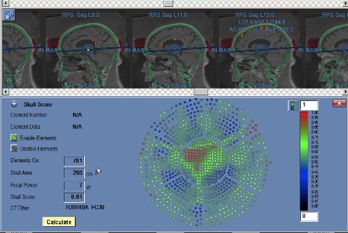How is the calculation of the bone tissue ultrasound conductivity coefficient performed Scull Score).#### **Our Unique Training Method**
In our medical center, we have developed and implemented a unique approach to preparing patients with low ultrasound conductivity of the skull bones. This is our know-how, based on scientific research and years of experience. Here’s how we do it:
**Accurate Diagnosis:**
We assess the condition of your bones using modern methods such as CT or MRI, and analyze the levels of vitamins and minerals in the body. This data helps us understand how to better prepare you for treatment.
**Individual Medication Preparation:**
If a vitamin deficiency is detected D or other substances, we prescribe medications that strengthen bones. For example, bisphosphonates like alendronate help increase bone density, making it more conducive to ultrasound.
**Research Approach:**
Our preparation method is based on the results of our own research, which we conduct in collaboration with leading research institutes, such as Lomonosov Moscow State University. We not only treat but also constantly seek new and more effective ways to help our patients.
In the photo, PhD in Physics and Mathematics, research associate at MSU [Alisa Aleksandrovna Krohmal](https://limu.msu.ru/people/alisa-aleksandrovna-krohmal-dorofeeva) presents our joint work on the densification of the cranial bone structure. **Team of Professionals:**
Our team includes endocrinologists, neurologists, and radiologists who work together to prepare you for treatment and make it as effective as possible.
Thanks to our preparation method, focused ultrasound becomes accessible even for those patients who previously had limitations due to the condition of their skull bones. After treatment, you will be able to return to your usual life, enjoying freedom of movement and control over your body.
Treatment of tremor with ultrasound in a patient after increasing the ultrasonic conductivity of the skull bones. Video published with the patient’s written consent.
#### **We are ready to help you!**
If tremor prevents you from living a full life, don’t delay in addressing this issue. Our center is not only a place where you are treated but also where new methods are developed to help you. We know how to prepare you for treatment and achieve the best results. [Contact us, and we will walk the path from diagnosis to successful treatment together.](https://buzaevclinic.com/services/mrgfus/) We are here to restore your confidence and freedom of movement!
#### For citation:
Sakharova G.M., Galimova R.M., Khatmullina A.N., Nabiullina D.I., Bikbulatova A.A., Buzaev I.V., Avzaletdinova D.Sh. Ultrasonic Conductivity of Skull Bones as a Limiting Factor in Performing Operations Using Focused Ultrasound for Central Nervous System Diseases: An Endocrinologist’s Perspective *FOCUS Endocrinology* . 2025;6(1):31-36. <https://doi.org/10.62751/2713-0177-2025-6-1-04>

Guzaliya Sakharova
Endocrinologist at V.S. Buzaev International Medical Centre. [See Full Bio](https://buzaevclinic.com/author/guzaliia-sakharova/)
**Categories:** Our Publications
---
### [At the conference, in honor of the 90th anniversary of the Department of Neurosurgery of RMANPO ](https://buzaevclinic.com/research/presentations/90-anniversary-rmanpo-2025/)
**Published:** May 20, 2025
**Author:** Igor Buzaev
**Content:**
Chief Freelance Neurosurgeon of the Ministry of Health of the Republic of Bashkortostan, Head of the Department of Neurosurgery at Bashkir State Medical University, Professor Shamil Makhmutovich Safin gave a presentation “**The Present and Future of Focused Ultrasound in Neurology and Neurosurgery** ” at the [scientific and practical conference dedicated to the 90th anniversary of the Department of Neurosurgery of the Russian Medical Academy of Continuous Professional Education in Moscow](https://scaf-spb.ru/spisok-konferencij/nauchno-prakticheskaya-konferenciya-posvyawennaya-90-letiyu-kafedry-nejrohirurgi-rmanpo/)

[Conference Program ](https://buzaevclinic.com/downloads/programma_konferencii_rmanpo_red_23_04.pdf).
**Categories:** Presentations and Reports
---
### [Tips for Managing Drooling in Parkinson's Disease ](https://buzaevclinic.com/conditions/parkinsons-disease/digestion-nutrition/sialorrhea-drooling/)
**Published:** May 19, 2025
**Author:** Dinara Nabiullina
**Categories:** Nutrition for Parkinson's Disease
---
### [How to deal with constipation and nausea in Parkinson's disease? ](https://buzaevclinic.com/conditions/parkinsons-disease/lifestyle-lifehacks/s1e4-constipation-nausea-management/)
**Published:** May 19, 2025
**Author:** Ekaterina Nadezhdina
**Content:**
In the latest episode of our podcast, gastroenterologist specializing in helping patients with Parkinson’s disease, Gulnaz Zagitova, answers questions.
> Do not take these answers as medical advice for your case and do not self-medicate! Each case is individual and requires specialist consultation, [You can easily get an online consultation here](https://buzaevclinic.com/telemedical-consultations/). Answers to frequently asked questions are provided for educational purposes.
In the latest episode of our podcast, gastroenterologist specializing in helping patients with Parkinson’s disease, Gulnaz Zagitova
## Briefly about what’s in the latest podcast episode:
### 1. How are Parkinson’s disease and the gastrointestinal tract connected?
The gastrointestinal tract is an amazing organ. It is the second brain in terms of the number of neurons. The vagus nerve emerges from the brain and innervates the entire gastrointestinal tract. There are theories that signals are transmitted from the gut microbiota to the brain via the vagus nerve, and changes in the brain affect the digestive organs.
### 2. How does a person’s emotional state affect the functioning of the gastrointestinal tract?
Your mood and feelings greatly affect the digestive organs. A significant part of a gastroenterologist’s consultation is dedicated to discussing this connection.
### 3. What gastrointestinal symptoms can manifest in Parkinson’s disease?
Swallowing difficulties, constipation, drooling — all these symptoms can be early manifestations of Parkinson’s disease.
### 4. Can constipation be an early symptom of Parkinson’s disease?
Yes, constipation is one of the first manifestations of Parkinson’s disease. The vagus nerve innervates the intestines, and if there is a problem, the intestines also suffer.
### 5. How can a gastroenterologist suspect Parkinson’s disease?
A gastroenterologist may suspect Parkinson’s disease if they observe changes in behavior, mood, slight hand tremor, as well as complaints of constipation, drooling, and difficulty swallowing.
### 6. What dietary recommendations are given to patients with Parkinson’s disease?
Increase fiber, water intake, and add supplements to the diet. Laxatives are also prescribed if dietary recommendations do not work.
### 7. What role does fiber play in the diet of patients with Parkinson’s disease?
Fiber helps improve bowel function, which is especially important for constipation characteristic of Parkinson’s disease.
### 8. Why is it important to drink enough water with Parkinson’s disease?
Water helps the intestines function properly and aids in the movement of food.
### 9. What medications are used to treat constipation in patients with Parkinson’s disease?
Laxatives from different groups are prescribed, which are selected individually for each patient.
### 10. How does a gastroenterologist help improve the quality of life for patients with Parkinson’s disease?
A gastroenterologist helps through dietary adjustments, prescribing medications, and addressing gastrointestinal issues such as constipation and swallowing difficulties.
### 11. Is it possible to prevent the deterioration of the gastrointestinal tract in patients with Parkinson’s disease?
Yes, through adherence to the principles of healthy eating, increasing fiber and water in the diet, and timely treatment of constipation.
### 12. How does the microbiota affect the health of patients with Parkinson’s disease?
The microbiota affects drug absorption, gut health, and the connection between the brain and the gastrointestinal tract.
### 13. What is microbiota transplantation and how can it help with Parkinson’s disease?
Microbiota transplantation is a method in which the purified stool of one person is transplanted into the intestine of another to improve health and gastrointestinal function.
### 14. What dietary features are important for patients with swallowing disorders?
It is recommended to consume thicker foods, such as pureed soups, to ease the swallowing process.
### 15. How to properly organize nutrition for a patient with Parkinson’s disease?
Follow the principles of healthy eating: increase fiber, drink more water, add a variety of vegetables and fruits.
### 16. Should the entire family’s diet be changed if one member has Parkinson’s disease?
Yes, a healthy diet for the whole family contributes to the improved health of all its members, including the patient.
### 17. How does the microbiota affect drug absorption in Parkinson’s disease?
The microbiota influences the absorption of medications, which is important for treatment efficacy.
### 18. What factors worsen bowel function in patients with Parkinson’s disease?
The intestine becomes sluggish and atonic against the backdrop of the disease, which worsens its function.
### 19. How to Properly Organize Daily Life for a Patient with Parkinson’s Disease?
Create a pleasant atmosphere, use convenient dining accessories such as napkins and comfortable footstools.
### 20. What devices can make life easier for a patient with Parkinson’s disease?
Special devices for the legs to change the angle between the femur and the spine, comfortable furniture and plumbing.
### 21. How does the aesthetics of eating affect the patient’s condition?
The aesthetics of eating, a calm atmosphere, and a clean table surface help improve the patient’s mood.
### 22. Why are pureed soups recommended for patients with swallowing disorders?
Pureed soups are easier to swallow, they don’t splash and don’t cause discomfort.
### 23. How to properly choose furniture and sanitary ware for a patient with Parkinson’s disease?
Patient comfort should be considered, for example, by using footrests and comfortable toilets.
### 24. What are the advantages of team-based medical work in treating Parkinson’s disease?
Teamwork allows for quick and effective improvement of a patient’s condition using a comprehensive approach.
### 25. How does studying the microbiota help in treating Parkinson’s disease?
Studying the microbiota helps to understand how to improve gut health and the connection between the brain and the gastrointestinal tract (GIT)
### 26. What are the prospects for microbiota transplantation in Russia?
The method is not yet approved in Russia, but it is actively being studied. In foreign countries, it is already widely used.
## Video podcast transcript:
– So, we continue. My interlocutor is another member of the V.S. Buzaev International Medical Centre team, gastroenterologist, dietitian, nutritionist, and ultrasound diagnostics doctor, Gulnaz Danisovna Zagitova. Hello!
– Good afternoon!
– We don’t know what time of day those who are dealing with Parkinson’s disease will be watching. Our podcast is dedicated to everything related to this disease, and as it turns out, almost everything is related. And I think today we will be exploring the connection between Parkinson’s disease and the gastrointestinal tract with you. Can you explain the connection?
– The gastrointestinal tract is an amazing organ. Although, probably every doctor who comes here says that their specialty is unique and “their organs” are the most interesting. But the intestine is truly an amazing organ in that it is considered the second brain due to the number of neurons. When we have any problems in the brain, whether functional or organic, the digestive organs suffer in one way or another. A very large nerve, the vagus nerve, emerges from the brain and innervates the entire gastrointestinal tract. There are even theories suggesting that signals are transmitted from the gut microbiota to the brain via the vagus nerve. In turn, changes from the brain via the vagus nerve affect the digestive organs.
– As a layperson, I can tell you that if there’s something unpleasant in my stomach or something “gurgling,” it extremely negatively affects the brain, mood, and everything else, really.
– In turn, your mood and feelings greatly affect the digestive organs. Therefore, asking if there is a connection—yes, undoubtedly, there is. I dedicate most of the consultation to this, emphasizing that your gastrointestinal tract is highly dependent on your mood.
– We are talking about the fact that Parkinson’s disease is not a sentence, but it is a long process that is much more pleasant to go through with specialists who, firstly, understand their narrow fields, but at the same time are ready to solve problems associated with this complex and quite challenging disease comprehensively. So, tell us how this is addressed in your field? Perhaps you could provide some examples. And, of course, we would be interested to know how it can be diagnosed if a patient comes to you specifically with gastrointestinal problems. Can you somehow determine if there are any prerequisites or primary symptoms of Parkinson’s disease?
– Parkinson’s disease is a nervous system disorder, and it manifests through the digestive organs as well. For example, impaired swallowing. A person might consult a gastroenterologist or a neurologist. Our task is to work together. If a neurologist knows or suspects Parkinson’s disease, I, as a gastroenterologist, can help improve the patient’s life through dietary measures or certain medications. Our goal is to enhance the patient’s quality of life and extend it without succumbing to the disease. Speaking about the possibility of a gastroenterologist suspecting the diagnosis during an appointment… There are cases where, before the onset of tremor, the patient suffered from constipation. There is even scientific evidence that constipation is one of the first manifestations of Parkinson’s disease. Because the vagus nerve, which I mentioned at the beginning of the dialogue, innervates the intestines. If there is a problem, the intestines suffer as well. Constipation is one of the most prominent manifestations. Continuing on how a gastroenterologist might suspect Parkinson’s disease… Constipation is generally a common problem among our population, especially when we are sedentary and have everything at our fingertips, making it difficult to suspect this particular disease. But if I notice changes in behavior, mood, or slight hand tremor, I naturally invite a neurologist for a consultation. This is the advantage of our clinic, where we can convene a council and examine the patient together. Therefore, yes, I believe a gastroenterologist can suspect Parkinson’s disease, not with a high probability, of course, but nonetheless, yes.
– So, am I correct in understanding that tremor can manifest in the digestive system earlier?
– Yes, yes, constipation, drooling, difficulty swallowing – all these symptoms can be early manifestations of Parkinson’s disease.
-I think it would be helpful to mention how Parkinson’s disease progresses from a gastroenterologist’s perspective and what actions should be taken: special nutrition, diet? What to recommend?
– In a team of specialists helping people with Parkinson’s disease, it is essential to include dietitians-nutritionists and gastroenterologists, as this issue is not only related to neurologists. We can ease the work of neurologists by addressing some of the patient care aspects, reducing the number of questions they face. Regarding “gastroenterological problems” in patients with Parkinson’s disease, there are specific medications. For example, constipation, one of the most prominent symptoms of Parkinson’s, can be treated with medications if dietary recommendations are ineffective. The intestines and stomach become “sluggish” and atonic due to this disease. Our task is to help them function. The gastrointestinal tract is a 9-meter muscular tube. Sounds impressive, right? Our goal is to ensure it contracts properly and aids in food movement. There are both dietary recommendations and medications. Some patients are prescribed laxatives from different groups for a long time, tailored individually with the neurology team. For others, dietary recommendations suffice: increasing fiber, for instance, increasing water intake, adding certain supplements… This work is always comprehensive and individualized for each patient. Some may experience Parkinson’s disease only through swallowing difficulties, while others may feel heaviness after eating, an unpleasant symptom that might lead them to avoid food altogether. Our relationship with food lasts from birth to death, making it one of the most important relationships. Regarding nutrition, we will emphasize adhering to the principles of a healthy diet.
– I can’t help but mention those people who encounter not as a patient, but as those who surround the patient – family and loved ones. So tell me, is there a need to create a personal diet for a patient with Parkinson’s disease, to prepare something special for them? Or can the whole family perhaps eat properly, and it will suit the entire family?
– It depends on the manifestation of the disease in a particular patient because they are all different. In general, we adhere to a basic diet, which includes the principle of a healthy plate, the “rainbow” principle among vegetables and fruits, and the principle of the number of ingredients per week. This ensures that a person receives all micro- and macronutrients from their diet. Yes, we can get everything from nutrition. Speaking of the environment, naturally, they need to follow the same recommendations. This way, they will also be healthier and work on their microbiota. By the way, microbiota is a separate organ. There are three kilograms of bacteria in you, just so you know. In me, there are about two to three kilograms of bacteria. Their quality depends on what we eat.
– Is mine bigger than yours? (laughs)
– Well, you’re bigger than I am. (laughs). But nevertheless, the ratio of bacteria in the gut… It’s like a separate state on which the degree of health and mood depends. By the way, the hormone of happiness, serotonin, is synthesized there. Therefore, a healthy diet for the whole family is a must. If we talk specifically about a patient with Parkinson’s disease… There may be nuances and features depending on what bothers them more.
– It would probably be strange to say that we do not recommend anyone to eat properly. But, nevertheless, have you tracked any dynamics? For example, you manage patients with Parkinson’s disease: one eats properly, and another ignores your recommendations. Is there any observable dynamic in maintaining quality of life? Positive in the case of proper nutrition and, let’s say, negative if a person eats improperly?
– In general, our population is not accustomed to eating vegetables and fruits. They are not accustomed to consuming enough protein and not accustomed to drinking water.
– What is this? This is due to…
– These are just dietary features and lack of awareness. Additionally, post-war eating habits, when it was cheaper and easier to eat soup, for example, than to add a lot of vegetables.
– I remember how our grandmothers used to say, “What kind of lunch is it if there’s no soup?” More bread makes it more filling, and here we have such an imbalance in the percentage on the plate. Now we have everything, and we can afford enough vegetables, enough fruits, and enough protein. Therefore, when we introduce small corrections to the patient’s diet, like “increase the amount of vegetables,” “add water,” the patient’s well-being improves. Some experience fewer cases of constipation, meaning better bowel function. Some feel less heaviness after eating. Even such small recommendations from a gastroenterologist-dietitian during a consultation with a patient with Parkinson’s disease improve the situation. The gut microbiota has a significant impact on the absorption of medications.
– Well, you said that every doctor loves their field of activity the most and considers it the best. But the V.S. Buzaev International Medical Centre is distinguished by the fact that the medical professionals work as a team. Tell us, with whom do you interact the most, who is your “dream team,” especially when working with patients with Parkinson’s disease?
– Certainly, we work here with a team of neurologists. They are the most advanced. In the case of Parkinson’s disease, the neurologist hands over the patient to me with complete information, carefully, from hand to hand. In turn, after making some adjustments, I can return the patient to the neurologist in the same manner. This “neurologist-gastroenterologist” tandem leads to more productive work and a faster improvement in the patient’s well-being.
– Our podcast is not only for patients but also for those who are dealing with this issue – relatives, close ones. Can you tell us how to adjust your daily life, how should those around approach a patient with Parkinson’s disease? Are there any practical tips? Yes, we know there are issues like nausea, drooling, constipation, heaviness in the stomach. What needs to be adjusted or maybe slightly corrected… Well, we understand about the diet. What about the daily routine or any special approach?
– In general, it’s important to approach the specific patient’s situation with understanding, as this illness significantly changes the life of our patient. Approach it with empathy, with understanding, immerse yourself, and help them through this journey. Because adjustments in daily life will happen one way or another. When it comes to nausea, create a pleasant atmosphere: a clean table surface, a calm environment, perhaps some relaxing music.
– Aesthetics, in general?
– Aesthetics, yes. That is, we must cultivate a culture not only among patients with the disease but also among all the people around them.
– I like everything you are telling me.
– I say that I love you in gastroenterology! (laughs). Yes, to create such a favorable atmosphere. Considering that swallowing is impaired – naturally, soups may not be suitable. Here, the food should be thicker, with some adaptations like napkins.
– What is a specific example of thicker food, for instance?
– For example, cream soups
– This is more adapted food that won’t spill, fall, and so on. Some special napkins to prevent the patient from getting dirty. Of course, it will still spoil the mood and be upsetting. Those who care for them must support the patient at all stages. Speaking of constipation, a comfortable toilet and a convenient stool near the feet will be necessary.
– This is very good, but by the way, I didn’t even think about it. Is there any specialized plumbing, or can you really just choose the one that suits best?
– I haven’t seen such toilets, of course, but there is a special device for the feet to change the angle between the femur and the spine. At home, we have a basin, and I have three children who put their feet on it. It’s a habit that will improve bowel movements and cause less discomfort because our toilets are actually a bit improperly designed. The angles are not right. And for patients who suffer from constipation, it’s even harder for them.
– Not orthopedic, right?
– Not orthopedic, well, you could say that. It’s simply determined through dialogue: is it comfortable or not? Observe, watch the patient. Talk. Communication is what quickly improves any situation. That’s why we talk, we tell patients to share everything, to open up, because the doctor might not remember or mention many things. Nevertheless, when there is a “patient-doctor” relationship, or even better, for example, “neurologist-gastroenterologist-patient.” This is the model at our V.S. Buzaev International Medical Centre, where we hand over the patient from one specialist to another.
– I would like to emphasize that when you hand over patients from one person to another, it doesn’t mean the patient will be going from office to office with a bunch of papers. Handing over means literally taking them by the hand and passing them to another. I’ve observed this, and I really want to clarify because it’s very important when a patient is left alone with their illness, wandering through endless medical corridors—it seems like a very discouraging situation. But in your bright medical center, in every sense, what he…
– Especially when a patient is vulnerable, they are left with their illness and don’t know what to do next. Our task is to show them that life goes on, just with some adjustments and a bit more interaction with certain doctors. Life will be just as bright and fulfilling. The main thing is to believe in it. That is, not to be alone with this illness, but to involve the patient’s loved ones and relatives, if they have any, of course.
– The V.S. Buzaev International Medical Centre has brought together doctors who are at the forefront of research and are constantly engaged in scientific activities. Is there something you can already share? What are you currently working on?
– There is currently a great interest in the state of the microbiota. The whole world is studying it. We are all trying to understand how it works, how we can influence it, and how we can study it. It consists of trillions of microorganisms—microbes that affect our entire body. One of the current interests, including in Parkinson’s disease, is the impact on the microbiota to improve health, medication absorption, and influence on the brain, the “brain-gut axis.” There is a method called fecal transplantation, where one person’s stool is taken, purified, studied, and transplanted into another person’s intestine.
– You must forgive the analogy, but it’s very similar to a blood transfusion, essentially, only related to feces.
– Yes, with the microbiota of the intestine.
– In principle, roughly speaking, the method is the same. But naturally, everything is purified, and everything is studied.
– What is the principle of this method?
– We take microbiota.
– No, I understood that. And what about…
– Through changes in the qualitative composition of the microbiota, our health improves, including in Parkinson’s disease. There are research studies, they are not very large, but they exist and show their effectiveness.
– That is, the “working mechanisms” of a healthy body are simply transferred to a body that lacks something. Right?
– Yes. And in this way, the condition improves. A series of such “intrusions” into the intestinal tract of the body is performed, several procedures. These are enemas or endoscopic procedures through which the necessary microflora—bacteria—are introduced.
– Is there practical application already in Russia?
– In Russia, this is not officially permitted yet, but we are studying this area.
– The method sounds quite strange, doesn’t it? But what difference does it make if it works in the end, right? If we set aside prejudices.
– The difference is huge because the number of side effects from chemical drugs is higher than from fecal transplantation. There are risks, of course, but this still needs to be studied, effectiveness proven, and implemented at the national level… However, for example, abroad, it is already practiced and used quite widely.
– I’m precisely talking about the fact that it doesn’t matter how it sounds if it works?
– Sounds a bit off, of course.
– I wish you success in your research activities and, of course, in your work with patients with Parkinson’s disease.
– Thank you, Damir!
## Additional resources:
For more information, read other articles on Parkinson’s disease treatment:
- [Strategy and Tactics on How to Overcome Parkinson’s Disease ](https://buzaevclinic.com/conditions/parkinsons-disease/beat-parkinsons-strategy-tactics-possibility-of-victory/)
- [On the Treatment of Tremor and Stiffness with Focused Ultrasound ](https://buzaevclinic.com/services/tremor-mrgfus-treatment/)
- [Our Mission and Vision ](https://buzaevclinic.com/values/mission-vision/)
- [Safe and Comfortable Home for Parkinson’s Disease ](https://buzaevclinic.com/conditions/parkinsons-disease/lifestyle-lifehacks/safe-home-environment/)
- [New and Future Treatment Methods ](https://buzaevclinic.com/conditions/parkinsons-disease/present-and-future-parkinsons-treatment/)
- [Parkinson’s Disease. Microflora and Intestinal Dysbiosis ](https://buzaevclinic.com/conditions/parkinsons-disease/digestion-nutrition/microbiota-dysbiosis/)
- [Constipation in Parkinson’s Disease ](https://buzaevclinic.com/conditions/parkinsons-disease/digestion-nutrition/constipation/)
## Subscribe to the podcast:
- [Apple Podcasts](https://podcasts.apple.com/ru/podcast/%D0%BE%D0%B4%D0%B8%D1%81%D1%81%D0%B5%D1%8F-%D0%B8%D1%81%D1%86%D0%B5%D0%BB%D0%B5%D0%BD%D0%B8%D1%8F-%D0%BC%D0%B5%D0%B4%D0%B8%D1%86%D0%B8%D0%BD%D0%B0-%D0%BC%D0%B5%D0%BD%D1%8F%D0%B5%D1%82%D1%81%D1%8F-%D1%87%D1%82%D0%BE%D0%B1%D1%8B-%D0%B8%D0%B7%D0%BC%D0%B5%D0%BD%D0%B8%D1%82%D1%8C/id1795050429)
- [CastBox](https://castbox.fm/channel/6530073?utm_campaign=ex_share_ch&utm_medium=exlink&country=ru)
- [Overcast](https://overcast.fm/itunes1795050429)
- [RSS](https://podcasts.buzaevclinic.ru/feed/podcast/healing-journey-odyssey/)
- [Vkontakte](https://vk.com/tremor_mrgfus_russia?z=podcast-177056040_456239059)
- [Yandex](https://music.yandex.ru/album/35482228)
- [Youtube](https://www.youtube.com/channel/UC_imLOoAVr2v69OTqskVX7Q)
## Podcast Producer

Ekaterina Nadezhdina
[See Full Bio](https://buzaevclinic.com/author/ekaterina-nadezhdina/)
Reviewed By:  Gulnaz Zagitova  Gulnaz Zagitova
Gastroenterologist, Dietitian
[See Full Bio](https://buzaevclinic.com/author/gulnaz-zagitova/)
**Categories:** Lifestyle and Tips for Parkinson's Disease
**Tags:** Podcast Parkinson's Disease , Video
---
### [Parkinson's Disease: The Power of a Loved One, Sex, Intimate Life ](https://buzaevclinic.com/conditions/parkinsons-disease/lifestyle-lifehacks/s1e3-finding-strength-in-love-sex-and-parkinsons/)
**Published:** May 19, 2025
**Author:** Ekaterina Nadezhdina
**Content:**
In the latest episode of our podcast “Odyssey of Healing ” answers to questions about sexual life and urological disorders in Parkinson’s disease from neuro-urologist Ivan Kuvin
> Do not take these answers as medical advice for your case and do not self-medicate! Each case is individual and requires a specialist consultation, [You can easily get an online consultation here](https://buzaevclinic.com/telemedical-consultations/). Answers to frequently asked questions are provided for educational purposes.
In the latest episode of our podcast “Odyssey of Healing ” answers to questions about sexual life and urological disorders in Parkinson’s disease from neuro-urologist Ivan Kuvin
## Briefly about what’s in the latest podcast episode:
### 1. What is the role of a neurourologist in treating Parkinson’s disease?
Ivan Kuvin: Our main mission is a comprehensive approach, as Parkinson’s disease affects many organs and systems. The urologist is responsible for the patient’s quality of life, including issues with urination and sexual life. We identify the root cause of problems and prescribe the correct diagnosis and treatment.
### 2. What urination problems do patients with Parkinson’s disease experience?
Ivan Kuvin: Problems with urination are often related to disorders of the nervous system. Our task is to determine what exactly causes these disorders and help the patient cope with them.
### 3. How does Parkinson’s disease affect the sexual life of patients?
Ivan Kuvin: Patients with Parkinson’s disease often experience changes in their sexual life. An active sex life helps reduce the risk of disease progression and improves psycho-emotional well-being.
### 4. Can erectile dysfunction be an early sign of Parkinson’s disease?
Ivan Kuvin: Yes, research shows that men with erectile dysfunction around the age of 40 have an increased risk of developing Parkinson’s disease, especially if there are comorbid conditions such as diabetes or hypertension.
### 5. Why is sexual life important for patients with Parkinson’s disease?
Ivan Kuvin: Sexual activity contributes to the improvement of emotional and physical well-being. Patients who lead an active sexual life reduce the risks of disease progression.
### 6. How to maintain an active sex life with Parkinson’s disease?
Ivan Kuvin: We talk with patients about what they need, how often to have sex to bring maximum pleasure and improve their condition.
### 7. How are the psycho-emotional state and sexual activity related?
Ivan Kuvin: Sexual life improves mood and self-esteem, which minimizes the impact of illness on quality of life.
### 8. How does a neuro-urologist help a patient overcome embarrassment and delicate issues?
Ivan Kuvin: We create a trusting atmosphere, discuss delicate issues in an “intimate setting,” so the patient can openly talk about their problems.
### 9. What role do relatives and sexual partners play in supporting the patient?
Ivan Kuvin: Relatives and partners help the patient not to withdraw into themselves, encouraging dialogue and problem-solving. Their support is extremely important.
### 10. What modern methods of treating erectile dysfunction exist?
Ivan Kuvin: Medications such as phosphodiesterase type 5 inhibitors (e.g., Viagra, Tadalafil) are used, as well as prostaglandin injections. In severe cases, penile prosthesis may be considered.
### 11. How does hormone therapy help patients with Parkinson’s disease?
Ivan Kuvin: Hormone replacement therapy improves the course of Parkinson’s disease, especially with low testosterone levels, which affect sexual desire and activity.
### 12. Why is it important to see a urologist in a timely manner, even if there are no symptoms?
Ivan Kuvin: Men over 45 are recommended to undergo an annual examination by a urologist to identify hidden issues such as prostate adenoma, prostate cancer, or inflammatory diseases.
### 13. How often should you have a urologist examination after the age of 45?
Ivan Kuvin: Even in the absence of complaints, men are recommended to visit a urologist once a year for prevention and diagnosis of possible diseases.
### 14. What risk factors influence the development of erectile dysfunction?
Ivan Kuvin: Among the risk factors are vascular disorders, hormonal changes, neurological problems, as well as comorbidities such as hypertension and diabetes.
### 15. What medications are used to treat sexual problems in Parkinson’s disease?
Ivan Kuvin: Drugs such as phosphodiesterase type 5 inhibitors (Viagra, Tadalafil), as well as injectable forms with prostaglandins, help manage erectile dysfunction.
### 16. How do specialists interact within the teamwork of a medical center?
Ivan Kuvin: Treating a patient is a team effort. The neurologist prescribes the main therapy, while the urologist helps address specific issues related to urination and sexual life.
### 17. How to adjust treatment to avoid hypersexuality in patients?
Ivan Kuvin: We collaborate with a neurologist to adjust therapy. For example, if a medication causes hypersexuality, we discuss the possibility of replacing it.
### 18. What surgical treatment methods are used for severe erectile dysfunction?
Ivan Kuvin: In severe cases, penile prosthesis surgery is performed to help restore sexual function in patients.
### 19. How does sexual activity prevent the progression of Parkinson’s disease?
Ivan Kuvin: An active sex life improves psycho-emotional and physical well-being, which helps slow the progression of the disease.
### 20. Why is a personalized approach to treatment important for patients with Parkinson’s disease?
Ivan Kuvin: Every patient is unique. We conduct diagnostics, including hormonal and cancer screening, to develop an individualized treatment plan, improve quality of life, and extend the patient’s youth.
## Video podcast transcript:
**Damir Lukmanov:** This is the podcast “Odyssey of Healing,” where we meet doctors from the V.S. Buzaev International Medical Centre and learn about advanced medicine. Opposite me is the pleasant-looking neuro-urologist and andrologist Ivan Alekseevich Kuvin. Hello!
**Ivan Kuvin:** Hello!
**Damir Lukmanov:** We have mentioned many times that the work at the V.S. Buzaev International Medical Centre is team-based. We will again discuss Parkinson’s disease, considering that our content is likely to be of greater interest to those who have already encountered this issue, their relatives and friends, and those who are about to begin some form of treatment. In front of me is a neuro-urologist. What is your role in this team of specialists addressing the problem of Parkinson’s disease?
**Ivan Kuvin** : What, so to speak, is our main mission. Let’s start with the fact that this disease is quite complex.
**Damir Lukmanov:** I want to note that some of your colleagues have already mentioned Parkinson’s disease as a kind of journey that simply needs to be overcome, preferably in the company of pleasant and highly qualified specialists.
**Ivan Kuvin** : Let’s start with the fact that Parkinson’s disease is quite a complex condition that requires a comprehensive approach, as it affects many organs and systems. Undoubtedly, the “captain” in treating this disease is the neurologist specializing in Parkinson’s. However, other specialists also assist. The urologist is responsible for the patient’s quality of life, with important aspects including issues with urination and sexual life. After all, these issues are fundamental to health. When we meet a patient with Parkinson’s disease at different stages, we can already anticipate which problem will prevail. A patient comes to me, I see that they have already been examined by a neurologist, and I already understand what stage they are at. Similarly, I can already imagine what complaints they will bring to me. But in any case, people aged 40 and older mainly suffer from this disease. And these people, even without Parkinson’s disease, may have various health deviations or comorbidities. Therefore, my task as a urologist is to determine the root cause of urination problems, issues in sexual life, and prescribe the correct diagnosis and treatment. And therefore, of course, a doctor of another specialty cannot look as deeply. This is where teamwork comes in, including the neuro-urologist.
**Damir Lukmanov:** Speaking of root causes and connections, Parkinson’s disease is equally… That is, are there any symptoms, any problems that consistently appear in those who suffer from Parkinson’s disease?
**Ivan Kuvin** : Very good question. Suppose today we talk about men’s erectile function, sexual life, sexual dissatisfaction: there is interesting data suggesting that people who have erectile dysfunction at around the age of 40 have a risk of developing Parkinson’s disease. If you add comorbidities such as diabetes and hypertension, the risk of developing Parkinson’s disease becomes even higher.
**Damir Lukmanov:** Listen, am I correct in understanding that a normal sex life and the absence of any urological problems are key to a healthy continuation of life?
**Иван Кувин** : Yes, absolutely. For both men and women, sexual activity brings significantly more positive emotions—both emotional and physical. Those with Parkinson’s disease who are sexually active significantly reduce their own risks of disease progression. This is very important!
**Damir Lukmanov:** This is extremely important. Here, I must immediately ask a question: do you think this is more of a physiological feature or a psychological dependency? That is, the person is fine with their sexual life, has a good mood, self-esteem, and consequently minimal, so to speak, impact on the quality of life
**Иван Кувин** : Yes. Men and women with Parkinson’s disease are generally not in the best emotional state when they first learn of their diagnosis. And, of course, everything related to movement within the paradigm of this disease requires special attention. Sexual intercourse is a huge, let’s say, “big dance.” Fine motor skills, sensitivity, and special skills are needed. And, of course, we calmly discuss with patients in our center how often they should have sex, what they enjoy in sex, what they need, and how we can help them achieve the maximum amount of pleasure to improve their physical and, naturally, psycho-emotional state.
**Damir Lukmanov:** The topic is quite delicate. You’ve probably encountered patients who are tense and shy to talk about it openly during appointments. How do you “draw them out” to learn all their existing problems and be helpful?
**Ivan Kuvin** : There are certain medical and personal nuances here. I find it quite easy to communicate with any patient. When a man or woman comes in, and sometimes they come together, we discuss certain aspects of their sexual life. Sometimes I am alone with the patient. And here, in a “private setting,” without any extra ears, the patient is able to share much more with the doctor, even things they haven’t shared in the presence of their partner.
**Damir Lukmanov:** You said that sometimes patients come in pairs.
**Ivan Kuvin** : They usually come in pairs.
**Damir Lukmanov:** For me, it’s surprising because I thought they withdraw into themselves, and that’s precisely the problem. First, help doesn’t come on time if the patient doesn’t speak up about it. How should the patient’s family and friends behave?
**Ivan Kuvin** : Of course, a person with Parkinson’s disease often withdraws, trying to understand and reevaluate themselves. Here, the task of relatives and close ones is to prevent them from retreating into themselves. They need to be encouraged to engage in dialogue. If it’s a spouse, then it’s essential to talk and motivate the patient to address the issues that might be “stuck” in their mind, hindering their life. It’s usually difficult to get anyone to visit a urologist’s office, even a healthy person. And if they have additional challenges, urology might not be their immediate concern. Yes, they often think about other things. But their sexual life continues nonetheless. If they notice changes in it, it’s important to support them, take them by the hand, bring them to a neuro-urologist, and solve the issues together. Certainly, only support.
**Damir Lukmanov:** I know that the principle of operation of the V.S. Buzaev International Medical Centre, which you represent, is teamwork. Can you tell us how you interact with your colleagues? Do you have a person who makes the main decisions? Or do you do it collectively, together?
**Иван Кувин** : Certainly. Here, it will be difficult for one specialist to bring a patient to recovery. Therefore, once again, our “flagship” is This is a neurologist who examines and prescribes therapy. After that, I consult. We discuss “sensitive” issues that the patient may not be ready to open up about, perhaps to a female doctor or other doctors. Here we discuss sexual life. I can say that there are certain medications prescribed by the doctor, but the patient experiences an effect such as hypersexuality. In this case, we need to interact with the neurologist to discuss this case and say: “Let’s try to change this therapy because this medication helps with Parkinson’s disease but slightly interferes with their sexual life.” Some patients don’t want to have sex, while others want it too often. This also affects the quality of life not only for the patient but also for their sexual partner. Therefore, these issues are resolved only through direct contact between specialists. In our center, we are capable of solving this problem!
**Damir Lukmanov:** Let’s touch on some issues of modern medicine. Are there any modern methods or medications for addressing problems in sex?
**Ivan Kuvin** : Erectile dysfunction is a multifactorial problem in itself. Here, issues related to the condition of blood vessels, the hormonal system, and neurological profile come into play, so it’s important to determine what is primary. Remember, at the beginning of our dialogue, we talked about the patient possibly having some predisposing factors? Now we like to talk about hormone replacement therapy. Let’s imagine the profile of our patient: a person over 40 years old. A man comes in with some complaints. Well, obviously, there is a high probability that he will have insufficient testosterone levels, which undoubtedly affects sexual desire and male activity. Therefore, it is very important to find this problem, even if it is accompanying, but we cannot exclude it. Find this problem and eliminate it. If necessary, prescribe hormone replacement therapy. By the way, it improves the course of Parkinson’s disease. The second point: let’s imagine that the patient has concomitant hypertension. How can we manage without a cardiologist? We can’t. So we involve a cardiologist to correct arterial hypertension. Now, the patient is “covered” from two sides, seemingly unrelated directly to Parkinson’s disease.
**Damir Lukmanov:** From the aspects that significantly affect its quality of life.
**Ivan Kuvin** : Of course! Medications for treating erectile dysfunction are standardized worldwide; the important factors are the dosage and the form in which they are administered: tablet or injection? My job here is to understand, analyze, evaluate, and prescribe medications for erectile dysfunction treatment, adjusting if necessary. This, of course, takes some time, but we are not in a hurry. For the Buzaev Medical Center, the main goal is for the patient to achieve the desired treatment results during this period of their condition. By the way, phosphodiesterase type 5 inhibitors, well-known medications like Viagra and Tadalafil, are effectively used for treating erectile dysfunction in Parkinson’s disease, but the dosage varies for everyone. There are injectable medications into the penis containing prostaglandins, which is the next stage of erectile dysfunction treatment. But, in principle, all this is possible. The patient needs to work with a neuro-urologist to understand how to use these properly. And all this helps prolong their youth with Parkinson’s disease—male, sexual youth, of course.
**Damir Lukmanov:** During a conversation with your colleagues, we found out that Parkinson’s disease is not a sentence, but we are still talking about alleviating symptoms, and the disease itself is not curable. In your case, patients suffering from Parkinson’s disease and having problems with urination and erectile dysfunction. Can you cure patients, at least within your specialty?
**Иван Кувин** : I would say that curing is very optimistic.
**Damir Lukmanov:** We are optimists, yes.
**Иван Кувин** : We can bring it to a perfect state. And we actively use this, using our knowledge and skills. Patient compliance is also important here, meaning adherence to therapy, as well as the involvement of their sexual partner. We understand that the primary dialogue between the sexual partner and the patient, the conversation “what do you want, how do you want it, what is needed for you to feel better?” should lead to the patient being in my office – the office of a urologist-andrologist, where we address these delicate issues.
**Damir Lukmanov:** So, do I understand correctly that once a patient contacts you, they become your charge, and you guide them throughout the illness? How often should regular check-ups be done? Or only as the condition worsens, when symptoms appear?
**Ivan Kuvin** : Let’s start with the fact that a man over 45 should see a urologist once a year.
**Damir Lukmanov:** So even if there are no problems?
**Ivan Kuvin** : Yes. In addition to erectile dysfunction, there is also prostate adenoma, prostate cancer, which is becoming more common in younger people, inflammatory reactions, urolithiasis, which hasn’t gone away, so a person should undergo a “check-up.” And here, if a person is already under active observation – yes, we schedule with them: you’ll come in a month, in two months. Not all patients are concerned about erectile dysfunction, especially at the onset of the disease. But we can already diagnose hidden risks, the same hormonal screening, the same cancer screening, ultrasound, blood tests, none of this has been canceled. Therefore, in general, it is individual. I don’t remember us ever treating anyone in a template manner at our center. A wonderful team. We work absolutely individually with each person.
**Damir Lukmanov:** Speaking of breaking the mold, can you mention anything within the framework of perhaps innovative methods or some interesting achievements in the world of medicine related to your field?
**Ivan Kuvin** : There is a surgical question. If a person has erectile dysfunction that does not respond to oral or injectable forms, then the person may be referred for penile prosthesis implantation. This is an interesting topic, but it will affect not only patients with Parkinson’s disease.
**Damir Lukmanov:** I suggest summarizing. Problems with erectile function and urination are often encountered in Parkinson’s disease, right?
**Ivan Kuvin** : Yes.
**Damir Lukmanov:** Sexual life is very important during the treatment of Parkinson’s disease.
**Ivan Kuvin** : Yes, it helps maintain psycho-emotional status and physical condition. We know that an active sex life prevents the progression of Parkinson’s disease.
**Damir Lukmanov:** Relatives of the patient, while providing all possible support, must ensure that the patient undergoes a complete examination.
**Ivan Kuvin** : Certainly. Because relatives and sexual partners are the first people who know almost everything about the patient. And in this “doctor-relative-spouse(-partner)-patient” connection, we can prevent many complications associated with Parkinson’s disease.
**Damir Lukmanov:** Regular check-up of the body. Age 45+ – this is a mandatory examination once a year.
**Ivan Kuvin** : Yes, a urologist examination, ultrasound, cancer screening, and a test for sex hormones.
**Damir Lukmanov:** I think we have covered this delicate topic comprehensively. Thank you very much for the interesting and informative conversation! Good luck!
**Ivan Kuvin** : Thank you very much!
## Additional resources:
For more information, read other articles on Parkinson’s disease treatment:
- [Strategy and Tactics on How to Overcome Parkinson’s Disease ](https://buzaevclinic.com/conditions/parkinsons-disease/beat-parkinsons-strategy-tactics-possibility-of-victory/)
- [On the Treatment of Tremor and Rigidity with Focused Ultrasound ](https://buzaevclinic.com/services/tremor-mrgfus-treatment/)
- [Our Mission and Vision ](https://buzaevclinic.com/values/mission-vision/)
- [New and Future Treatment Methods ](https://buzaevclinic.com/conditions/parkinsons-disease/present-and-future-parkinsons-treatment/)
- [Parkinson’s Disease. Microflora and Intestinal Dysbiosis ](https://buzaevclinic.com/conditions/parkinsons-disease/digestion-nutrition/microbiota-dysbiosis/)
- [Constipation in Parkinson’s Disease ](https://buzaevclinic.com/conditions/parkinsons-disease/digestion-nutrition/constipation/)
## Subscribe to the podcast:
- [Apple Podcasts](https://podcasts.apple.com/ru/podcast/%D0%BE%D0%B4%D0%B8%D1%81%D1%81%D0%B5%D1%8F-%D0%B8%D1%81%D1%86%D0%B5%D0%BB%D0%B5%D0%BD%D0%B8%D1%8F-%D0%BC%D0%B5%D0%B4%D0%B8%D1%86%D0%B8%D0%BD%D0%B0-%D0%BC%D0%B5%D0%BD%D1%8F%D0%B5%D1%82%D1%81%D1%8F-%D1%87%D1%82%D0%BE%D0%B1%D1%8B-%D0%B8%D0%B7%D0%BC%D0%B5%D0%BD%D0%B8%D1%82%D1%8C/id1795050429)
- [CastBox](https://castbox.fm/channel/6530073?utm_campaign=ex_share_ch&utm_medium=exlink&country=ru)
- [Overcast](https://overcast.fm/itunes1795050429)
- [RSS](https://podcasts.buzaevclinic.ru/feed/podcast/healing-journey-odyssey/)
- [Vkontakte](https://vk.com/tremor_mrgfus_russia?z=podcast-177056040_456239059)
- [Yandex](https://music.yandex.ru/album/35482228)
- [Youtube](https://www.youtube.com/channel/UC_imLOoAVr2v69OTqskVX7Q)
## Podcast Producer

Ekaterina Nadezhdina
[See Full Bio](https://buzaevclinic.com/author/ekaterina-nadezhdina/)
Reviewed By:  Ivan Kuvin  Ivan Kuvin
[See Full Bio](https://buzaevclinic.com/author/ivan-kuvin/)
**Categories:** Lifestyle and Tips for Parkinson's Disease
**Tags:** Podcast Parkinson's Disease , Video
---
### [Levodopa: Toxic or Safe? Answers from a Parkinsonologist ](https://buzaevclinic.com/conditions/parkinsons-disease/treatment/tips-and-levodopa-treatment-faq/)
**Published:** May 18, 2025
**Author:** Ekaterina Nadezhdina
**Content:**
In the latest episode of our podcast “Odyssey of Healing ” answers to questions about levodopa and Parkinson’s disease treatment from neurologist-Parkinsonologist, PhD Gulnara Nailyevna Akhmadeeva
> Do not take these answers as medical advice for your case and do not self-medicate! Each case is individual and requires specialist consultation, [You can easily get an online consultation with a Parkinson’s specialist here](https://buzaevclinic.com/telemedical-consultations/). Answers to frequently asked questions are provided for educational purposes.
Parkinson’s Disease. Should You Be Afraid of Taking Levodopa? Season 1, Episode 2 of the Podcast ‘Odyssey of Healing. Medicine is changing to change your life ‘. Video. ## Briefly about what’s in the latest podcast episode:
### What is levodopa and how is it used to treat Parkinson’s disease?
Levodopa is a prodrug that is a precursor of dopamine, a substance that is deficient in the brain in Parkinson’s disease. Levodopa is used for replacement therapy to compensate for this deficiency. It is considered the most effective medication for treating the symptoms of Parkinson’s disease.
### Why was the life expectancy of patients with Parkinson’s disease short before the advent of levodopa?
Before the introduction of levodopa into medical practice, patients with Parkinson’s disease had a short life expectancy—on average, 5 to 7 years after the onset of the first symptoms. This is explained by the lack of effective treatments that could slow the progression of the disease and alleviate its symptoms.
### How is levodopa related to dopamine in the brain?
Levodopa is a precursor of dopamine. When introduced into the body, it converts into dopamine, compensating for its deficiency in the brain, which helps improve the condition of patients and reduce the symptoms of Parkinson’s disease.
### Why is levodopa compared to insulin in the treatment of diabetes?
The comparison is based on a similar principle of action: just as insulin compensates for the deficiency of this hormone in patients with diabetes, levodopa compensates for the deficiency of dopamine in patients with Parkinson’s disease. Both medications help normalize the body’s condition and improve the quality of life for patients.
### What myths and fears are associated with the use of levodopa?
There is a myth that levodopa is toxic and causes many side effects. However, numerous studies conducted over the past 50 years have proven that levodopa does not have a toxic effect on brain neurons and does not contribute to the progression of Parkinson’s disease.
### Is it true that levodopa is toxic to brain neurons?
No, this is a misconception. Research has shown that levodopa does not destroy brain neurons and does not accelerate disease progression. It is safe when prescribed and used correctly under a doctor’s supervision.
### Why does Parkinson’s disease cause an increased need for levodopa as it progresses?
Over time, patients experience a decrease in the neurons’ storage capacity. This means that neurons lose the ability to retain levodopa for extended periods, leading to the need for increased doses of the medication to maintain its effect.
### What is “reduction of neurons’ cumulative ability ” and how does this affect the action of levodopa?
Reduction in the storage capacity of neurons is a process in which neurons lose the ability to retain dopamine synthesized from levodopa. This results in the medication’s effect becoming less prolonged, requiring patients to take it more frequently or in larger doses.
### What is a phenomenon “dose depletion ” and how does it manifest in patients?
Phenomenon “dose depletion ” occurs when the effect of a single dose of levodopa becomes insufficient, and the symptoms of the disease return before the next dose. This is related to the progression of the disease and the reduced ability of neurons to retain levodopa.
### Why is it important to prescribe levodopa at the right time, not too early or too late?
Starting levodopa too early can lead to a faster development of side effects, such as the “wearing-off” phenomenon. Therefore, doctors use a “delayed-start” strategy, prescribing less effective medications in the early stages of the disease to postpone the initiation of levodopa.
### At what stages of Parkinson’s disease is levodopa treatment usually started?
Levodopa is prescribed at later stages of the disease, for example, in cases of balance disorders, falls, or other serious manifestations characteristic of the third stage of Parkinson’s disease.
### Why is levodopa not always prescribed in the early stages of Parkinson’s disease?
In the early stages, antiparkinsonian drugs from other groups are more commonly used. This allows delaying the need for levodopa and avoiding side effects associated with its long-term use.
### What symptoms of Parkinson’s disease require the rapid prescription of levodopa?
Levodopa is prescribed for symptoms such as stiffness, slowness of movement, balance disorders, and frequent falls, which are characteristic of the third stage of the disease.
### What alternative medications are used in the early stages of Parkinson’s disease?
In the early stages, antiparkinsonian drugs from other groups are used, which are less effective but allow delaying the start of levodopa treatment.
### What is a strategy “delayed start ” and how does it help in the treatment of Parkinson’s disease?
The “delayed start” strategy involves using less effective drugs in the early stages of the disease to postpone the initiation of levodopa. This helps avoid the rapid development of side effects and prolong the effectiveness of treatment.
### Which groups of patients most often need levodopa in old age?
Patients over 70 years old with risks of dementia, hallucinations, and somatic diseases most often need levodopa, as it is a safer option compared to other medications.
### How does levodopa help patients over 70 with risks of dementia and other diseases?
Levodopa causes fewer side effects than other antiparkinsonian drugs and can be used in smaller doses, reducing risks for elderly patients.
### What surgical treatment methods are used in addition to levodopa?
Surgical methods include the implantation of brain stimulators and MRI-guided focused ultrasound. These methods help reduce levodopa doses and improve patients’ quality of life.
### How does the implantation of brain stimulators help improve the quality of life for patients?
This procedure allows for a reduction in the doses of levodopa taken, prolongs the beneficial effects of treatment, and improves the overall quality of life for patients.
### What are the advantages of MRI-guided focused ultrasound for treating Parkinson’s disease?
[Focused ultrasound is a non-invasive method suitable for patients over 70-80 years old ](https://buzaevclinic.com/services/tremor-mrgfus-treatment/). It helps improve quality of life without the risks associated with surgical intervention.
### How long can patients take levodopa?
Patients can take levodopa for more than 20 years. Their condition is maintained through combined treatment methods, including modern surgical technologies.
## Video Transcript
**Damir Lukmanov:** Opposite me is a neurologist specializing in Parkinson’s at the V.S. Buzaev International Medical Centre, a specialist in movement disorders, PhD Gulnara Nailevna Akhmadeeva. Hello!
**Gulnara Akhmadeeva:** Hello, Damir, nice to meet you!
**Damir Lukmanov:** Nice to meet you, yes. Unlike people who have encountered some problems and are currently facing a choice: which treatment methods to choose? We are talking about this from the perspective of modern medicine. And I would like to ask you about Levodopa. It does decline, doesn’t it?
**Gulnara Akhmadeeva:** Declines, yes.
**Damir Lukmanov:** I hope so. In conversations with the buzaevclinic team doctors, I learn a lot of new things, and our podcast is dedicated not to doctors, but to those who are currently addressing these issues. We have already found out that Parkinson’s disease is not a verdict. Let’s put it in a few words: what is it, and most importantly, why are Levodopa medications so feared?
**Gulnara Akhmadeeva:** Levodopa medications were discovered and approved for use in patients with Parkinson’s disease about 50 years ago or even longer. Before these medications, patients with Parkinson’s disease had a very limited lifespan, living only about 5-7 years from the onset of the first symptoms. Why is Levodopa actively used and applicable? The reason is that it is a prodrug—a precursor of dopamine, which is deficient in the brain in Parkinson’s disease. A simple analogy can be drawn with diabetes and insulin. In diabetes, the amount of insulin decreases, causing all the problems. Patients with diabetes can inject insulin to bring themselves, conditionally, back to normal, stabilizing their carbohydrate metabolism. The same happens here. We use the most effective medication—the substance lacking in the brain, we administer it to patients, and, accordingly, they feel better. Essentially, it is replacement therapy. That’s why it is so effective.
**Damir Lukmanov:** We understand that it is effective. But why does Levodopa cause some fear? Could it be some misinformation, maybe a small amount of quality information available?
**Gulnara Akhmadeeva:** What is the fear in patients? The fear is that Levodopa is considered toxic and has a huge number of side effects. In fact, this is not the case, to get ahead. Over these 50 years, many studies have been conducted on many thousands of patients, proving that it has no toxic effect on brain neurons and, accordingly, does not contribute to their destruction or the progression of Parkinson’s disease. As for the myth of increased or new side effects, they certainly exist, but this is not related to Levodopa itself. It is related to the natural progression of Parkinson’s disease, with a decrease in the number of cells that remained at the onset of the disease, and this progression is increasing. Accordingly, a property of the brain manifests, such as an increased need for Levodopa.
**Damir Lukmanov:** Is this addiction?
**Gulnara Akhmadeeva:** This is not habituation, but a decrease in the cumulative capacity, the buffering capacity of neurons. If at the beginning of the disease we give a certain amount of Levodopa to a patient, and the neurons can capture and hold that one tablet for a day or two, in principle, the patient can be in good condition for those days. However, over time, these neurons become “leaky,” and through a certain number of “holes,” the amount of Levodopa gradually decreases, it “seeps out.” And over time, due to the progression of the disease, the number of “holes” simply increases. This leads to the need for Levodopa, the need for multiple doses throughout the day. So, it is no longer enough to take Levodopa once, because the dose depletion occurs. Patients, not knowing that this is caused by the progression of the disease and thinking that it is due to Levodopa itself, of course, are mistaken. Our task, the task of doctors, Parkinson’s specialists, is to explain that Levodopa is not toxic, it does not shorten life expectancy, and it does not have pronounced side effects. There are limitations to its use, of course, but again, it is up to the doctors who can adjust the regimen to improve the patient’s condition and, most importantly, add Levodopa when it is necessary for a particular patient, because there are specific indications. It is important not to miss that critical point for a particular patient when Levodopa is needed. This is the doctor’s task.
**Damir Lukmanov:** So, Levodopa is not a panacea, not everyone is prescribed it, am I understanding correctly?
**Gulnara Akhmadeeva:** In fact, all patients with Parkinson’s disease eventually turn to Levodopa, one way or another. It’s just that each person does so at their own stage, depending on when they see a doctor and how their disease progresses.
**Damir Lukmanov:** But nevertheless, this medication is not always necessary, right?
**Gulnara Akhmadeeva:** Levodopa is not always necessary. For a patient in the early stages of the disease, for example, if one hand is trembling, if symptoms are predominantly on one side, we usually do not start with Levodopa. I can explain when we do start with Levodopa. For example, a patient comes to the doctor with hand tremors, stiffness, slowness, and complains of instability when walking, balance issues leading to falls. These symptoms are characteristic of the third stage of the disease, and there are five stages in total. The manifestation of the third stage in the form of falls dictates the need for the quickest possible prescription of the most effective medications. In this case, to improve the patient’s condition and reduce the risk of falls and injuries, we will prescribe Levodopa immediately. But this is not the only example. In fact, there is a group of patients in the so-called age group, 70+, especially those with high risks of dementia, possible hallucinations, and risks of somatic diseases. These are patients to whom we cannot prescribe medications from other antiparkinsonian groups because they have higher risks of other side effects. Other medications, compared to Levodopa, may cause more hallucinations, lower blood pressure, and more swelling. For such patients “70+” with high risks, prescribing Levodopa is safer. Since Levodopa is generally more effective than other antiparkinsonian drugs, we can prescribe it in lower doses, thereby reducing the risk of side effects. This must be understood. So, this is the age group with a high number of potential side effects and patients in the third stage.
**Damir Lukmanov:** If Levodopa is so effective, then why mention any alternative medication options at all?
**Gulnara Akhmadeeva:** Due to the fact that with age and as the disease progresses, there is a greater need for Levodopa, which unfortunately also depends on the dosage. Often, patients come to the appointment who were prescribed Levodopa at an early stage when it was not yet indicated. It was prescribed, and patients have been taking the drug for, say, 7 years, only Levodopa. During this time, they have reached a higher dose of Levodopa, and of course, they have developed the “wearing-off” phenomenon, the “on-off” phenomenon—the very “side effects” we fear. All of this could have been avoided if we had used the “delayed start” strategy from the beginning. What is this? It means that at the first stage, we usually use (for middle-aged patients, especially younger ones) antiparkinsonian drugs from other groups, not Levodopa. They are, of course, slightly less effective. But their use over a certain period, for example, 2-5 years, can improve the quality of life for the patient and delay the use of Levodopa. Of course, we will eventually come to Levodopa for this patient, but it will be a little later. Therefore, there are different strategies for patients, and it is important to understand that starting with Levodopa is not always necessary due to the higher risk of “side effects,” which we also do not need. Levodopa is needed at the right time: neither too late nor too early.
**Damir Lukmanov:** It seems to me that nothing scares a person as much as the lack of alternatives. We say that sooner or later a patient facing Parkinson’s disease will turn to Levodopa. But it is only necessary to explain that it is safe. And everything that happens regarding side effects – unfortunately, that’s how our body is structured. Right?
**Gulnara Akhmadeeva:** In reality, the body is such an interesting thing. For example, if we take two patients comparable in age, gender, education level, and number of comorbidities… We might give Levodopa to one and not to the other, and we will get completely different effects. And even if we give the same amount of medication, we will still likely get different effects because each patient has their own social interactions, level of physical activity, habits, hobbies, and communication level—all of which influence how the patient ultimately lives with their Parkinson’s disease. Everything is very individual, of course.
**Damir Lukmanov:** Based on your experience at the V.S. Buzaev International Medical Centre, how long have you been observing a patient who was prescribed Levodopa, and how many years has it lasted?
**Gulnara Akhmadeeva:** Do you mean from the start of taking Levodopa or in general?
**Damir Lukmanov:** I’m interested in the duration of the “distance,” how long is it? 5 years, 10 years, 15 years?
**Gulnara Akhmadeeva:** No, definitely, it’s more than that. I’ve been working with such patients for almost 20 years. So, all this time I’ve been observing patients to whom I once prescribed Levodopa, and they still live with this disease, using Levodopa. Naturally, progress does not stand still. Along with Levodopa, we definitely resort to more advanced treatment methods, meaning neurosurgical operations. It must be understood that when a patient starts taking Levodopa at some point, we are preparing them for the fact that a side effect will arise sooner or later. But this is a feature of the disease. It’s just important to be ready for the moment when side effects occur, we will have the very alternative you mentioned. An alternative of additional treatment, radical treatment with the help of neurosurgery. And then, depending on the age and condition of the patient when the need for the same neurosurgery arises. If the patient is “preserved,” if the patient is not 70 years old, for example, if they do not have hallucinations, serious diseases such as diabetes, heart problems, coronary heart disease, etc. – we can recommend them for the installation of brain stimulators. Yes, this operation is invasive, yes, this operation involves craniotomy, with the installation of electrodes. But this method allows reducing the amount of Levodopa taken, extending their good quality of life, that is, not only life expectancy but also quality. If we mean that the patient, to some extent, “could not preserve” until this age, or they have crossed the threshold of 70 years, for example, and then there is a need for a more progressive method of treatment – then we can recommend a non-invasive method. For example, operations conducted in our center – MRI-guided focused ultrasound, and then we are not limited by the age of the patients. We have a patient, for example, 75+ or 80+, when we have exhausted all possibilities of drug therapy, we take them for our operation, which is called an operation, but in fact, it is non-invasive, and we similarly improve their quality of life and extend life precisely in good quality.
**Damir Lukmanov:** I wanted to touch on your collaboration, your partnership, your interaction within the medical center. All doctors work as a single coordinated organism, and if a patient comes to you, you solve all tasks together. Tell us, how does the interaction occur? With doctors of which specialties do you also conduct consultations?
**Gulnara Akhmadeeva:** In fact, we have a very cool collegial decision-making process at our center, and I’ve always liked it. First and foremost, we collaborate with the neurosurgeon, Rezida Maratovna Galimova, who makes decisions in her capacity as a neurosurgeon. The decision to operate on each specific patient is made based on all available data, including MRI, together with the neurosurgeon. We definitely interact with our MRI specialist, Dmitry Konstantinovich Kreotin, because making a decision for such a neurosurgical operation is impossible without assessing risks and possible contraindications. This requires knowledge of MRI and CT, so the MRI doctor is essential. But we also necessarily work with specialists in therapeutic profiles. I have three “best friends” – a psychiatrist who helps with elderly patients, with hallucinations or dementia, which, if present, are contraindications for our surgery, and we continue to manage such patients together. The second excellent doctor who helps navigate all the “intricacies” of the patient, all symptoms, and comorbidities is, of course, our therapist-cardiologist-nephrologist. With her, we decide: to take the patient or not? Most likely to take, of course, but after we adjust medication doses, correct antihypertensive measures, and so on, I mean preoperative preparation activities. The third specialist who usually joins our consultations is, of course, our neurourologist Ivan Alekseevich Kuvin, who helps men with libido issues, male and female problems, the latter often presenting with frequent urination urges, incontinence – the problem known as overactive bladder. These are the three “friends” without whom I, as a neurologist specializing in Parkinson’s, do not see the most effective treatment for my patients. We always work as a team of four where necessary. Plus, two more doctors, as I mentioned, when we make the decision about “to operate or not to operate.”
**Damir Lukmanov:** As far as I know, the medical center also conducts research and educational activities. Are you involved in educational work?
**Gulnara Akhmadeeva:** We conduct clinical research, observe patients, and write papers. Rezida Maratovna defended her Doctor of Medical Sciences degree, and we have PhD theses being written in several areas. We constantly hold educational events for doctors. We are engaged not only in research but also in clinical work. Among the moments we are proud of is that in the fall, we held the second session of our in-person educational course for doctors, where physicians even from other countries come to see, integrate into our educational process, and observe our clinical process, including how we perform our operations.
**Damir Lukmanov:** Patients, their relatives, and doctors are watching us. I know that you are open to collaboration and that medical professionals often reach out to you when they encounter unusual situations that you have already faced, worked through, and are ready to share your knowledge about. Yes, what specifically can you share with the professional world?
**Gulnara Akhmadeeva:** Well, all I have is my clinical experience over many years. I work with such patients, and among them, of course, there are not only patients with typical classical variants of Parkinson’s disease or Alzheimer’s disease, but also interesting dementias, interesting Parkinsonism, and Parkinsonism plus. These are interesting patients with functional disorders who seem to come with tremor or suspicion of another disease, and it turns out to be some psychiatric condition. And here we have a big advantage – we solve problems with patients collegially. And that’s why we often come to the right decision. We now have really complex patients coming to us so that we can help them diagnose and prescribe treatment. Such cases are becoming more and more frequent. I confirm your words that our center is open to cooperation, open to learning, to exchanging experiences, because such interesting and complex patients, of course, are encountered in the life of every doctor, and certainly, such interesting patients should be shared. We are ready for this and very happy.
**Damir Lukmanov:** As I look at you, you’ve used the adjective “interesting” several times. I see a doctor with bright eyes who is interested in what they do. That’s great, that’s wonderful. This, by the way, is a distinctive feature of the entire buzaevclinic team. I wish you success in your work!
**Gulnara Akhmadeeva:** Thank you very much, Damir! It was a pleasure working with you.
## Additional resources:
For more information, read other articles on Parkinson’s disease treatment:
- [Strategy and Tactics on How to Overcome Parkinson’s Disease ](https://buzaevclinic.com/conditions/parkinsons-disease/beat-parkinsons-strategy-tactics-possibility-of-victory/)
- [On the Treatment of Tremor and Stiffness with Focused Ultrasound ](https://buzaevclinic.com/services/tremor-mrgfus-treatment/)
- [Our Mission and Vision ](https://buzaevclinic.com/values/mission-vision/)
- [Safe and Comfortable Home for Parkinson’s Disease ](https://buzaevclinic.com/conditions/parkinsons-disease/lifestyle-lifehacks/safe-home-environment/)
- [New and Future Treatment Methods ](https://buzaevclinic.com/conditions/parkinsons-disease/present-and-future-parkinsons-treatment/)
## Subscribe to the podcast:
- [Apple Podcasts](https://podcasts.apple.com/ru/podcast/%D0%BE%D0%B4%D0%B8%D1%81%D1%81%D0%B5%D1%8F-%D0%B8%D1%81%D1%86%D0%B5%D0%BB%D0%B5%D0%BD%D0%B8%D1%8F-%D0%BC%D0%B5%D0%B4%D0%B8%D1%86%D0%B8%D0%BD%D0%B0-%D0%BC%D0%B5%D0%BD%D1%8F%D0%B5%D1%82%D1%81%D1%8F-%D1%87%D1%82%D0%BE%D0%B1%D1%8B-%D0%B8%D0%B7%D0%BC%D0%B5%D0%BD%D0%B8%D1%82%D1%8C/id1795050429)
- [CastBox](https://castbox.fm/channel/6530073?utm_campaign=ex_share_ch&utm_medium=exlink&country=ru)
- [Overcast](https://overcast.fm/itunes1795050429)
- [RSS](https://podcasts.buzaevclinic.ru/feed/podcast/healing-journey-odyssey/)
- [Vkontakte](https://vk.com/tremor_mrgfus_russia?z=podcast-177056040_456239059)
- [Yandex](https://music.yandex.ru/album/35482228)
- [Youtube](https://www.youtube.com/channel/UC_imLOoAVr2v69OTqskVX7Q)
## Podcast Producer

Ekaterina Nadezhdina
[See Full Bio](https://buzaevclinic.com/author/ekaterina-nadezhdina/)
Reviewed By:  Gulnara Akhmadeeva  Gulnara Akhmadeeva
[See Full Bio](https://buzaevclinic.com/author/gulnara-akhmadeeva/)
**Categories:** About the Treatment of Parkinson's Disease
**Tags:** Podcast Parkinson's Disease , Video
---
### [Special offer for Russians on tremor treatment with ultrasound ](https://buzaevclinic.com/prices/promo/offer-mrgfus-tremor-treatment-for-russians/)
**Published:** December 26, 2024
**Author:** Igor Buzaev
**Content:**
We support Russian citizens and offer them a discount of 720 000 rubles, the cost of treatment for Russians is **980 000 rubles**
For CIS citizens, a discount is provided and the cost is – 1 500 000 rubles
Without a discount, the cost of MRgFUS treatment for tremor at our center is – 1 700 000 rubles.
For comparison, abroad the price of MRgFUS treatment is 18 000 – 40 000 euro
#### Included in this price
- MRgFUS procedure
- medical examinations at our clinic before and after surgery according to our protocol
- hotel accommodation
- nutrition
- transportation of the patient and one accompanying person within Ufa
- after the surgery, we monitor the patient for 12 months according to our protocol, both via telemedicine and in person at the clinic
- in case of symptom recurrence within 12 months after the first MRgFUS procedure, we perform a repeat procedure **free of charge**, provided that a positive outcome is expected and it is safe for the patient.
[Here you can read how it was formed ](https://buzaevclinic.com/services/mrgfus/faq/pricing-explained-costs-breakdown/ "Here you can read how it is formed ") cost of MRgFUS treatment for tremor
**Categories:** Special Offers
---
### [Where to study to become a neurologist specializing in Parkinson's disease? ](https://buzaevclinic.com/education/healthcare-professionals/where-to-learn-to-parkinsons-neurologist/)
**Published:** March 5, 2025
**Author:** Dinara Nabiullina
**Excerpt:** Course "Current Issues in the Diagnosis and Treatment of Parkinson's Disease" by the team from the Advanced Center for Modern Treatment of Movement Disorders April 18–25, 2025
**Categories:** To doctors and nurses
**Tags:** Training Event Announcements
---
### [Elimination of tremor in Parkinson's disease ](https://buzaevclinic.com/services/mrgfus/parkinsons-cases/feedback-kazakhstan-kz/)
**Published:** September 13, 2023
**Author:** Aleksej Savelev
**Content:**
ҚA patient from Kazakhstan is talking about being treated at the Buzaev clinic. Doctors helped him overcome the tremor in his hand and stiffness in his leg, which greatly hindered his life, in three hours. Now he is ready to return to a full life.
[Review of tremor treatment at the V.S. Buzaev Clinic ](https://buzaevclinic.com/services/mrgfus/parkinsons-cases/feedback-kazakhstan-ru/) (на русском языке)
**Categories:** Reviews and Treatment Results of Parkinson's Disease with Ultrasound
**Tags:** Video
---
### [In the "Productivity Lab" about lean management ](https://buzaevclinic.com/research/presentations/performance-lab-lean-in-tourism/)
**Published:** December 22, 2024
**Author:** Dinara Nabiullina
**Content:**
[20 December in Ufa hosted the intensive workshop “Productivity Lab” ](https://ufa-news.net/society/2024/12/20/435354-amp.html), where key issues of strategic planning, efficiency improvement, sustainable tourism, and optimization of logistics processes were discussed
As part of the “Tourism” section, Dinara Ilgizovna Nabiullina, the chief physician of our medical center, delivered a report. She spoke about the principles of lean management successfully applied in the center to attract foreign patients and shared experiences in developing medical tourism at a high level.
> In my presentation, I wanted to emphasize the importance of the fact that the principles of lean management truly work, but only if they are based on sincerity and openness! The main principle we apply is transparency in all processes.
>
> Dinara Nabiullina, Chief Physician of V.S. Buzaev International Medical Centre
We are confident that only honest and respectful treatment of patients and their loved ones can build trust and ensure high quality.
• [The open results of our 4-year work experience can be read in our book ](https://buzaevclinic.com/kniga-fokusirovannyj-ultrazvuk-pod-kontrolem-mrt-v-nevrologii/);
• Literally [an open operating room allows relatives and patients to be part of the treatment: doctors without secrets can consult and make decisions together](https://buzaevclinic.com/services/mrgfus/faq/relatives-family-presence-on-procedure/);
• We take responsibility for our treatment and regularly monitor our patients throughout the year.


**Categories:** Presentations and Reports
**Tags:** Past Training Events
---
### [Dystonia. Doctor of Medical Sciences Rezida Galimova on TVC ](https://buzaevclinic.com/conditions/dystonia/tvc-doctor-i-interview-dystonia-treatment/)
**Published:** December 5, 2024
**Author:** Igor Buzaev
**Content:**
## Cervical Dystonia. Interview with Doctor of Medical Sciences Rezida Galimova on the Program “Dr. I… ” on TVC
In the latest episode of the program [“Doctor I…”](https://www.tvc.ru/brands/28/episode/78061) from September 20, 2022, the first Russian [MRgFUS neurosurgeon Rezida Maratovna Galimova](https://buzaevclinic.com/team/doctors/rezida-galimova/)
Today we are excited to present an engaging discussion centered on the treatment of cervical dystonia. This topic has caught our attention for a reason: in the past, we became [pioneers in the use of MRI-guided focused ultrasound for the treatment of movement disorders](https://cyberleninka.ru/article/n/pervyy-opyt-provedeniya-talamotomii-metodom-fokusirovannogo-ultrazvuka-pod-kontrolem-magnitno-rezonansnoy-tomografii-v-rossii)
Cervical dystonia, also known as “torticollis “, for centuries was an unsolvable problem for doctors. However, today, thanks to innovative techniques and the development of research, we are one step closer to ensuring a healthy and comfortable life for everyone. [In this episode of the program “Doctor I…”](https://www.tvc.ru/brands/28/episode/78061), neurosurgeon [Rezida Galimova](https://buzaevclinic.com/team/doctors/rezida-galimova/) will talk about modern methods that help patients gain freedom from this disease.
The focus is particularly on focused ultrasound — a technology that has already [proven itself in our clinic as an effective method](https://annaly-nevrologii.com/journal/files/journals/1/articles/1020/public/1020-34291-1-PB.pdf). Rezida Galimova, with her team, is turning the dream of painless and precise treatment into reality.
Let’s embark on this inspiring journey together to new heights in medicine, and perhaps we can see beyond the horizon a shared dream of a world where medicine is safe, humane, and successful.
**Irina Pershina:** In antiquity, cervical dystonia was described as «”stiff neck syndrome.” At that time, the only method of treatment there was cutting of muscles and tendons. Of course, this method did not solve the problem and was terribly dangerous. What modern treatment methods are used today? And will they help the patient get rid of this disease forever? Let’s discuss. Today we have a neurosurgeon, PhD, as our guest – Rezida Galimova. Good morning!
**Rezida Galimova :** Good morning, hello!
**Irina Pershina:** What is cervical dystonia why is this disease also commonly called torticollis?
**Rezida Galimova:** The basis of this disease is a disruption of the impulses sent from the brain to the neck muscles or other trunk muscles. And when these impulses are sent uncoordinatedly – there is multidirectional muscle work, leading to head turns – to hyperkinesis, such as we call them, involuntary movements from the head. And therefore, it is often popularly called spasmodic torticollis because they see spasticity from certain muscles, they see torticollis, which is why the synonym “spasmodic torticollis” has formed
**Kirill Radzig :** At what age is this usually begins and who suffers more from it: men or women?
**Rezida Galimova : For dystonia there is no specific age. There are children in whom we observe cervical dystonia or other forms dystonia. Of course, Bloom – it’s 30-60 years, but it should not be ruled out that this disease и occurs in children and gradually develops. The feature of the disease is that in women this is observed much more frequently. A lot of genes have currently been identified Groups DYT, which are detected in certain forms of dystonia. Therefore, now the attitude towards dystonia has somewhat changed with the development of genetics: the disease more often associated with genetic pathology
**Irina Pershina:** How does this disease start and progress?
**Rezida Galimova : Each patient describes the onset of the disease in their own unique way. Many patients such disease symptoms there is even in childhood. Some have tics, some have squinting, “sniffing,” shoulder shrugging. Then adults forget these symptoms. And in adulthood one of the very first symptoms is appearance of muscle sensation either on the right or on the left on the left. They clearly say: “I started feel the muscle”, “I started feeling tension in this muscle” This may appear suddenly or when performing certain actions. Over time, muscle tension increases, leading to twisting or tilting heads may appear. May appear movements, we call them hyperkinesias. Some call it tremor or tremor, that is, turning of the head. To the right, left, forward, or backward.
**Irina Pershina:** Hyperkinesias can vary. From What does this diversity depend on? Is there a clear characteristic of who has which hyperkinesias can they be?
**Rezida Galimova : We distinguish different forms of hyperkinesias. When we observe the head turning to the right or left, it is called torticollis. When turns with a slight tilt to the right or left, we call it Laterocollis. And, naturally, when a person tilts their head forward – this is anterocollis, backward – Retrocullis.
**Irina Pershina:** Does it depend on the foci of the impaired neurons, am I understanding correctly? On the location where they disrupt their function?
**Rezida Galimova : Most likely, the genetic disorders we find in patients encode transmission impairment neurotransmitters
**Irina Pershina:** Here in certain places?
**Rezida Galimova : Yes, in certain places. And these neurotransmitters are directed differently and regulate these impulses At the base of the brain – basal ganglia extrapyramidal system. And depending on where it occurred, we see different muscle groups in our patients.
**Kirill Радциг :** Can these movements be controlled?
**Rezida Galimova : At the first stage, many patients do indeed control these movements.
**Irina Pershina:** So, somehow by willpower, right?
**Rezida Galimova : Yes, they sometimes initially arise from some emotional excitement, surge. Then patients develop points, pressing on which they can stop the movement. Patients find these points in an amazing way because they start pressing and suddenly find a point that stops the forward movement of the head. These points we called in neurology «”zero points” since they «reset these movements. In addition, patients always try to make some corrective movements, support their head. At most often during my consultations, patients sit and hold their head with both hands. That is, indeed, if they fix the back of the head to the chair, if they support head, if they make some tilts, then these hyperkinesias or tremors may decrease or almost disappear
**Irina Pershina:** I know that there are several methods for treating this disease. Could you please tell us about them
**Rezida Galimova : Currently, we categorize all treatment methods into conservative and surgical methods. Conservative methods – this is drug therapy, in case of dystonias – botulinum therapy Conservative treatment therapy with medications includes various drugs aimed at reducing either the emotional background the patient, or to reduce these movements, to relax muscles. Unfortunately, data medications have many side effects: drowsiness, development of dependence. Therefore, patients are recommended to definitely add therapy to the treatment of cervical dystonia botulinum, or botulinum toxin, when the doctor selects specific muscles, and into the muscles where noted spasticity, where we see by means electroneuromyography increased activity, entered given toxin And this leads to muscle relaxation, and these movements simply disappear The effect of this therapy lasts from 6 to 9 months. However, unfortunately, the human body has a unique characteristic – it can develop a tolerance to the toxin, begins to produce antibodies and stops responding to it And at the moment when the patient stops responding to botulinum therapy (after 5-6 therapies) – this is the moment when you need to think about surgery. Surgical methods are divided into surgical and non-surgical, for example, like our focused ultrasound.
**Irina Pershina:** About this method please tell me Read more.
**Rezida Galimova : MRI-guided focused ultrasound allows a neurosurgeon to penetrate the brain through the skin, subcutaneous tissue, and bones и «Remove Plot brain, where these neurons «sit and produce this impulse We are just technically «remove this area, and these pathological impulses cease apply. And the patient experiences a reduction in symptoms clinical presentation
**Irina Pershina:** How do you find these specific areas in the human brain?
**Rezida Galimova : Unfortunately, modern MRI development does not allow us to see these areas, but we we assume approximately where they can be localized We know theoretically and calculate mathematically where this area is located in this patient. The feature of focused ultrasound therapy is that we direct a certain amount of ultrasound beams, heat a specific area of the brain to a certain temperature, and observe the patient. If we see that hyperkinesias have decreased in the patient on the table after our preliminary treatment – means we «стоим» on the correct area, we are already conducting therapeutic sonication – ultrasound treatment and conduct monitoring already after treatment, and then we finish the procedure
**Irina Pershina:** Rezida Maratovna, thank you very much for your work, for restoring people’s health, and bringing them back to normal life. I really hope that our program currently viewed by those who who needs in your help, and they will definitely reach out to you
**Rezida Galimova : Thank you!
**Irina Pershina:** Stay with us, we’ll be back after the break.
## Read about dystonia:
[Video of results before and after ultrasound treatment of cervical dystonia ](https://buzaevclinic.com/services/mrgfus/dystonia-cases/cervical-dystonia-ultrasound-driver/)
[MRgFUS for Dystonias (Chapter of Our Research Monograph) ](https://buzaevclinic.com/kniga-fokusirovannyj-ultrazvuk-pod-kontrolem-mrt-v-nevrologii/mr-fuz-pri-distoniyah/)
[Mission and Vision of V.S. Buzaev International Medical Centre ](https://buzaevclinic.com/values/mission-vision/)

Igor Buzaev
Doctor of Medical Sciences, Professor of the Department of Hospital Surgery at Bashkir State Medical University, one of the creators of the first center in Russia for non-invasive neurosurgical treatment of tremor and Parkinson’s disease using MRI-guided focused ultrasound. [See Full Bio](https://buzaevclinic.com/author/igor-buzaev/)
[LinkedIn](https://linkedin.com/in/igor-buzaev)
**Categories:** About Dystonia
**Tags:** We in the Media
---
### [How can a neurologist learn to read MRI scans? ](https://buzaevclinic.com/education/healthcare-professionals/events/mri-neurodegeneration-course/)
**Published:** March 5, 2025
**Author:** Dinara Nabiullina
**Categories:** Events
**Tags:** Training Event Announcements
---
### [School Preparation. Video School for Parents ](https://buzaevclinic.com/conditions/peadiatric/get-ready-to-the-school-video/)
**Published:** August 16, 2021
**Author:** Aleksej Savelev
**Content:**
Recommendations from the neuropsychologist and pediatrician at V.S. Buzaev International Medical Centre.
Liana Rafistovna Agzamova, Clinical Psychologist, Neuropsychologist at the V.S. Buzaev International Medical Centre, together with the Center’s Pediatrician, Yazgul Rakhimovna Ishkinina, discuss issues of preparing a child for school. Behavior control, ability to communicate with adults, child’s independent skills, conflict resolution.
## Preparing for School
Getting Ready for School. Video for Moms.
## Heading to kindergarten!
What parents need to know before taking their child to kindergarten for the first time – discussed by a pediatrician and psychologist from the V.S. Buzaev International Medical Centre
Preparing for Kindergarten, Video for Moms
**Categories:** About Children's Health
**Tags:** Enlightenment , Video
---
### [Video Before and After Treatment of Tremor and Rigidity with Ultrasound ](https://buzaevclinic.com/services/mrgfus/parkinsons-cases/video-quick-tremor-and-rigidity-relief/)
**Published:** December 12, 2024
**Author:** Rezida Galimova
**Content:**
## How Innovations Are Changing Medicine for Parkinson’s Disease
In our quest to improve the quality of life for patients aiming to [overcome the physical limitations of Parkinson’s disease](https://buzaevclinic.com/conditions/parkinsons-disease/), we successfully apply [MRI-guided focused ultrasound.](https://buzaevclinic.com/services/tremor-mrgfus-treatment/) Each case is not just treatment but a transformation of the patient’s life, as demonstrated by our recent experience.
## Step by Step to Freedom of Movement
### In two hours, without blood and the need for anesthesia, we eliminated the patient’s tremor and stiffness, allowing him to write and return to an important beloved activity in life – management!
Our patient, who had long suffered from tremor and stiffness in the left hand, managed to get rid of these symptoms in just 160 minutes of the procedure!
This became possible thanks to our developments in the field of focused ultrasound, which allowed us to reduce symptoms by more than 80% after the first treatment.
Further sessions allowed for the elimination of residual tremor and restored the patient’s ability to confidently sign with their own hand.
## Patient History
The patient had been suffering from Parkinson’s disease since 2017 and, being left-handed, experienced significant difficulties with writing. It was critically important for him to eliminate the tremor to be able to sign freely—an important part of his professional life as a project manager. Our innovations allowed him to achieve this desire, and now he leads an active life without symptoms.
> The patient has been ill since 2017, with the disease starting with stiffness in the fingers of the left hand, later developing into severe tremor. The patient is left-handed and could not write. He consulted a neurologist and was diagnosed with Parkinson’s disease. The treatment was ineffective; to achieve even a slight reduction in tremor, he took 8 tablets of 250 mg levodopa. After a year, dyskinesias developed as a complication of the medication.
>
> **At that very moment, he was appointed as the project manager and had to sign a lot.**
>
> When he arrived at the V.S. Buzaev International Medical Centre, to the question: “what is the greatest pleasure he would experience without tremor and dyskinesias? ” he replied: “**I want to sign the logbook at the entrance myself without hesitation “**.
We performed the treatment on June 19, 2024, with a duration of 160 minutes. After the treatment, there is no tremor, stiffness, slowness, or dyskinesia UPDRS III part before treatment 64 – > 34 after treatment.
Last examination on December 09, 2024 (in 6 months): Condition is good, no return of symptoms. Takes 3-4 tablets of levodopa, no dyskinesias. The entire month after treatment, couldn’t stop and signed everything.
Video story about how in two hours, without blood and anesthesia, we eliminated a patient’s tremor and stiffness: he was able to write and return to his favorite activity in life—working as a manager! The video is posted with the patient’s written consent. There are indications and contraindications for treatment, requiring consultation with a neurologist and neurosurgeon. ## Our Achievements and Contributions to Medicine
This case reflects another of our achievements on the path to making disease treatment painless and effective. We are proud that our innovative focused ultrasound treatment is entering a new era, helping patients [from more than 29 countries](https://buzaevclinic.com/global/countries/)
You can learn more about our achievements on our website by visiting the [section of research publications by our staff.](https://buzaevclinic.com/research/)
Video with patient consent
## Conducted treatment for Parkinson’s disease
[Rezida Maratovna Galimova – co-founder of the center, neurosurgeon,
Doctor of Medical Sciences ](https://buzaevclinic.com/team/doctors/rezida-galimova/)
[Akhmadeeva Gulnara Nailevna – neurologist-parkinsonologist, specialist in movement disorders, PhD ](https://buzaevclinic.com/team/doctors/gulnara-akhmadeeva-neurologist-parkinsonologist/)
[Ksenia Nikolaevna Meshchanova – MRI Radiologist ](https://buzaevclinic.com/team/doctors/kseniia-meshchanova-mri-imaging/)
[Denis Albertovich Akhmetdinov – MRI-technician ](https://buzaevclinic.com/team/nurses/denis-akhmetdinov/)
## Read more
[Our Mission and Vision ](https://buzaevclinic.com/values/mission-vision/)
[More about focused ultrasound ](https://buzaevclinic.com/services/tremor-mrgfus-treatment/)
[Book a consultation right now ](https://buzaevclinic.com/telemedical-consultations/)
[List of Our Research Publications and Presentations to Physicians ](https://buzaevclinic.com/research/publications/)
[They talk about us in the media and award us ](https://buzaevclinic.com/news/in-the-media/)
[The doctor and patient share their experience of treating essential tremor with MRI-guided focused ultrasound on TVC in the program “Doctor I… “](https://buzaevclinic.com/services/mrgfus/essential-tremor-cases/tvc-post-operation-interview-doctor-and-patient/)
[We have published a book with long-term results of Parkinson’s disease treatment using ultrasound, and it is available online for free! ](https://buzaevclinic.com/kniga-fokusirovannyj-ultrazvuk-pod-kontrolem-mrt-v-nevrologii/mr-fuz-pri-bolezni-parkinsona/)
Video story about how we eliminated a patient’s tremor and stiffness in two hours without blood and anesthesia: he was able to write and return to his favorite activity in life – working as a manager! The video is posted with the patient’s written consent. There are indications and contraindications for treatment; consultation with a neurologist and neurosurgeon is required.

Rezida Galimova
The first female neurosurgeon in MRgFUS, one of the founders of the first non-invasive ultrasound tremor neurosurgery center in Russia, Doctor of Medical Sciences, MBA (John Hopkins University), Associate Professor of the Department of Neurosurgery at BSMU. [See Full Bio](https://buzaevclinic.com/author/rezida-galimova/)
[TikTok](https://www.tiktok.com/@galimovarezida)
**Categories:** Reviews and Treatment Results of Parkinson's Disease with Ultrasound
**Tags:** Video
---
### [MRI with Claustrophobia. Is It Possible? ](https://buzaevclinic.com/services/expert-mri-imaging/mri-with-claustrophobia/)
**Published:** December 4, 2024
**Author:** Igor Buzaev
**Content:**
When you suffer from claustrophobia, undergoing an MRI scan can seem daunting. But for us, it’s not an obstacle! Using approved methods, the V.S. Buzaev International Medical Centre is changing the approach in medicine for patients with such challenges.
## Is it possible to have an MRI with claustrophobia?
Claustrophobia varies. However, quite often, in more than 80% of cases among patients who consider themselves claustrophobic, we can conduct an MRI examination.
This review is the first to [systematically summarize the literature on the prevalence of claustrophobic reactions during MRI](https://www.sciencedirect.com/research/article/pii/S1078817414001576). The frequency of claustrophobic reactions ranges from 0.46% to 5.29% in the included studies, with a combined effect size of 1.18%. The frequency of claustrophobic reactions may be influenced by the examination conducted, gender, and scanner design.
## Claustrophobia should not be an obstacle on your path to health
In our clinic, we have developed a unique approach to conducting MRI, focused on your comfort and peace of mind.
### We will dedicate time to you
Working in a fruitful synergy of psychology and medicine, we apply advanced psychological support methods. Our specialists will prepare you using techniques of [neuro-linguistic programming (NLP)](https://academic.oup.com/bjr/article-abstract/83/986/113/7449673), [meditation](https://www.frontiersin.org/journals/medicine/articles/10.3389/fmed.2023.1253824/full), and [cognitive-behavioral therapy (CBT)](https://www.sciencedirect.com/research/article/abs/pii/S0005789422001101), making it easier for you to undergo the scan.
### We use special techniques
Remember how great magicians make objects disappear on stage right [in front of the audience using mirrors](https://www.wikihow.com/Make-a-Person-Disappear)?
We take an example and also use a mirror that creates the illusion of open space, which significantly eases the stay in the machine.
### Let someone hold your hand!
Professional training is just part of our approach, but it is the love for our work that drives us to transform medicine for your comfort and health.
#### Is it important to feel **support from loved ones**?
You can bring someone with you for the examination to ensure your journey to health is safe and as comfortable as possible. [Even when we operate, we allow a loved one to be present if the patient desires](https://buzaevclinic.com/services/mrgfus/faq/relatives-family-presence-on-procedure/)!
#### Are you alone?
To create a safe space for you, we will support you both emotionally and physically. Our staff is always ready to assist and provide moral support.
> Your loved one can accompany you throughout the entire procedure. Our staff can also stay with you and hold your hand
Here is a video for example. You can see our nurse holding the patient’s hand. [Dinara Ilgizovna](https://buzaevclinic.com/team/doctors/dinara-nabiullina-neurologist/) is conducting psychological preparation. The mirror creates an illusion of space. And most importantly, the team’s love for the patient and their work makes us change approaches and attitudes in medicine. By the way, the nurse in the video has already graduated from university and become a doctor!
## What we do not use
### Open-type MRI machine
We do not use open MRI; we use a machine with a spacious large opening **expert class GE Optima 450w GEM 1.5 Tesla** **with the widest aperture among scanners of this class – 70 cm** Such an aperture is maximally comfortable even for larger patients (up to 130 kilograms). In addition, we have noise reduction programs if noise concerns you.
Any open-type device technologically has a lower magnetic field strength in comparison. This raises serious concerns about the quality of the images.
### Anesthesia or sedation
We have the option to provide you with sedation, but inside the MRI machine, you will be difficult to monitor. What if, as a described side effect of the sedative, [respiratory arrest](https://journals.lww.com/anesthesiaclinics/fulltext/2003/41020/anesthesia_for_magnetic_resonance_imaging.5.aspx) occurs? This would be difficult to detect and could be dangerous.
Even when we perform MRI on infants, we place them in the MRI with their mother, who breastfeeds them, and they peacefully sleep together without medication. We strive to avoid sedation as much as possible during MRI examinations. And our non-sedation techniques work.
## Conclusion
We firmly believe in a world where taking care of your health becomes a simple and pleasant process, and treatment is safe and compassionate. These unique opportunities combining technology and a human approach to treatment shape the new face of medicine that we are building at the [V.S. Buzaev International Medical Centre](https://buzaevclinic.com/values/). Our [mission and vision](https://buzaevclinic.com/values/mission-vision/) are filled with humanism and modern achievements aimed at creating safe and error-free treatment for all. This also applies to conducting MRI examinations for patients with claustrophobia, which we make possible with a good machine and without the need for anesthesia.
Our Whatsapp, to make an appointment or ask a question

Igor Buzaev
Doctor of Medical Sciences, Professor of the Department of Hospital Surgery at Bashkir State Medical University, one of the creators of the first center in Russia for non-invasive neurosurgical treatment of tremor and Parkinson’s disease using MRI-guided focused ultrasound. [See Full Bio](https://buzaevclinic.com/author/igor-buzaev/)
[LinkedIn](https://linkedin.com/in/igor-buzaev)
**Categories:** MRI diagnostics of diseases
**Tags:** Video
---
### [Essential tremor treated with ultrasound. Review on TVC ](https://buzaevclinic.com/services/mrgfus/essential-tremor-cases/tv-series-feedback/)
**Published:** December 18, 2024
**Author:** Igor Buzaev
**Content:**
## Essential tremor treated with ultrasound. Review on TVC
Essential tremor treated with ultrasound. Video review on the TVC television channel in the program “Doctor I… “Review, video before and after treatment of essential tremor with ultrasound in the program “Doctor I… ” on TVC. The patient was able to sew again and returned to her favorite activity.
About us [in the media](https://buzaevclinic.com/news/in-the-media/) and awarded, but most importantly, it’s how our patients recover and what they say about the results. It’s inspiring to see the outcomes!
A series of broadcasts about the treatment of tremor and dystonia on the federal TV channel TVC. Program “Doctor I…”
- [Interview with one of the founders of the medical center, Professor ](https://www.tvc.ru/channel/brand/id/28/show/episodes/episode_id/75141) from BSMU [Igor Vyacheslavovich Buzaev](https://buzaev.ru)
- [Interview with Neurosurgeon Rezida Galimova on Cervical Treatment dystonias ](https://www.tvc.ru/channel/brand/id/28/show/episodes/episode_id/78061) (beginning at the twelfth minute)
- [Interview ](https://www.tvc.ru/channel/brand/id/28/show/episodes/episode_id/76149)[TV Centre Channel ](https://www.tvc.ru/channel/brand/id/28/show/episodes/episode_id/76149)[neurologist Yulia Alexandrovna Sidorova and a patient before treating tremor with focused ultrasound ](https://www.tvc.ru/channel/brand/id/28/show/episodes/episode_id/76149)
- [Interview with a patient and her feedback after treating tremor using the MRgFUS method on TVC channel ](https://buzaevclinic.com/services/mrgfus/essential-tremor-cases/tvc-post-operation-interview-doctor-and-patient/)
Other videos before and after on our [Youtube](https://www.youtube.com/@mrfustreatmenttremor) channel, [Rutube,](https://rutube.ru/u/buzaev/) [VKontakte](https://vk.com/tremor_mrgfus_russia)
Essential tremor treated with ultrasound. Video review in a program on the television channel dream come true
**Categories:** Reviews and Treatment Outcomes of Essential Tremor with Ultrasound
**Tags:** Patient Reviews and Stories , Video
---
### [TV Show "Happy Hour" about tremor and ultrasound treatment ](https://buzaevclinic.com/conditions/tremor/tremor-treatment/tv-interview-happy-hour-tremor-causes-and-mrgfus/)
**Published:** January 15, 2024
**Author:** Aleksej Savelev
**Categories:** About the treatment of tremor
**Tags:** Video , We in the Media
---
### [What kind of anesthesia is used in the treatment of tremor with ultrasound? ](https://buzaevclinic.com/services/mrgfus/faq/is-parkinsons-ultrasound-treatment-painful/)
**Published:** January 16, 2020
**Author:** Aleksej Savelev
**Content:**
## What type of anesthesia is used for treating tremor with ultrasound?
For testing effectiveness and safety, the patient must be conscious and able to report their sensations, as well as follow instructions and commands. For example, as soon as the final stage of treatment begins, we can assess its effectiveness by asking the patient to do things like write their name or draw a spiral. As the tremor gradually disappears, the doctor and patient can see the results immediately during therapy. When the tremor is fully controlled, the treatment is complete. The patient can dress and go home.
Therefore, we use local anesthesia at the fixation points of the stereotactic frame (frontal and occipital protuberances), which does not affect the severity of the tremor. The brain does not have pain receptors, so ultrasound exposure rarely causes significant pain sensations. Short-term neurological manifestations may occur, such as a sensation of flipping or swaying of the body in space. With high bone density of the skull vault, higher ultrasound energy is required, which may cause nausea. In this case, antiemetic drugs are used (as needed), and an electric suction device is always ready.
Some patients report a feeling of pressure from the silicone membrane that encloses the space where water circulates. This factor cannot be eliminated, as a loose fit of the membrane would cause leaks.
Thus, the procedure is quite tolerable, although it requires psychological preparation from the patient, as with any surgery.
[More about the treatment of tremor and Parkinson’s disease with focused ultrasound ](https://buzaevclinic.com/services/tremor-mrgfus-treatment/)
**Categories:** Frequently Asked Questions
**Tags:** Video
---
### [Why is the cost of treating tremor and Parkinson's with ultrasound so high? ](https://buzaevclinic.com/services/mrgfus/faq/pricing-explained-costs-breakdown/)
**Published:** September 16, 2024
**Author:** Igor Buzaev
**Content:**
# How is the price of MRgFUS treatment for tremor formed?
The invaluable team of people who have studied all their lives uses equipment and supplies to assist you before, during, and after surgery.
## The treatment price includes:
- Your hotel stay during treatment
- Nutrition While You Are Undergoing Treatment
- Preparation for Surgery (MRI and Consultations with Required Specialists)
- Procedure for Treating Tremor with Focused Ultrasound
- The price includes patient monitoring according to a special protocol for one year
- If symptoms return within 12 months after the first MRgFUS treatment at our facility, we consider the possibility of a free repeat procedure based on available resources. We weigh the patient’s safety and the expected effect. We are interested in long-term results; the recurrence rate and outcomes of repeat treatments have been analyzed and [officially published in a research article](https://cyberleninka.ru/article/n/reoperatsiya-s-ispolzovaniem-fokusirovannogo-ultrazvuka-pod-kontrolem-magnitno-rezonansnoy-tomografii-u-patsientov-s-essentsialnym), as well as in a research book “[MRI-guided focused ultrasound in neurology ](https://buzaevclinic.com/kniga-fokusirovannyj-ultrazvuk-pod-kontrolem-mrt-v-nevrologii/)“.
## The cost of the operation consists of:
- depreciation of high-end MRI (only expensive top-tier machines are compatible with the operating table)
- prices of the operating table from the world’s only manufacturer (monopolist)
- consumables, especially a membrane sized for the patient’s head with an MRI coil
- expenses for renting premises, electricity, water, and utilities
- the cost of work by a team of rare specialists who have studied this their entire lives
For comparison, abroad the price of such a procedure ranges from [18,000](https://neurologyopen.bmj.com/content/6/1/e000582), [18,000 Euro](https://us-uk.bookimed.com/clinics/country=spain/procedure=mri-fus/) to 40,000 dollars.
## How much does surgery for tremor or Parkinson’s disease with ultrasound cost?
[The current price and special offers for tremor treatment with ultrasound can be found here ](https://buzaevclinic.com/prices/mrgfus-tremor-parkinsons-treatment/).
**Categories:** Frequently Asked Questions
---
### [Driving a Car with Parkinson's Disease ](https://buzaevclinic.com/conditions/parkinsons-disease/lifestyle-lifehacks/car-driving/)
**Published:** October 21, 2022
**Author:** Aleksej Savelev
**Content:**
# Is it possible to drive a car if diagnosed with “Parkinson’s Disease”?
The presence of Parkinson’s disease does not automatically mean that you need to stop driving. However, when taking certain medications, symptoms may arise that could make driving dangerous.
When taking certain antiparkinsonian medications, **daytime sleepiness** may occur, in severe cases reaching the level of **episodes of sudden, uncontrollable sleep onset**.
Most often, this occurs **when taking medications from the group of dopamine receptor agonists – pramipexole (Mirapex), piribedil (Pronoran), ropinirole (Requip Modutab, Sindranol).**
Daytime sleepiness in mild cases may naturally occur after eating, while watching TV in a horizontal position, or when reading a book or newspaper while sitting.
More pronounced and dangerous can be considered episodes of falling asleep in a more active state – for example, at the table while eating, or sitting in a car as a passenger during a conversation with a companion
The most severe cases are when a person literally falls asleep on the go, standing, in public places – this is called “narcolepsy” and can lead to injury.
Therefore, if driving is an integral part of the patient’s life, it must be taken into account when prescribing dopamine receptor agonists. It is essential to mention during the doctor’s visit that the patient drives a car and to be more cautious and attentive during the stage of selecting the effective dosage of medications.
**If problems with daytime sleepiness arise, either stop driving or consider changing the medication with your doctor.**
In addition to daytime sleepiness, more advanced stages of Parkinson’s disease may exacerbate movement and cognitive disorders—general slowness, periodically occurring stiffness and “off” phases, as well as slowed reactions.
All of this must be considered and sensibly evaluated when planning trips to minimize potentially dangerous situations for yourself and others.
Stay healthy!
Neurologist-Parkinsonologist at V.S. Buzaev International Medical Centre®, PhD,
Akhmadeeva Gulnara Nailevna.
**Categories:** Lifestyle and Tips for Parkinson's Disease
---
### [How to correctly diagnose "Parkinson's disease"? ](https://buzaevclinic.com/conditions/parkinsons-disease/diagnostic/diagnose-criteria/)
**Published:** September 23, 2022
**Author:** Aleksej Savelev
**Content:**
# Diagnosis “Parkinson’s disease ” (БП).
How to put it correctly?
This is the prerogative of a neurologist, a specialist in movement disorders (Parkinsonologist)
The diagnosis of “PD” is made clinically, meaning the diagnosis can be made based solely on the presence of certain symptoms. This is a triad of movement symptoms –
**The first and main symptom (consists of three types of movement disorders)**
*– Bradykinesia* (slowness)
*– hypokinesia* (reduction of movement amplitude)
*– oligokinesia* (slowing of movements)
May manifest as reduced facial expressions (“mask-like face”), a quiet voice, changes in handwriting (small, bead-like handwriting), decreased arm movement activity while walking, in rhythm with steps.
**Second important symptom** – resting tremor. This is the classic “parkinsonian” tremor that occurs in a relaxed position (or while walking) and resembles “pill rolling” or “coin counting” “.
**Third symptom** – muscle rigidity, manifested by increased muscle tone and increased stiffness
**These symptoms form the syndrome of parkinsonism.**
Each syndrome has its own CAUSE, and there can be many causes for the development of parkinsonism syndrome (as well as conditions that mimic parkinsonism) – tumor, stroke, hypothyroidism, depression, rheumatic and joint diseases, normal pressure hydrocephalus
Therefore, in the diagnosis of Parkinson’s disease, a thorough neurological examination, medical history collection, and identification of comorbidities and medications are necessary, along with neuropsychological testing (clock drawing test, depression and anxiety questionnaires), laboratory tests (complete and biochemical blood tests, thyroid hormones, ceruloplasmin, and others), neuroimaging (CT and MRI); consultations with other specialists (cardiologist, endocrinologist, etc.)
Currently, there is no specific diagnostic test or examination capable of confirming the diagnosis. Neuroimaging methods – single-photon emission computed tomography (SPECT or SPECT, DaTscan) and positron emission tomography (PET) with fluorodopa have maximum specificity and sensitivity, close to 100%, and are practically unavailable here in the Russian Federation
More accessible are ultrasound examination of the substantia nigra and MRI of the brain with nigrosome 1 study. The result of the studies may depend on the quality of the equipment and the specialist’s experience.
It is becoming less necessary to conduct a diagnostic levodopa test – a single dose of Nakom (taking 1 tablet of standard levodopa), or an extended levodopa test (taking levodopa for 2-3 weeks), with an assessment of the drug’s effect – IMPROVEMENT in motor condition (reduction of tremor, reduction of stiffness and slowness) while taking the drug.
Sometimes, for diagnosis, dynamic observation over several months or years is necessary—often cases of atypical parkinsonism (diseases from the parkinsonism-plus group) in the early stages can be very similar to the onset of Parkinson’s disease.
Overall, it can be concluded: if there are complaints of tremor, stiffness, impaired fine motor skills, or uncooperative hand movements, it is always better to consult a neurologist and determine the cause of the ailment, as early diagnosis and early treatment prolong life and improve quality of life.
In our Extrapyramidal Center, it is possible to conduct a comprehensive examination – consultation with a Parkinson’s specialist, laboratory tests (if necessary), MRI with nigrosome assessment, ultrasound of the substantia nigra. We will be happy to help. Health to you and your loved ones!
Akhmadeeva Gulnara Nailevna, PhD, neurologist specializing in Parkinson’s disease
V.S. Buzaev International Medical Centre
**Categories:** About the Symptoms and Diagnosis of Parkinson's Disease
---
### [We are among the first in the world to treat epilepsy with ultrasound! ](https://buzaevclinic.com/conditions/epilepsy/seasures-mrgfus-treatment-pioneers/)
**Published:** April 3, 2025
**Author:** Renata Safiullina
**Content:**

Renata Safiullina Редактор научных новостей
Студентка Башкирского государственного медицинского университета, стажер ММЦ им. В.С. Бузаева. [See Full Bio](https://buzaevclinic.com/author/renata-safiullina/)
### We are among the first in the world to treat epilepsy with focused ultrasound!
In the prestigious journal **cell.com** ( Heliyon) review released *“*[Therapeutic use of transcranial ultrasound for epilepsy: A review”](https://www.cell.com/heliyon/fulltext/S2405-8440(25)01382-9). [Among the 5 main publications reviewed in this article ](https://www.cell.com/action/showFullTableHTML?isHtml=true&tableId=tbl2&pii=S2405-8440%2825%2901382-9) mentioned [Rezida Maratovna Galimova](https://buzaevclinic.com/team/doctors/rezida-galimova/) with the [team](https://buzaevclinic.com/team/), who performed a [unique successful surgery for the treatment of involuntary laughter and crying caused by a rare tumor — hypothalamic hamartoma](https://buzaevclinic.com/conditions/hypothalamic-hamartoma/) (see the last line R. Galimova).
At the time of the operation, this tumor was treated using the non-invasive method of **MRgFUS** (MRI-guided focused ultrasound) by only three centers in the world, including ours. We are proud to be among the pioneers in the treatment of epilepsy with MRgFUS (sometimes referred to in Russia as MRI-guided focused ultrasound).
Citation in a literature review in a journal of such global standing is success and recognition.
---
### What is epilepsy?
Epilepsy is a brain disorder in which a person experiences seizures or fits. This occurs because the electrical signals in the brain start to malfunction. For some people, epilepsy is treated with medication, but for about **40% of patients**, medications do not help. This is called **drug-resistant epilepsy**
If medications do not work, doctors look for other treatment methods. Sometimes complex brain surgeries are necessary, but such operations can be risky, especially if the issue is deep within the brain tissue. This is why scientists are searching for new, less dangerous treatment methods.
---
### MRI-guided focused ultrasound — a new method for treating epilepsy
Focused ultrasound is a technology that allows treating the brain without incisions. Ultrasound waves are directed to a specific point in the brain to affect the tissues. This procedure is performed under **MRI** guidance to see everything happening in the brain.
There are two types of focused ultrasound:
1. **High-intensity ultrasound ( HiFU)** — is used for heating and destroying tissues, such as tumors
2. **Low-intensity ultrasound ( LiFU)** — is used to alter brain function (neuromodulation) to reduce the frequency of epileptic seizures.
---
### Why is this important?
Focused ultrasound is a **non-invasive method**, meaning it does not require surgery or incisions. Ultrasound can pass through the skull bones and reach deep areas of the brain where epilepsy foci are located. This method is already used to treat other conditions, such as Parkinson’s disease, pain, brain tumors, and even depression. Now it has begun to be used for the treatment of epilepsy, and our clinic has become one of the first in the world to use this technology.
---
Work in the MRgFUS Operating Room
### How does it work?
During the procedure, ultrasound waves are precisely directed to the desired point in the brain. They can:
- **Opening the blood-brain barrier** — this is a protective layer that usually does not allow medications to enter the brain. Ultrasound temporarily opens this barrier so that medications can reach the exact target location
- **Modify neuron function** — it helps reduce the number of seizures in patients with epilepsy
- **Destroy tumors** — High-intensity ultrasound heats tissues and destroys them.
---
> Quote [from the article](https://www.cell.com/heliyon/fulltext/S2405-8440(25)01382-9): Recently, Galimova and co-authors published a case of a patient who underwent nine sessions of ultrasound treatment on a hamartoma, after which he immediately got rid of episodes of involuntary laughter and crying for up to seven months after treatment … Galimova and co-authors reported positive changes in social and professional activities.
---
### A Unique Case in Our Clinic
[We performed surgery on a patient with a rare tumor — hypothalamic hamartoma ](https://buzaevclinic.com/research/publications/jnsburdenko-successful-mrgfus-treatment-hypothalamic-hamartoma/). This tumor causes unusual symptoms such as uncontrolled laughter and crying. Treatment of such tumors used to require complex surgery, but we used **MRgFUS** to eliminate it “epileptic signals “. At the time of the operation, this method was used by only three centers in the world, including ours. This makes us pioneers in the use of focused ultrasound for the treatment of epilepsy.
---
### What next?
Research shows that focused ultrasound may become one of the most promising methods for treating epilepsy. It is safe, precise, and does not require a long recovery period after the procedure. We continue to work on improving this technology to help patients with epilepsy who do not respond to medication.
If you or your loved ones have questions about epilepsy treatment, we are always ready to help. Our team constantly studies new treatment methods and applies the most advanced technologies. We are proud to improve the lives of patients with epilepsy!
**Categories:** About Epilepsy
**Tags:** Research News
---
### [Review 2 Years After Ultrasound Treatment for Parkinson's Disease ](https://buzaevclinic.com/services/mrgfus/parkinsons-cases/orchestra-conductor/)
**Published:** April 18, 2025
**Author:** Dinara Nabiullina
**Content:**
Our patient, a 71-year-old man with Parkinson’s disease (tremor-dominant form, predominantly affecting the right side), successfully underwent surgery using MRgFUS technology.
## Before the procedure and video of the ultrasound treatment process
MRgFUS Procedure. Elimination of tremor with focused ultrasound. ## Video and review two years after ultrasound treatment of tremor in Parkinson’s disease
Two-Year Long-Term Outcome of Tremor Elimination in a Conductor with Parkinson’s Disease
Conducting an orchestra again became possible after a non-invasive brain surgery.
The result of tremor elimination using MRI-guided focused ultrasound at the V.S. Buzaev International Medical Centre in 2021. Long-term result in 2023.
**\*The patient gave voluntary informed consent for filming and publishing the video.**
**Categories:** Reviews and Treatment Results of Parkinson's Disease with Ultrasound
**Tags:** Video
---
### [Dmitry Krektin – neurologist who became the best MRI-technician ](https://buzaevclinic.com/team/career-success-stories/dmitiri-krekotin-to-be-the-best/)
**Published:** April 28, 2025
**Author:** Guzel Ziyazetdinova
**Excerpt:** Dmitry Krektin developed a superpower in the buzaevclinic team by participating in the non-invasive neurosurgery of Parkinson's disease. He performed the first MRgFUS operation in Russia
**Content:**
[Dmitry Krekotin, radiologist ](https://buzaevclinic.com/team/doctors/dmitrii-krekotin-mri-imaging/)
Dmitry Krektin changed five MRI centers before settling on the Buzaev Centre.
Dmitry Krekotin developed a superpower with the buzaevclinic team by participating in [non-invasive MRI-guideded focused ultrasound neurosurgery](https://buzaevclinic.com/services/tremor-mrgfus-treatment/) for Parkinson’s disease. He performed the first MRgFUS operation in Russia.
Deep knowledge of anatomy and visualization of the nervous system structures, excellent command of all the capabilities of an expert MRI scanner, and the ability to describe studies in detail, patient consultation after the study, and the ability to correctly interpret the obtained data…
All this is about Dmitry Krekotin. Many doctors and patients insist on having MRI scans interpreted specifically by Dmitry.
Dmitry is a true star, shining brightly at the V.S. Buzaev International Medical Centre!
Preparation of Equipment for Treating Tremor with Focused Ultrasound
**Categories:** Stories of Success
---
### [Dinara Nabiullina: I want to work right here! ](https://buzaevclinic.com/team/career-success-stories/dinara-nabiullina-work-only-here/)
**Published:** April 28, 2025
**Author:** Dinara Nabiullina
**Excerpt:** From clinical resident to PhD, chief physician, and co-author of a scientific monograph on focused ultrasound treatment in two years! "Is it difficult to work here? Oh yes!"
**Content:**

### Quickly realized that I want to work here!
Dinara Nabiullina, neurologist
After completing my residency, I asked myself: where should I work?
Working in a government structure (clinic, neurology department) frankly scared me for a number of reasons:
- Inability to give the patient enough time and attention
- Huge amount of paperwork
- A large influx of patients whom you cannot lead to results due to workload, and as a result, you quickly burn out
Having decided that I want to work in private medicine, I began searching for a medical center.
When I learned about the V.S. Buzaev International Medical Centre, I studied the team composition and quickly realized that I wanted to work here. But I was so scared that I sent my resume to all the clinics in Ufa except this one
Still, gathering my courage, I ended up at an interview with Rezida Maratovna and Igor Vyacheslavovich, and they believed in me. In 1.5 years of working at the center, I completed many additional educational courses, participated in writing about 10 research publications, started my own dissertation, and have already passed the pre-defense. At the same time, I attended to my beloved patients. The team of doctors here is so motivating for development that you simply can’t fall behind! But most importantly, I believe, is the support and mutual assistance! Joint discussion of clinical cases, organization of consiliums — all that helps solve any patient problems!
> Is it difficult to work here? Oh yes! But if you’re not afraid of challenges, eager to develop for the benefit of patients, and work surrounded by the most talented doctors — WELCOME
**Categories:** Stories of Success
---
### [The Story of Gulnara Akhmedeeva in the Buzaevclinic Team ](https://buzaevclinic.com/team/career-success-stories/gulnara-akhmadeeva-personal-development/)
**Published:** April 28, 2025
**Author:** Guzel Ziyazetdinova
**Excerpt:** Gulnara Akhmadeeva is a neurologist and specialist in Parkinson's disease, PhD. She loves her work and is deeply dedicated to her challenging profession. At the Buzaev Clinic, Gulnara has fully realized her potential. With endless charm and incredible work ethic, she is always at the forefront of research achievements and discoveries. Gulnara Nailyevna helps our patients significantly ease the burden of Parkinson's disease!
**Content:**
## In Love with the Profession
Gulnara Akhmadeeva — neurologist and Parkinson’s disease specialist, PhD.
She loves her work and is deeply committed to her challenging profession.
At the Buzaev Clinic, Gulnara is realizing her potential. With endless charm and incredible work ethic, she is always at the forefront of research achievements and discoveries. Gulnara Nailyevna helps our patients significantly alleviate the burden of Parkinson’s disease!
**Categories:** Stories of Success
---
### [Video of the first MRgFUS operation for Parkinson's disease in Russia ](https://buzaevclinic.com/services/mrgfus/parkinsons-cases/the-first-in-russia-procedure/)
**Published:** May 6, 2020
**Author:** Aleksej Savelev
**Content:**
Video about the first successful treatment of tremor in Russia using MRI-guided focused ultrasound. Ufa. May 5, 2020 Today, on May 5, 2020, a very brave and responsible team in our clinic performed the first MRI-guided focused ultrasound treatment in Russia for a patient with Parkinson’s disease.
I am especially grateful to each member of our team for working together like one big, well-coordinated mechanism.
I want to express immense gratitude to everyone who supported us today. I would like to extend special thanks to Professor Sergey Nikolaevich Illarioshkin, who was remotely connected with us, and Professor Safin Shamil Makhmutovich, who also joined us multiple times during both the training and treatment stages.
I am very happy, as is our entire team, that we were able to do this today.
**Categories:** Reviews and Treatment Results of Parkinson's Disease with Ultrasound
**Tags:** Video , We in the Media
---
### [Is anesthesia needed for treating tremor with ultrasound? ](https://buzaevclinic.com/services/mrgfus/faq/general-anaestesia-tremor-mrgfus-treatment/)
**Published:** February 16, 2024
**Author:** Aleksej Savelev
**Content:**
 ## Is anesthesia needed for treating tremor with ultrasound?
Elimination of tremor with focused ultrasound is performed without anesthesia. The patient must be conscious and able to report what they are experiencing, as well as follow instructions and commands. For example, as soon as the final stage of treatment begins, we can assess its effectiveness by asking the patient to do things like write their name or draw a spiral.
As the tremor gradually disappears, the doctor and patient can see the results immediately during therapy. When the tremor is completely eliminated, the treatment is concluded. The decision to end the procedure is made together with the patient and their companion after a joint evaluation of the achieved result.
[More about the treatment of tremor and Parkinson’s disease with focused ultrasound ](https://buzaevclinic.com/services/tremor-mrgfus-treatment/)
**Categories:** Frequently Asked Questions
---
### [Preparation for Urological Examinations ](https://buzaevclinic.com/services/urology-andrology-mens-health/get-ready-to-examination/)
**Published:** October 17, 2022
**Author:** Aleksej Savelev
**Content:**
1. Arrive with a moderately full bladder (because there is a possibility that an ultrasound examination of the bladder, kidneys, or prostate may need to be performed during the appointment). Drink 2-3 glasses of water one and a half hours before the appointment.
2. If there are results from previous urine and blood tests, as well as protocols from earlier ultrasound studies, please bring them with you (you can have photos of them on your phone)
3. Do not go to the bathroom for 2 hours (as you may need to have a urethral swab taken)
## Preparation before Cystoscopy or Ureteral Stent Removal
Have with you a **REFERRAL** for cystoscopy or removal ureteral stent, or **HOSPITAL DISCHARGE SUMMARY** , where is it indicated that this procedure needs to be performed at a specific period of time
1. To reduce discomfort during cystoscopy or ureteral stent removal, if there are no contraindications or allergic reactions, we recommend taking the following tablets 2 hours before the procedure: Phenazopyridine 100 mg – 2 tablets.
It is also recommended to take any NSAID pain reliever 2 hours prior: Ketoprofen 100 mg, or Nimesulide 100 mg, or Ibuprofen 200 mg
Directly before the procedure, if there are no contraindications, the doctor will insert a gel with Lidocaine into your urethra, which additionally reduce possible discomfort
## Preparation before Ultrasound of the kidneys
No specific preparation is required.
It is recommended to exclude for a day gas-producing foods: cabbage, legumes, dairy products, grapes, watermelon, white bread, carbonated drinks.
## Preparation before Ultrasound Scrotum
No specific preparation required
## Preparation before Ultrasound prostate (TRUS)
1. 4 hours before the examination, have a bowel movement or use a small cleansing enema.
2. Arrive with a moderately full bladder. Drink 2-3 glasses of water an hour and a half before the appointment.
## Preparation before Ultrasound urinary bladder
1. Come with a moderately full bladder. Drink 2-3 glasses of water an hour and a half before the appointment.
**Categories:** Urology, Andrology, and Men's Health
**Tags:** Preparation for Procedures
---
### [MRgFUS procedure for hypothalamic hamartoma ](https://buzaevclinic.com/services/mrgfus/hypothalamic-hamartoma/first-case/)
**Published:** December 10, 2023
**Author:** Rezida Galimova
**Content:**
## The operation for hypothalamic hamartoma that was performed
in Bashkortostan, previously only done in the USA
Treatment of Involuntary Laughter and Crying in Hypothalamic Hamartoma with Focused Ultrasound At the V.S. Buzaev International Medical Centre in Ufa, a clinical study has begun: the first patient had a tumor near the hypothalamus removed using ultrasound. This is the very center of the skull, where surgery is very difficult, and ultrasound allows for non-contact procedures.
Condition before surgery:
32 года
Ill since age 4 (gelastic seizures), at age 16 dacrystic and bilateral tonic-clonic seizures developed.
At the time of consultation in April 2022, a 32-year-old patient with a hypothalamic hamartoma three types of seizures were registered despite taking oxcarbazepine (1200 mg/day) and levetiracetam (500 mg/day):
1)isolated gelastic and dacrystic with preserved consciousness (up to 5-6 times a day);
2)focal hypomotor seizures with loss of consciousness, accompanied by gelastic
with dacryocystic manifestations, oral-alimentary automatisms, bimanual automatisms,
speech perseverations; (up to 2-4 times a month)
3)focal with evolution into bilateral tonic-clonic (once every 2 months)
Despite ongoing medication therapy, the frequency of seizures remained high: gelastic – up to 5-6 times a day, focal hypomotor – 2-4 times a month, bilateral tonic-clonic –
once every 2 months
MRgFUS perifocal ablation of hypothalamic hamartoma was performed.
Treatment of involuntary laughter and crying in hypothalamic hamartoma with focused ultrasound. MRI after 6 months. Treatment of Involuntary Laughter and Crying in Hypothalamic Hamartoma with MRI-guided Focused Ultrasound. MRI Control video-EEG monitoring with 2-hour sleep deprivation was performed 6 months after MRgFUS dissection of the GPi: during sleep and wakefulness, regional epileptiform activity was recorded in the right temporal region in the form of single sharp-slow wave complexes, with a low representation index. Compared to the video EEG before the surgery, there is a clear positive dynamic in the form of a significant decrease in the representation index of epileptiform activity in the right hemisphere of the brain.
Since the dissection of the HH using MRgFUS, the patient has not experienced gelastic and dacrystic seizures for 6 months.
The frequency of tonic-clonic seizures, according to the seizure diary and the patient’s account, has decreased to once a month. The epileptologist is conducting dynamic monitoring of the patient. The patient continues taking medications without changes.
Overall, she notes obvious positive changes in daily life,
social and professional activities: confidently leads meetings at work, actively communicates with relatives and friends,
decided to quit smoking
For inquiries regarding planning MRgFUS for hypothalamic hamartoma, please contact
by phone [ +7 967 740-60-99](tel:+79677406099) Neurosurgeon, Doctor of Medical Sciences, Rezida Maratovna Galimova

Rezida Galimova
The first female neurosurgeon in MRgFUS, one of the founders of the first non-invasive ultrasound tremor neurosurgery center in Russia, Doctor of Medical Sciences, MBA (John Hopkins University), Associate Professor of the Department of Neurosurgery at BSMU. [See Full Bio](https://buzaevclinic.com/author/rezida-galimova/)
[TikTok](https://www.tiktok.com/@galimovarezida)
**Categories:** Hypothalamic hamartoma
---
### [Preparation for Ultrasound ](https://buzaevclinic.com/services/ultrasound-imaging-diagnostics/get-ready-to-sonography/)
**Published:** October 17, 2022
**Author:** Aleksej Savelev
**Content:**
# Preparation for Ultrasound
Preparation for Ultrasound (Ultrasonography)
### Preparation for Liver Ultrasound Elastography
- hunger 3-4 hours
- in case of significant bloating (flatulence, distension), take Espumizan 40 mg – 2 capsules 3 times a day the day before the examination and 2 capsules on the day of the examination
### How to Properly Prepare for an Abdominal Ultrasound
Doctor’s instructions on how to prepare for an ultrasound of the liver and pancreas, stomach with spleen, and gallbladder are identical:
- Follow a diet excluding fatty and gas-producing foods for three days before the examination;
- Refrain from eating and drinking eight hours before the ultrasound
- Take only doctor-approved medications
- Refrain from chewing gum and smoking on the day of the examination.
### What can and cannot be eaten before an abdominal ultrasound?
Diet before an abdominal ultrasound allows foods that promote normal digestion: beef, chicken breast, white turkey meat, river fish, steamed or stewed vegetables, boiled eggs.
When preparing for an abdominal ultrasound, you can eat other dishes: soups, rice porridge cooked in water, boiled potatoes, etc. The main thing is that the foods in your diet do not cause a feeling of heaviness, bloating, intestinal disorders, or other problems.
It is recommended to maintain a healthy drinking regimen—at least 1.5 liters of water per day, excluding tea and coffee. Preparation for an abdominal ultrasound examination begins 3 days prior.
### What should not be eaten before an abdominal ultrasound
Gases produced by certain products interfere with ultrasound waves penetrating the area under study and seriously impair the view, distorting the projection of organs on the screen and images.
Exclude the following products:
- milk and any dairy products (soft cheese, cottage cheese, ice cream, etc.)
- Carbonated drinks
- alcohol;
- legumes
- cabbage, radish, turnip
- pasta and flour products
- all grains except rice
- fresh apples, pears, grapes;
- products containing artificial sweeteners
Preparation for an abdominal ultrasound includes the mandatory exclusion of foods from the diet that provoke an individual reaction—fermentation in the stomach, gas.
## Is it possible to eat before an abdominal ultrasound
Eating before the examination is allowed no later than 8 hours prior. If the diagnosis is scheduled for 6:00 PM, you need to eat by 10:00 AM. Usually, the examination is conducted in the morning, so breakfast will be possible only after the ultrasound. Fish and meat are not suitable as dishes for dinner or breakfast before the ultrasound.
Many patients are interested in whether they can drink coffee before an abdominal ultrasound. Unfortunately, you will have to do without your morning cup of coffee on the day of the examination. Tea, juice, and other colored drinks are also not allowed.
## Is it possible to drink before an abdominal ultrasound?
The answer to the question of whether you can drink water before an abdominal ultrasound is negative. Complete abstinence from food and any liquids, including tea, coffee, and water, is a necessary measure. The stomach must be empty; otherwise, there is a chance of distorting the image obtained with the ultrasound sensor.
The last intake of pure water is allowed 2 hours before the examination. To obtain the most objective data on the functioning of the gallbladder and liver, it is advisable to refrain from drinking before the ultrasound for a longer period: 8 hours.
## How to Prepare for an Abdominal Ultrasound for Adults: Approved Medications
A few days before the examination, you can start taking enzyme preparations that aid in comfortable digestion, such as “Creon” or “Mezim.” If there are issues with increased gas formation, carminative agents like “Espumizan” are taken.
The day before the procedure, you need to take an adsorbent—Polysorb or another suitable medication. Bowel cleansing before the ultrasound is also important. If necessary, you should have an enema or take a mild laxative—Duphalac or its equivalent.
Any medications you take during preparation for an ultrasound and on the day of the examination should be coordinated with your healthcare provider.
## Preparation for Ultrasound with Test Meal
Such a gallbladder examination requires dietary restrictions for the preceding three days, similar to an abdominal ultrasound.
You need to bring food with you. The procedure is performed twice: the first time on an empty stomach, the second approximately half an hour later. For a trial breakfast, sour cream, yogurt, or cream with a fat content of up to 20%, and 2 egg yolks, either boiled or raw, are allowed.
## Preparation for Abdominal Ultrasound for Women and Men
Preparation for an abdominal ultrasound in men and women is the same.
Exception to the rules — pregnant women. Recommendations on how to prepare for an abdominal ultrasound for a woman expecting a child exclude the use of an enema for bowel emptying. In case of constipation, only mild herbal laxatives approved by a gynecologist can be taken.
## Unscheduled or emergency ultrasound
In the case of unscheduled or emergency ultrasound examinations, it is permissible to conduct the examination without prior preparation. However, the patient should be aware that some parameters cannot be determined by the doctor due to physiological reasons (for example, the volume and wall thickness of the gallbladder cannot be accurately assessed if food was consumed less than 5 hours before the examination).
## What else do you need to know
It is not advisable to perform an abdominal ultrasound after an EGD due to impaired visualization.
## Preparation for Pelvic Ultrasound in Women
Preparation for a pelvic ultrasound procedure includes general aspects common to all types of research, and specific ones:
- following a specific diet regimen for two days
- to prevent bloating, take Espumizan for 2-3 days before the examination in the dosage prescribed by your doctor
- drinking water immediately before an ultrasound (only for abdominal);
- quitting smoking and alcohol
- bowel cleansing for transrectal examination
## Is it possible to eat before a pelvic ultrasound
Before an ultrasound of the uterus and appendages, it is advisable not to eat and to conduct the examination on an empty stomach. If it is conducted during the day, the last meal should be 3-4 hours before the ultrasound.
## What should not be eaten before a pelvic ultrasound
Diet before pelvic ultrasound for 2–3 days prior should consist of foods that are easily digestible, do not cause heaviness or gas. This list includes:
| lean meat and fish |
|---|
| Groats |
| Hard cheeses |
| Boiled eggs |
| yeast baking |
| fresh vegetables and fruits |
| Legumes |
| Fermented dairy products |
| Carbonated and alcoholic beverages |
To prevent bloating, take Espumizan 2-3 days before the examination in the dosage prescribed by your doctor.
## How much water to drink before a pelvic ultrasound?
Before a transabdominal ultrasound, you need to drink about 1 liter of fluid and avoid going to the bathroom. Ultrasound waves do not pass through areas filled with air, which complicates diagnosis, so the bladder should be full, and the intestines should be free of gas.
During a transvaginal ultrasound, the bladder does not need to be filled.
If you are unsure about the type of examination you will undergo and whether you need to drink before a pelvic ultrasound, consult your doctor in advance.
## On which day of the menstrual cycle should a pelvic ultrasound be done
The day of the cycle depends on the purpose of the study:
- 5–7 day of the cycle — scheduled ultrasound to assess the uterus and appendages, detect diseases
- 3 times throughout the cycle — 8-10/15-16/23-24 days for dynamic observation of follicle development and maturation (determination of folliculogenesis)
- 5–10 day, 16–20 — if fibroid is suspected
- from the 25th to the 28th day of the cycle, if the cycle is 28–30 days respectively — for endometriosis
If there is a cycle disruption, ultrasound can be performed regardless of the specific period. Any day is suitable for assessing the condition of organs after surgery or abortion.
## Is it possible to have an ultrasound during menstruation
During menstruation, an ultrasound will be less informative because blood and clots obstruct the view of the uterine cavity and make it difficult to assess the condition and thickness of the endometrium.
## Is it possible to have a gynecological ultrasound before menstruation
Not recommended. In most cases, the examination is more informative in the first half of the cycle.
Ultrasound examination before menstruation is indicated if endometriosis is suspected. During this period, the inner layer of the uterine walls is the thickest, making endometrial tissues more visible.
## Preparation for Pelvic Ultrasound for Girls
Preparation for children is the same as for adults. The difference is that the examination is conducted only transabdominally—the doctor moves the sensor over the lower abdomen. For teenage girls who have started menstruating, the procedure is performed on the 5th–7th day of the menstrual cycle with a full bladder. It is necessary to drink 0.5–1 liter of fluid 2 hours prior.
## How to Prepare for a Pelvic Ultrasound for Men
The pelvic organs in men are located close to each other, so an inflammatory process that begins in one organ quickly spreads to others. The examination is conducted using an abdominal or transrectal method. Additionally, a Doppler study of the nearby vessels may be required.
Regarding the diet to follow for three days before the procedure, it involves abstaining from fatty foods and those that cause gas. Smoking and alcohol consumption should be avoided
The examination is conducted on an empty stomach with a full bladder. If a transrectal examination is scheduled, you need to take bowel-cleansing medications the day before and perform an enema 2-3 hours prior.
## How often can an ultrasound be done?
Undergoing examinations is permissible as many times as prescribed by the doctor. There are no contraindications to the study. Ultrasound radiation poses no danger to the body.
**Categories:** Ultrasound Diagnostics
**Tags:** Preparation for Procedures
---
### [How to prepare for an appointment with a Parkinson's specialist? ](https://buzaevclinic.com/conditions/parkinsons-disease/lifestyle-lifehacks/get-ready-to-parkinsons-doctor-appointment/)
**Published:** February 16, 2024
**Author:** Gulnara Akhmadeeva
**Content:**
Gulnara Akhmadeeva, neurologist specializing in Parkinson’s disease at the V.S. Buzaev International Medical Centre.
### 1. Collecting information about the condition
- **Medical History** : Prepare a brief description of your medical history, including symptoms, their onset, and progression. Consider and describe specific issues and symptoms you want to discuss with the doctor:
- nature of motor symptoms: tremor, slowness, stiffness, balance disorder/falls
- nature of non-motor symptoms: changes in emotional sphere, thinking, dizziness, low blood pressure, drowsiness, constipation, or pain
- determine the dynamics of changes since the last appointment
- have there been any cases of hospitalization or visits to the emergency department?
- **Medications** : Make a list of all the medications you are currently taking, including dosages and frequency. Bring the list where you recorded the medications you are taking. [Tips for organizing the intake of antiparkinsonian drugs are described in this article](https://buzaevclinic.com/conditions/parkinsons-disease/treatment/how-to-take-parkinson-pills-properly/)
- **Surgery:** Have any surgeries been performed since the last appointment? If a deep brain stimulation surgery was conducted DBS) regarding Parkinson’s Disease — is clarification required for individual symptoms? The doctor may recommend electrode replacement based on indications
- **Rehabilitation:** Have you noticed changes in gait, exercise tolerance, voice volume, or swallowing difficulties? Consult regarding the need for a rehabilitation specialist (physiotherapist or speech therapist).
- Discuss the **optimal regimen of physical activity throughout the day.** Our team has created an entire Community for patients with Parkinson’s disease, where specialists from the therapeutic exercise center help patients improve and consolidate motor skills (link).
### 2. Preparing questions
- Write down the questions you want to ask the doctor. For example:
- What symptoms may indicate disease progression?
- What tests or examinations will be necessary?
- What are the possible treatment options?
### 3. Symptom recording
- Keep a symptom journal to track their changes and impact on daily life.
### 4. Support
- Consider bringing a loved one for moral support and help in remembering information.
### 5. Documents
- Make sure you have all the necessary medical documents, including the results of previous examinations.
### 6. Time and Place
- Make an appointment at a convenient time for you and your close relative.
- Make sure you know the time and place of the appointment, as well as how to get to the clinic.
By following these simple steps, you will prepare for your appointment with the Parkinson’s specialist in a way that benefits both you and your treating physician!

Gulnara Akhmadeeva
[See Full Bio](https://buzaevclinic.com/author/gulnara-akhmadeeva/)
[YouTube](https://www.youtube.com/channel/UCLJMTPWarzceRoAglfcogKw)
**Categories:** Lifestyle and Tips for Parkinson's Disease
**Tags:** Preparation for Procedures
---
### [Preparation for the Hydrogen Breath Test ](https://buzaevclinic.com/services/endoscopy-gastroenterology/prepare-to-hydrogen-breath-test/)
**Published:** July 25, 2024
**Author:** Aleksej Savelev
**Content:**
Hydrogen breath test – a research method that allows determining the concentration of hydrogen in exhaled air, used to identify small intestinal bacterial overgrowth syndrome (SIBO).
**Duration** 60-120 minutes
**Device** Lactofan
In a healthy person, hydrogen and/or methane are absent in exhaled air on an empty stomach, as they are not produced during metabolism.
The release of these gases with breathing occurs when carbohydrates are broken down by anaerobic bacteria in the intestines, the majority of which are located in the large intestine. In the small intestine, they are present only in small amounts.
In the case of bacterial overgrowth, the number of anaerobic bacteria in the small intestine can significantly increase, leading to elevated production of hydrogen or methane. This condition is called Small Intestinal Bacterial Overgrowth (SIBO).
## Indications:
Patients with digestive organ diseases:
• chronic gastritis and functional dyspepsia
• dysfunction of the gallbladder and bile ducts
• Diverticulosis intestine
• chronic pancreatitis (in including symptoms of exocrine pancreatic insufficiency)
• Gallstone disease
• cirrhosis of the liver
• diabetes mellitus
• functional constipation
• irritable bowel syndrome
• Lactase insufficiency (in т.ч. Hypolactasia adults)
• fructose intolerance
## Hydrogen breath test should be performed if these symptoms are present
• bloating or abdominal distension
• Stool disorders (loose stools or constipation)
• Bitterness and unpleasant taste in the mouth
## Additional Indications
• with long-term use of drugs that suppress the production of hydrochloric acid in желудке(т. new group of proton pump inhibitors: Omeprazole rabeprazole pantoprazole, esomeprazole Lansoprazole and others)
• after stomach and intestinal surgeries
Absolute contraindications for performing the test are:
• Existing fasting hypoglycemia or suspicion of it
• Allergy to Lactulose (medication that the patient takes during the study)
• Availability Ileostomies (artificial opening in the abdominal cavity for stool diversion)
## How to Prepare for the Test
• Plan your time so that you do not eat for at least 14 hours before the examination. During this time, you can only drink water. The last meal before the test should be light.
• For 24 hours before the test, do not eat onions, garlic, cabbage, legumes, and pickled vegetables, and do not drink milk and juices. You should avoid any foods high in fructose, sweeteners, ketchup, honey, mayonnaise, and nuts.
• Avoid alcohol consumption two days before the test.
• stop taking laxatives three days before the test
• Do not smoke or chew gum for 12 hours before the examination.
• It is recommended to discontinue any antibiotics 2 weeks before the test. Proton pump inhibitors should be discontinued a week before the procedure – omeprazole pantoprazole rabeprazole, enzymes, non-steroidal anti-inflammatory drugs
## On the day of the test:
• Cancel taking vitamins.
• Do not use adhesives for dental prostheses
• Brush your teeth in the morning.
• Limit physical activity.
• Conduct the test in a calm seated position.
• Smoking before and during the procedure теста запрещено!
• Test duration – approximately 2 hours.
• During the study, you may experience discomfort in the abdomen, bloating, rumbling. In rare cases – headache, allergic reaction (to лактулозу).
Doctor who conducted the research issues a conclusion
In the case of a positive result, i.e., detection of small intestinal bacterial overgrowth syndrome, consultation with a gastroenterologist is necessary to determine the treatment plan and possible additional investigations.
**Categories:** Endoscopy and Gastroenterology
**Tags:** Preparation for Procedures , Publications of the Gastroenterology Team
---
### [Протокол КТ для предварительного планирования МР-ФУЗ ](https://buzaevclinic.com/services/mrgfus-treatment-en/ct-preplan-for-mrgfus/)
**Published:** September 29, 2024
**Author:** Igor Buzaev
**Content:**
# CT protocol for MRgFUS preplaning
- [Patient information](#aioseo-for-patient)
- [Imaging specialist information](#aioseo-imaging-specialist-information)
- [Compatible CT Scanners](#aioseo-compatible-ct-scanners)
- [CT Protocol to Set](#aioseo-ct-protocol-to-set)
- [Patient Position and Slices](#aioseo-patient-position-and-slices)
- [How to set up your CT machine for MRgFUS preplan CT?](#aioseo-how-to-set-up-your-ct-machine-for-mrgfus-preplan-ct)
- [Philips CT machine: how to perform CT scan for MRgFUS preplan?](#aioseo-philips)
- [General Electric (GE) CT machine: how to perform CT scan for MRgFUS preplan?](#aioseo-general-electric-ge)
- [Siemens CT machine: how to perform CT scan for MRgFUS preplan?](#aioseo-siemens)
- [Toshiba CT machine: how to perform CT scan for MRgFUS preplan?](#aioseo-toshiba)
## Patient information
1. Show this page to the X-ray technician. Please note that you need a **SPECIAL CT scan.** The scanning mode should provide the necessary information without distortion to plan your MRgFUS tremor treatment with focused ultrasound!
2. Create a folder on your desktop, copy all the contents of the disk with the CT scan results into it.
3. Then the folder should be archived (compressed) with a zip or 7-zip archiver and sent to us by email <mri.imcb@gmail.com> or via a file-sharing service such as [Google Drive](https://drive.google.com) or [Dropmefiles](https://dropmefiles.com/).
## Imaging specialist information
### Compatible CT Scanners
- General Electric (All models): **BONEPLUS**
- Toshiba (All models): **FC30**
- Siemens (Somatom Sensation, Siemens Somatom Emotion, Siemens Somatom Scope, Siemens Definition AS): **H60s or H60f**
### CT Protocol to Set
**DO NOT apply any enhancement filters** on CT scans intended for use in MRgFUS neurosurgical procedures.
The CT Gantry should have **NO TILT** applied to it.
**Slice thickness:** Between 0.625 and 1.25 mm
Uniform Slice Thickness Spacing: 0 mm
**Resolution**: 512×512
**Contrast Agents**: none.
**Coverage**: Full
Prescription **MUST cover entire skul**l (not limited to target area).
Please ensure that the top slice is above the skull (in the air)
and the lowest slice is all the way to the base of the skull.
**Orientation**: It is favourable to align the axial CT images with the AC-PC plane and perpendicular to the midline.
### Patient Position and Slices
Computer tomography protocol for MRgFUS preplan: referred patient positioning on the CT tableComputer tomography protocol for MRgFUS preplan: referred patient positioning on the CT tableComputer tomography protocol for MRgFUS preplan: AC-PCComputer tomography protocol for MRgFUS preplan: AC-PC planeComputer tomography protocol for MRgFUS preplan: AC-PC with slice orientationComputer tomography protocol for MRgFUS preplan: AC-PC plane with slice linesComputer tomography protocol for MRgFUS preplan: MRI AC-PCComputer tomography protocol for MRgFUS preplan: MRI plane AC-PC
### How to set up your CT machine for MRgFUS preplan CT?
#### Philips CT machine: how to perform CT scan for MRgFUS preplan?


#### General Electric (GE) CT machine: how to perform CT scan for MRgFUS preplan?


#### Siemens CT machine: how to perform CT scan for MRgFUS preplan?


Siemens Syngo SAFIRE disable!
Siemens Syngo (new)
#### Toshiba CT machine: how to perform CT scan for MRgFUS preplan?
1. All actions are performed on the scanning monitor (operator console). The first step is selecting the required patient.

2\. The second step is to click ‘Raw-data’ (raw data processing). I have marked with the cursor where to click in each photo.

3\. If necessary, change the slice thickness.


4\. Then we change the Kernel
Finally, we press “Reconstruction” (bottom left corner of the screen).
**Categories:** MRI-guided Focused Ultrasound Treatment
**Tags:** Preparation for Procedures , Video
---
### [Dialogue between a doctor and a patient with a chronic condition ](https://buzaevclinic.com/conditions/mental-health/chronical-disease-doctor-and-patient-dialogue/)
**Published:** January 11, 2020
**Author:** Aleksej Savelev
**Content:**
When a person learns about their chronic incurable disease, their usual worldview collapses. The beliefs and perceptions about themselves and the surrounding world, formed over a lifetime to meet the need to live in a safe environment, change. The person and their loved ones experience a strong emotional shock. In a difficult situation where the patient wants to have a complete understanding of the disease, interaction with the doctor is an important aspect. At this moment, the patient needs emotional support. Having comprehensive information helps the patient form an adequate attitude towards the disease. Sparse data from the attending doctor about the illness (for example, only the diagnosis is given), evasion of questions regarding treatment, condition during therapy, results, increase anxiety, create fears, and can create or worsen symptoms of the disease. If the doctor provides adequate information not only about the diagnosis but also discusses the treatment plan with the patient, informs them about the goals of the research and procedures, it creates a sense of involvement in the treatment and control of the situation. All this contributes to the person understanding their motives for recovery and forming a responsible attitude towards treatment and their life. The patient gains strength and meaning in solving important problems for them. When informing the patient, I take into account their individual characteristics: age, gender, social status, family situation, specifics of the disease, upcoming treatment, personality type. The principle of competence is important to me: to speak with each patient in a language they understand so that they can comprehend what is being discussed. My conversation with the patient always has key significance. It touches on important life issues, so I care about the place and time of the conversation. No one should interfere with our conversation, the intimacy of the conversation is very important. I choose a convenient time for the patient to talk. There is a rule that if the doctor does not have enough time, they should inform the patient at the beginning of the conversation so as not to abruptly interrupt it later. In my case, I allocate time correctly so that there is enough time and no reason to interrupt it. In the first stage, I usually provide just as much information as the patient can accept. Only then do I lead them to understand the serious change in their condition. Chronic illness often poses a real threat, and I do everything to prevent the patient from underestimating the importance of their behavior in treatment. On the other hand, excessive details can only worsen the patient’s severe emotional state. In today’s world, many patients want to receive complete and detailed information about their illness: they use special literature, the internet, and critically evaluate everything doctors tell them. Now I always ask what the person already knows about the disease and what they want to learn. Often patients can tell many myths and erroneous stories, which are also important to discuss to dispel them. I do everything to give the patient hope for new medicine, treatment, and the latest scientific discoveries. Hope is an internal resource for the patient to exist in a difficult situation of illness. Thanks to our new technologies, even a bold ambitious plan to defeat the disease becomes quite realistic!
Author: Luiza Nailevna Raztdinova, medical psychologist, consultant psychotraumatologist.
**Categories:** About Dementia and Mental Health
---
### [Visit to the Ultrasound Laboratory of Lomonosov Moscow State University ](https://buzaevclinic.com/collaborations/visit-msu-limu-visit-june-2024/)
**Published:** June 28, 2024
**Author:** Igor Buzaev
**Content:**
## Visit of the Laboratory of Medical and Industrial Ultrasound of Lomonosov Moscow State University to the first medical center of ultrasonic neurosurgery in Russia
With great joy and enthusiasm, we announce that on June 25-26, 2024, our Center was visited by esteemed guests from Lomonosov Moscow State University — staff members [Alisa Alexandrovna Krochmal](https://limu.msu.ru/people/darya-dmitrievna-chupova) and [Ekaterina Maksimovna Ponomarchuk](https://limu.msu.ru/people/ekaterina-maksimovna-ponomarchuk/), as well as PhD student [Darya Dmitrievna Chupova](https://limu.msu.ru/people/darya-dmitrievna-chupova)
 respected guests from Lomonosov Moscow State University — staff members Alisa Alexandrovna Krochmal and Ekaterina Maksimovna Ponomarchuk, as well as graduate student Daria Dmitrievna Chupova. [This visit became a real event for our team ](https://limu.msu.ru/content/vizit-limu-v-pervyy-v-rossii-medicinskiy-centr-ultrazvukovoy-neyrohirurgii), because we are proud to be pioneers in Russia, using high-intensity focused ultrasound since 2020 HIFU) for non-invasive transcranial impact on brain areas. The progressive treatment methods we use are aimed at combating complex diseases such as essential tremor, Parkinson’s disease, dystonia, dyskinesia, and other movement disorders
MRI-guided focused ultrasound allows for effective and gentle treatment of patients without incisions and pain, which is a true breakthrough in medicine. The method, developed by the company Insightec, approved in Europe and the USA, and now in Russia. To date, about 5000 patients worldwide have been successfully treated with it
Our guests from MSU had a unique opportunity to observe this innovative treatment method in action and discuss with our doctors current medical issues that require new solutions.
We thank our colleagues from Lomonosov Moscow State University for their visit, engaging conversation, and desire to jointly develop medical research. We are confident that this is the beginning of a fruitful collaboration that will bring new achievements and successes in the fight for people’s health.
Video: Treatment of Parkinson’s Disease with Ultrasound in Collaboration with the Medical Ultrasound Laboratory of MSU
**Categories:** Collaboration
**Tags:** Video
---
### [How to suspect thyroid problems? ](https://buzaevclinic.com/conditions/endocrinology-hormones/how-to-check-thyroids/)
**Published:** January 9, 2024
**Author:** Gulnaz Khakamova
**Content:**

Gulnaz Khakamova
[See Full Bio](https://buzaevclinic.com/author/gulnaz-khakamova/)
Despite the variety, all thyroid diseases can be divided into three groups. Explained by [Gulnaz Albertovna Khakamova](https://buzaevclinic.com/team/doctors/gulnaz-khakamova-endocrinologist/), PhD, endocrinologist at the V.S. Buzaev Clinic®.
## How to Suspect Thyroid Problems
> Video Analysis: thyroid gland — when to sound the alarm and when it’s enough to just monitor
### 3 disease groups by function
1. **Thyrotoxicosis** — excessive hormone production
2. **Hypothyroidism** — insufficient hormone production
3. **Euthyroid changes** — function is preserved, but the volume increases or nodes appear
---
### Symptoms of Thyrotoxicosis («a lot of hormones)
- Increased basal metabolic rate
- Nervousness, irritability, anxiety
- Tremor of the hands or the whole body
- Rapid heartbeat
- Weight loss with a good appetite
- Subfebrile temperature ≈ 37 ° C
- Hot moist skin, increased sweating
- Unstable stool
- Menstrual cycle disorder
- Possible “bulging eyes” (ophthalmopathy)
---
### Symptoms of Hypothyroidism («(“hormones are low”)
- Metabolism slowdown, constant fatigue feeling
- Lethargy, drowsiness, impaired memory and concentration
- Swelling, puffiness of the face and limbs
- Weight gain
- Menstrual Cycle Disorder
- Often – depressive states
---
### When the gland structure suffers (nodules, goiter)
- Discomfort or “lump” in the throat
- Difficulty swallowing
- Feeling of shortness of breath
- Visible neck enlargement (cosmetic defect)
---
## What to do if you recognize your symptoms?
1. **Do not self-medicate.**
2. Consult a qualified endocrinologist for:
- palpation of the thyroid gland,
- ultrasound examination,
- blood test for TSH, free T4, etc.
3. If necessary, undergo additional examinations (scanning, node biopsy)
---
### Results
The thyroid gland can “remain silent” for years, so it’s important to listen to your body and have regular check-ups. Detailed articles on diagnosis, treatment, and prevention are available on our website.
> Take care of your health, seek help in a timely manner, and stay informed!
**Categories:** About Hormones
**Tags:** Video
---
### [MRgFUS vs Deep Brain Stimulation (DBS) ](https://buzaevclinic.com/services/mrgfus-treatment-en/mrgfus-vs-dbs/)
**Published:** September 13, 2024
**Author:** Igor Buzaev
**Content:**
MRgFUS is **no-incisions-treatment** and [HAS NOT the 1.2% risk of symptomatic intracranial haemorrhage as DBS](https://www.tandfonline.com/doi/full/10.1080/02688697.2021.1958150#d1e1074), but any surgery with incisions could complicate with [haemorrhage](https://www.sciencedirect.com/research/article/abs/pii/S0303846715300482), [infection, festering](https://karger.com/sfn/article-abstract/92/4/227/293366/The-Factors-Involved-in-Deep-Brain-Stimulation?redirectedFrom=fulltext).
- DBS is famous for bilateral treatment, but **modern [MRgFUS can treat both sides](https://jamanetwork.com/journals/jamaneurology/article-abstract/2821255)**, too, in many individual cases
- MRgFUS treament does **not require implants**, [DBS is implantation of a stimulator](https://www.medtronic.com/uk-en/healthcare-professionals/therapies-procedures/neurological/deep-brain-stimulation.html) and electrode.
- DBS stimulator should be [replaced every 37.4±17.3 months \[range: 4–93 months\]](https://www.prd-journal.com/article/S1353-8020(06)00261-6/abstract).
- DBS lead [can fracture (break)](https://onlinelibrary.wiley.com/doi/full/10.4061/2010/409356).
> This information does not apply to everybody and is for informational purposes only. A doctor’s consultation is necessary for individual advice in any case. [Book the appointment.](https://buzaevclinic.com/telemedical-consultations/)
**Categories:** MRI-guided Focused Ultrasound Treatment
---
### [Video Review and Treatment Story of Parkinson's Disease with Ultrasound ](https://buzaevclinic.com/services/mrgfus/parkinsons-cases/result-tajikinstan-farmer/)
**Published:** May 3, 2024
**Author:** Dinara Nabiullina
**Content:**
What makes the Buzaev Clinic unique? **The attending neurologist can accompany their patient for non-invasive focused ultrasound treatment** and even be present during the procedure!
Our patient is a native of the sunny country of Tajikistan, works as a foreman on a collective farm with hundreds of heads of cattle, has a 2-hectare fishing pond, and is raising four children.
And quite unexpectedly, at the age of 50, his right hand, and then his leg, stop obeying him, starting to tremble involuntarily – over time, the tremor in the hand and leg does not subside, but rather intensifies. Gradually, slowness of movement sets in.
**Just yesterday, a stable and quite happy life with a clear and predictable future suddenly became dangerous and unpredictable. Worry about his health and family well-being only worsened the overall condition. At such a young, active age, the sudden development of the disease was a real shock for him and his loved ones.**
## **Medication Therapy**
In 2022, our patient visited the Lukmoni-Khakim Clinic in Khujand, where Tajik doctors, together with specialists from the Buzaev Clinic, diagnosed “Parkinson’s Disease” and prescribed treatment.
After starting medication for tremor, he experienced a significant improvement in his condition. Control over the tremor was maintained for two years, but by 2024, the shaking in his hand no longer decreased after taking the medication.
Doctors decided to change the treatment strategy and offered surgery using the modern MRgFUS method.
Learning more about the new non-invasive treatment method using MRI-guided focused ultrasound, our patient decided to come to Russia for treatment.
## **Online Consultation**
After several joint online consultations with a Parkinson’s specialist and a neurosurgeon from the Buzaev clinic, doctors from Tajikistan prepared the patient for treatment, and on April 24, 2024, the patient flew to the V.S. Buzaev International Medical Centre ®️.
## **Non-invasive elimination of tremor and stiffness in Parkinson’s disease at the Buzaev Clinic**
The entire treatment took about three hours. The patient’s head was shaved smoothly (to allow better passage of ultrasound waves to the brain), a stereotactic frame was placed and securely fixed on the head, and now he is following the neurologist’s instructions on the MRI table: quickly bringing fingers of the outstretched arms together and apart, holding palms straight in front with fingers spread… After this, the neurosurgeon performs targeted ultrasound beam exposure on the thalamic nucleus of the brain, and the stiffness and tremor check is repeated again.
And the right spot is found! In the presence of a loved one and the attending physician, right on the operating table, the hand and leg became obedient again, as if by the wave of a magic wand, stiffness and tremor disappeared.
Neurologist Abdurakhmon Erkinovich, the attending physician of our patient, was very impressed with the result. He is very pleased with the outcome and is confident that patients in his clinic, for whom medications for tremor and stiffness have stopped working, will now be able to return to normal life.
## **Rehabilitation**
Despite the fact that treatment with focused ultrasound is well tolerated by patients and does not require staying in the intensive care unit (usually they go home by the evening of the next day), they all have access to a rehabilitation program on the [**Semitsvetik online community**](https://buzaevclinic.com/conditions/parkinsons-disease/lifestyle-lifehacks/parkinsons-disease-community/) platform, where they can communicate with other people with Parkinson’s disease, receive advice from a Parkinsonologist, and regularly participate in a motor rehabilitation program with other Community members.
Watch these and other [results of Parkinson’s disease treatment with ultrasound on our YouTube channel.](https://youtu.be/DfURm5EcKZ8?si=dQoYL7FAKgctPAQ9)
**Categories:** Reviews and Treatment Results of Parkinson's Disease with Ultrasound
**Tags:** Video
---
### [Buzaevs and Stent // Evening Ufa. 31.03.2009 ](https://buzaevclinic.com/values/vyacheslav-buzaev/vechernaya-ufa-newpaper-buzaevs-and-stent/)
**Published:** April 19, 2020
**Author:** Viktor Tremonov
**Content:**
Dynasties in the medical field are not uncommon. But for a father and son to, figuratively speaking, share one heart is rare. Why do you operate as a team with Igor Vyacheslavovich? After all, this is, no matter what, double the responsibility—as a father and as a teacher.
Ignatenko, Nadezhda Buzaevy and Stent // Evening Ufa. 2009. March 31. P. 1
**Categories:** Patients about Vyacheslav Buzaev
---
### ["I still remember his eyes," – a patient about V.S. Buzaev ](https://buzaevclinic.com/values/vyacheslav-buzaev/patient-gratitude-cava-filter/)
**Published:** April 19, 2025
**Author:** Rezida Galimova
**Content:**
Patient about Vyacheslav Buzaev
I want to talk about how fate brought me together with Vyacheslav Stepanovich Buzaev. I am currently at the Buzaev Clinic. We learned about it at the RCH, where my husband was undergoing treatment, and they diagnosed him. Additional examination was needed, which they couldn’t conduct there because they didn’t have the necessary equipment. So, they suggested this clinic to me. When I heard the name Vyacheslav Stepanovich, I was, of course, very surprised that I hadn’t heard or known about this clinic, but I didn’t know anything at the time.
I then remembered the words of a philosopher, a Tibetan scholar, who was asked after a lecture why people didn’t know about his existence. And then he said, “I don’t advertise myself, but those who need me will find me themselves.” That’s how it happened. It means we needed this very clinic. The name of Vyacheslav Stepanovich Buzaev is very dear to me. He is a savior, a person who gave me life. The first was given to me by the Lord God, the second by Vyacheslav Stepanovich.
In 2004, I started coughing very severely. I experienced a spontaneous cough. I struggled with this cough for 4 months. I was hospitalized in the 6th hospital and observed in clinics. No one could give me an accurate diagnosis. They suspected cancer and tuberculosis. Already, you could say, on the brink of life and death, my son brought me to a cardiology center, where I was immediately taken for examination. I regained consciousness in the intensive care unit. As I was told later, a thrombus had detached, got stuck in the iliac vein, and from there shot small pieces, causing a pulmonary embolism, which is why I had this cough.
After some time, while still in the hospital, I found out that the operation was performed by Dr. Vyacheslav Stepanovich Buzaev. Of course, my first thought was to thank him. I was on the sixth floor, and his department was on the fifth. My son and I went down to his department before discharge. I entered the office, and of course, I was trembling like a leaf in the wind. First of all, I didn’t know what had happened to me, what this filter was, how I would live with it. I had many…
I was really hoping the doctor would explain everything to me. When I entered, I saw a large man, and I still remember his eyes. Open eyes, very stern, seeing my tense state, apparently anxious. When I explained to him that I came to thank him, he said, you know, don’t worry, don’t be afraid, you will forget about this filter, you will live a full normal life, but only if you don’t forget all the doctor’s instructions. And thanks to him, I have been living for 19 years, I raised my third grandchild, I raised a great-granddaughter, I am very grateful that he gave me this life. And I won’t be wrong if I say that it was at the cost of his own life that he gave life to patients like me.
And hearing that this is specifically Buzaev’s clinic, you know, it also instilled in us a great hope for recovery here as well. We were given a final diagnosis, where the doctor analyzed everything so thoroughly and clearly, you know, and so competently that there were no questions left. Neither about the diagnosis nor the treatment methods. And so I only had one question. How to proceed further? Rezida Maratovna explained this to me. I am very grateful to her. She explained everything to me step by step, what we should do next.
And we hope that we will overcome this disease as well. We have lived with it for about 58 years, and hopefully, God willing, we will live several more years. I want to say that this clinic will help everyone.
I hope we will also come to this clinic. I recommend it to all my friends and acquaintances. And most importantly, you know, I am proud of the dynasty, that in the person of Vyacheslav Stepanovich’s son and Dr. Rezida Galimova, they will continue the work started by the doctor with a capital letter, as I call Vyacheslav Stepanovich, bringing all his ideas and plans to life.
I am very grateful to all the clinic staff, I wish prosperity, good health, and creative success to everyone.
**Categories:** Patients about Vyacheslav Buzaev
---
### [I did all this thanks to your father, I bow before him ](https://buzaevclinic.com/values/vyacheslav-buzaev/professor-selivanov-about-vs-buzaev/)
**Published:** April 19, 2025
**Author:** Rezida Galimova
**Content:**
Review by Professor Sergey Grigorievich Selivanov, author of the first book on innovation in the Russian Federation.
Igor Vyacheslavovich, against the background of your father, whom I know **as the best cardiologist of the Republic of Bashkortostan** since the time you came from Novosibirsk, right?
And which cured me of hypertension for 12 years. Those were my best creative years, and I am also a professor, the author of 25 books, the only first textbook on innovation in Russia, he cured me for this.
I did all this thanks to your father, I bow to him and thank you, as his son, who, together with Rezida Maratovna, created a magnificent center.
This is the best medical facility in the city of Ufa that I have ever visited, and I have been to many facilities, so I know what it’s like.
As a professor, I give you an A+ and wish you every success
Thank you!
**Categories:** Patients about Vyacheslav Buzaev
---
### [Video review after bilateral treatment of tremor with ultrasound ](https://buzaevclinic.com/services/mrgfus/essential-tremor-cases/video-patient-story-bilateral/)
**Published:** September 13, 2023
**Author:** Aleksej Savelev
**Content:**
Review of Bilateral Treatment of Essential Tremor in Both Hands with Ultrasound In 2022, he revisited the clinic and had the tremor in his right hand eliminated. In both cases, to eliminate hand tremors, doctors used MRI-guided focused ultrasound (MRgFUS). The result is stable, and the patient is satisfied with the treatment.
## Video transcription
> I’ve been ill for 8 years. Two years ago found on the website announcements “Clinic Buzaeva “, called, arrived, did the first operation
>
> Here it is, my hand, after two years of surgery
>
> I was thinking of doing (the other side) in six months, but not got it
>
> In two years, I visit the Buzaev Clinic for the second time and we treat the other side.
>
> Here it is, my other side!
>
> Satisfied with the surgery, everything went successfully
>
> When I was ill, I didn’t know where to turn or where to find a solution, but I realized that I found it in this clinic.
>
> The atmosphere of the clinic is very pleasant, good, you come here as if to your own home
>
> They follow you, do everything, well, exclusively everything!
>
> The operation lasted two hours, was 9 sonications
>
> The operation can be confidently maintained, no need to be afraid
>
> And the effect is colossal!
>
> Here they are, my two hands!
>
> I am satisfied, the day before yesterday this hand was shaking like this…
>
> And this one was also trembling, and all of me like this so that’s why it was trembling
>
> Now I’m like this (shows both hands).
>
> So, dear ones, who has such a problem, contact the Buzaev Clinic,
>
> My advice to you All!
**Categories:** Reviews and Treatment Outcomes of Essential Tremor with Ultrasound
**Tags:** Video
---
### [Review of tremor treatment at the V.S. Buzaev International Medical Centre ](https://buzaevclinic.com/services/mrgfus/parkinsons-cases/feedback-kazakhstan-ru/)
**Published:** September 11, 2023
**Author:** Aleksej Savelev
**Content:**
A patient from Kazakhstan talks about his treatment at the Buzaev Clinic. The doctors helped him eliminate hand tremor and leg stiffness caused by Parkinson’s disease in three hours, which greatly affected his life. Now he is ready to return to a full life again.
\#tremor #shaking\_hands #hands\_shaking #how\_to\_treat\_stiffness #Buzaev\_clinic #focused\_ultrasound #treatment\_in\_Russia
**Categories:** Reviews and Treatment Results of Parkinson's Disease with Ultrasound
**Tags:** Video
---
### [For the first time in Russia, ultrasound was used to eliminate tremor in both hands simultaneously ](https://buzaevclinic.com/services/mrgfus/essential-tremor-cases/first-in-russia-bilateral/)
**Published:** December 11, 2023
**Author:** Dinara Nabiullina
**Content:**
Video review of bilateral treatment of essential tremor with ultrasound on both hands At the V.S. Buzaev International Medical Centre®, it was the first time in Russia that tremor in both the right and left hands was non-invasively eliminated in stages. Two operations using the MRgFUS method allowed a 32-year-old patient with essential tremor to fully regain control of his hands.
\*The video is posted with the patient’s consent
**Categories:** Reviews and Treatment Outcomes of Essential Tremor with Ultrasound
**Tags:** Video
---
### [Hypertension / Arterial Hypertension Under Control! ](https://buzaevclinic.com/conditions/heart-and-vessels/arterial-hypertension/)
**Published:** September 10, 2020
**Author:** Nailya Mukhamadeeva
**Content:**
https://youtu.be/Egx8UdruHNc?si=OVGfxwZYlKqjuVM7
Cardiologist Nailya Rifatovna Mukhamadeeva from the V.S. Buzaev International Medical Centre discusses high blood pressure, the prevention of arterial hypertension, and shares useful tips.
**Categories:** About the Heart and Blood Vessels
**Tags:** Video
---
### [Video: Before, During, and After Treatment of Tremor with Ultrasound ](https://buzaevclinic.com/services/mrgfus/essential-tremor-cases/video-et-sidorova/)
**Published:** February 6, 2024
**Author:** Aleksej Savelev
**Content:**
Essential Tremor. MRI-guided focused ultrasound restores control over the hand
V.S. Buzaev International Medical Centre®. Neurosurgeon Galimova R.M., Neurologist Sidorova Y.A.
\*The video is posted with the patient’s consent
**Categories:** Reviews and Treatment Outcomes of Essential Tremor with Ultrasound
**Tags:** Video
---
### [Vaccination of Children at the Buzaev Clinic ](https://buzaevclinic.com/conditions/peadiatric/vaccination-children/)
**Published:** February 15, 2024
**Author:** Aleksej Savelev
**Content:**
Vaccination of Children at the Buzaev Clinic (photo) Child Vaccination
At the V.S. Buzaev International Medical Centre®, you can get children’s vaccinations. The cost includes a pediatrician consultation, which helps prevent complications.
**Categories:** About Children's Health
**Tags:** Video
---
### [News of the American College of Cardiology Congress ](https://buzaevclinic.com/collaborations/congress-acc-news/)
**Published:** April 17, 2024
**Author:** Aleksej Savelev
**Content:**

Professor Naufal Zagidullin talks about Sofya Sovetova, who presented her poster report at the congress.
**Research STEP-HFpEF DM.** Patients with heart failure with preserved ejection fraction related to obesity and type 2 diabetes ashowed significant improvement in symptoms and exercise tolerance after 1 year of semaglutide therapy despite less weight loss compared to patients without diabetes
In a study of patients with atherosclerotic cardiovascular diseases, the strategy **using Inclisiran** led to a reduction in LDL compared to standard therapy without adjustments for statin safety.
**Research ULTIMATE-DAPT:** most patients with ACS can safely discontinue aspirin 1 month after coronary intervention
Monotherapy with ticagrelor 1 month after the start of dual antiplatelet therapy DAPT) showed advantages over the 12-month DAPT (aspirin + ticagrelor) of intracoronary intervention in reducing significant bleeding without increasing the risk of thrombosis.
**Double-blind study TACT2:** Chelation (using EDTA) does not improve outcomes after MI in patients with type 2 diabetes during 48 months of observation
**In the research REDUCE-AMI** in patients with myocardial infarction and preserved ejection fraction, the reduction in the use of beta-blockers (bisoprolol or metoprolol) did not lead to a decrease in all-cause mortality or future myocardial infarctions compared to standard therapy
**Categories:** Collaboration
**Tags:** Video
---
### [Interview from the ACC Congress with cardiologist Maya Safarova ](https://buzaevclinic.com/collaborations/prof-zagidullin-and-safarova/)
**Published:** April 17, 2024
**Author:** Aleksej Savelev
**Content:**
Interview with Professor Naufal Zagidullin and cardiologist, head of the lipidology clinic Maya Safarova
At the recent Congress in Atlanta, Professor Naufal Zagidullin conducted an interview with cardiologist and head of the lipidology clinic, Maya Safarova. The colleagues discussed the latest news in lipidology.
**Categories:** Collaboration
**Tags:** Video
---
### [Ultrasound "tuned" the musician to a life without tremor ](https://buzaevclinic.com/services/mrgfus/parkinsons-cases/result-musician-jacob-papikyan/)
**Published:** April 23, 2024
**Author:** Aleksej Savelev
**Content:**
https://youtu.be/sni9i_KkPN8
Jakob Papikyan at a consultation with neurologist Dinara Ravilyevna Teregulova at the Buzaev Clinic.
Musician Jakob Papikyan, originally from Armenia, lived his entire life in Libya. Parkinson’s disease gradually deprived him of the ability to play his favorite instrument.
An optimistic, smiling, calm, and very charming musician came to Russia with his wife so that the specialists at the Buzaevclinic could help him overcome his ailment.
He sang while we eliminated the tremor and checked the accuracy of the target selection in the brain—and after the surgery, just a couple of hours later, he played for us in the clinic hall. Sounds in the ultrasound range helped Jakob return to music. Isn’t it great?
**Categories:** Reviews and Treatment Results of Parkinson's Disease with Ultrasound
**Tags:** Video
---
### [We Can Together! Conference on Breastfeeding ](https://buzaevclinic.com/conditions/peadiatric/breastfeeding-together/)
**Published:** July 28, 2024
**Author:** Aleksej Savelev
**Content:**
## We Can Do It Together! Breastfeeding with Illnesses in Babies and Mothers
## Online Conference
2 In August 2021, we held a mini-conference,
dedicated to World Breastfeeding Week “We Can Do It Together! Breastfeeding with Illnesses in Babies and Mothers “.
Welcome Address from the General Director of the V.S. Buzaev International Medical Centre, Rezida Galimova
Opening Remarks by Rezida Galimova at the Conference on Breastfeeding Good day, my name is Rezida Galimova, I am the director of the clinic. On August 1, World Breastfeeding Week began. Breastfeeding is deeply, physiologically ingrained in every mother. To breastfeed a child means to give them your warmth and care. As a result of this simple process, a delicate bond is formed between mother and baby, connecting them for many, many years. Ultimately, breastfeeding is economically advantageous and epidemiologically safer. Despite many positive factors, many mothers choose not to breastfeed.
And that is why this week exists, when the whole world tells mothers, young and not so young, that breastfeeding is simple and easy. It may be our main biological function. In support of mothers, we are holding a conference today where our experts will explain that it is simple and presents no problems. And if any arise, we will always be there to help you. Breastfeeding consultant Olga Vladimirovna Nodvikova will discuss the health-related challenges mothers may face that lead to reduced breast milk production.
She will explain how to manage these conditions and when it is necessary to consult a specialist. Our neurologist, Gaisina Dilbar Shavkatovna, will dispel many myths about tone disorders in infants and discuss what to do with children who have high tone, low tone, and whether there are changes in this tone and how it affects breastfeeding. Pediatrician Olesya Raisovna Zakirova will talk about how to feed infants if they are born with a heart defect. We all know that with heart defects, we observe not only structural and functional changes in the heart but also in many other organs and systems.
This is the topic of her presentation, and she will explain that it is not a contraindication or a refusal for mothers. When I became a mother, the question of whether to breastfeed or not did not arise for me. As a pediatrician, it was just natural for me. And I remember that amazing moment in my life when the baby was first placed on my breast. It’s an amazing moment. I wish every mother to experience it and remember it during the most challenging times. I wish all participants of our conference success and invite you to join it via the link on our website buzaevclinic.com Thank you so much!
For many years, we wait for some kind of magic, some gifts from the Universe, from loved ones, from those we cherish. We don’t even realize that we can give amazing gifts ourselves, like mothers to their little ones. Take care of yourself and your loved ones.
Conference: <https://www.youtube.com/watch?v=0lughmXA17w>
Answers to questions can be found at the link: [https://www.youtube.com/watch?v=78uJYkK\_2-E](https://www.youtube.com/watch?v=78uJYkK_2-E)
#### Presentation Schedule
1. Congenital Heart Defects and Breastfeeding. // Pediatrician A. R. Zakirova and Breastfeeding Consultant O. V. Nodvikova
2. Muscle Tone Disorders in Infants and Breastfeeding // Neurologist D. Sh. Gaisina and Breastfeeding Consultant O. V. Nodvikova.
3. Mother’s Illnesses Affecting Milk Supply and How to Help // O. V. Nodvikova, Breastfeeding Consultant
**Categories:** About Children's Health
**Tags:** Enlightenment , Past Training Events , Video
---
### [The buzaevclinic team organized a research section at Neuroforum 2024 ](https://buzaevclinic.com/research/presentations/neuroforum-2024-report/)
**Published:** July 3, 2024
**Author:** Aleksej Savelev
**Content:**
Report from Neuroforum – 2024
The buzaevclinic team creatively approached organizing a section at Neuroforum 2024: a barista offered invigorating coffee to neurologists, and the presentation on focused ultrasound was given by neurosurgeon Rezida Galimova and two neurologists, Dinara Teregulova and Gulnara Akhmadeeva.
After the presentation, an online quiz was held, and the winners were awarded monographs on the application of focused ultrasound in neurology.
The announcement of the upcoming [course on the diagnosis and treatment of Parkinson’s disease](https://buzaevclinic.com/education/healthcare-professionals/events/parkinsons-disease-for-neurologists-course-2024/), starting this fall in Ufa, has generated great interest among neurologists. A unique feature of the courses at the Buzaev clinic is not only the joint examination of patients, differential diagnosis, and treatment strategy selection, but also the opportunity to attend a unique MRI-guideded focused ultrasound operation.
Neurologists Dinara Ravilievna Teregulova, Sergey Anatolyevich Klyushnikov, Rezida Maratovna Galimova, Academician Sergey Nikolaevich Illarioshkin, Gulnara Nailievna Akhmadeeva, and Evgenia Alexandrovna Golchenko at Neuroforum 2024
**Categories:** Presentations and Reports
**Tags:** Past Training Events , Video
---
### [III Eurasian Congress: Innovation and Leadership in Medicine ](https://buzaevclinic.com/research/presentations/the-third-eurasian-neuro-congress/)
**Published:** March 31, 2025
**Author:** Igor Buzaev
**Content:**
## III Eurasian Neurological Congress: Transforming Medicine with the Alchemy of Intelligence and Compassion
**Date:** 28 March 2025
**Venue:** Ufa
**Website:** [eurasia-neuro.ru](https://eurasia-neuro.ru/)
[Download the program III Eurasian Neurological Congress in pdf.](https://buzaevclinic.com/downloads/eurasia-neuro-2025.pdf)
[Order of the Ministry of Health on the Congress ](https://buzaevclinic.com/downloads/eurasia-neuro-2025-mz-order.pdf)
III The Eurasian Neurological Congress became a unique platform for exchanging advanced knowledge and inspiring young doctors. Our team from the [V.S. Buzaev International Medical Centre](https://buzaevclinic.com/services/tremor-mrgfus-treatment/) organized a special section on the topic “[Focused Ultrasound in the Treatment of Nervous System Diseases](https://buzaevclinic.com/services/tremor-mrgfus-treatment/),” moderated by Doctor of Medical Sciences [Rezida Maratovna Galimova — the first neurosurgeon in Russia to use ultrasound for tremor treatment](https://buzaevclinic.com/team/doctors/rezida-galimova/ "Rezida Maratovna Galimova — the first neurosurgeon in Russia to use ultrasound for tremor treatment")
## Inspiring Future Leaders
We are one of the few private medical centers actively [engaged in the scientific and leadership development of young doctors](https://buzaevclinic.com/education/buzaevs-fellowship-award/). Participation in this congress demonstrated our [commitment to transforming medicine and implementing innovations in patient treatment](https://buzaevclinic.com/values/mission-vision/)
buzaevclinic team at III Eurasian Neurological Congress: Transforming Medicine with the Alchemy of Intelligence and Compassion ## Our Presentations
- **16:10-16:15:** Welcome Message. [Rezida Maratovna Galimova](https://buzaevclinic.com/team/doctors/rezida-galimova/), founder of the V.S. Buzaev International Medical Centre, neurosurgeon, MD, PhD
- **16:15-16:30:** «Result of 5-Year Experience in MRI-guided Focused Ultrasound. Parkinson’s Disease — [Dinara Ilgizovna Nabiullina](https://buzaevclinic.com/team/doctors/dinara-nabiullina-neurologist/), neurologist, General Director of V.S. Buzaev International Medical Centre, PhD
- **16:30-16:45:** «5-Year Experience with [MRI-guided focused ultrasound](https://buzaevclinic.com/services/tremor-mrgfus-treatment/). Essential Tremor — [Dilara Ramilevna Kuznetsova](https://buzaevclinic.com/team/fellows/dilara-ramilevna-kuznetsova-neurologist/), resident physician, V.S. Buzaev Fellowship Intern.
- **16:45-16:55:** «Result of 5-Year Experience with MRI-guided Focused Ultrasound. Dystonia — [Rezida Maratovna Galimova](https://buzaevclinic.com/team/doctors/rezida-galimova/), neurosurgeon, MD, PhD
- **16:55-17:05:** «”Predicting Target Points for Maximum Effect in Treating Patients with Essential Tremor Using Focused Ultrasound” — [Khatmullina Alsu Narkisovna](https://buzaevclinic.com/team/doctors/alsu-khatmullina-neurologist/)
- **17:05-17:15:** «Ultrasound Conductivity of the Skull. Solving the Issue Limiting Surgery Using Focused Ultrasound. Series of Clinical Cases — [Guzaliya Sakharova](https://buzaevclinic.com/team/doctors/guzaliia-sakharova-endocrinologist/), endocrinologist at V.S. Buzaev International Medical Centre.
- **17:15-17:25:** «”Evolution of Ablation Foci After MRgFUS Treatment” — [Margarita Maratovna Salikhova](https://buzaevclinic.com/team/fellows/margarita-maratovna-salikhova-mri-imaging/), radiologist, V.S. Buzaev Fellowship Intern.
- **17:25-17:35:** «”First Experience of Dissecting Hypothalamic Hamartoma Using MRgFUS in Russia” — [Alina Ayratovna Bikbulatova](https://buzaevclinic.com/team/fellows/alina-ayratovna-bikbulatova-mri-imaging/), radiologist, V.S. Buzaev Fellowship Intern.
- **17:35-17:45:** «Modern Achievements and Key Directions in the Treatment of Movement Disorders: A Comparative Analysis of the Safety and Effectiveness of Focused Ultrasound and Other Surgical Approaches — [Olga Valeryevna Kachemaeva](https://buzaevclinic.com/team/doctors/olga-kachemaeva-neurologist/), Associate Professor of the Department of Neurology at Bashkir State Medical University, PhD
- **17:45-17:55:** «Long-term observation of patients in outpatient settings after undergoing MRgFUS treatment — [Golchenko Evgenia Alexandrovna](https://prodoctorov.ru/orenburg/vrach/609314-golchenko/), Head of the Neurodegenerative Diseases and Cognitive Disorders Department at the V.I. Voinov Orenburg Regional Clinical Hospital.
## Participant Impressions
> **Alina Bikbulatova:** «My emotions at the Eurasian Neurological Congress are new to me. I was proud to represent our clinic’s team, to realize the full power of the work done, and to see the results»
“First Experience of Hypothalamic Hamartoma Dissection Using MRgFUS in Russia” — Bikbulatova Alina Ayratovna, radiologist, V.S. Buzaev Fellowship Intern.
> **Guzaliya Minvazykhovna Sakharova:** «When planning focused ultrasound, it is necessary to consider phosphorus-calcium metabolism, as it affects bone density and ultrasound conductivity. Our approach has already helped 16 patients and opens up new possibilities for personalized therapy»
«”Evolution of Ablation Foci After MRgFUS Treatment” — Salikhova Margarita Maratovna, radiologist, V.S. Buzaev Fellowship Intern.
> **Dilara Kuznetsova:** «III Eurasian Neurological Congress — my first large-scale conference where I was a speaker! It was incredible: new knowledge, experience, and a sea of inspiration. Special thanks to my mentors, whose faith and support helped me overcome fear and successfully present my work
«Result of 5-Year Experience with MRI-guided Focused Ultrasound. Essential Tremor — Kuznetsova Dilara Ramilievna, resident doctor, internship scholarship holder
V.S. Buzaev.
> **Alsou Khatmullina:** Recently, I had the opportunity to present a report at the Eurasian Neurological Congress on one of the most important topics in the treatment of essential tremor using MRgFUS ( MRgFUS) – calculation accuracy
> In my presentation, I aimed to clearly demonstrate the importance of precisely targeting ultrasound energy on target structures. I shared examples from our practice, showing that even small errors can affect treatment outcomes and patient quality of life. It was very gratifying to feel the audience’s genuine interest in this topic. For me, participating in such events is not only an opportunity to share my experience but also an important step in professional development, allowing me to stay updated on the latest achievements in neurology and neurosurgery and, of course, apply this knowledge for the benefit of our patients.
“Predicting Target Points for Maximum Effect in Treating Patients with Essential Tremor Using Focused Ultrasound” — Khatmullina Alsu Narkisovna
## About Our Center
**V.S. Buzaev International Medical Centre** — builds a world where maintaining a healthy and happy life is easy, and treatment becomes painless, error-free, and humane. We implement innovative methods such as MRI-guideded focused ultrasound and prepare future medical leaders through research and education. [V.S. Buzaev Fellowship Award recipients](https://buzaevclinic.com/education/buzaevs-fellowship-award/) are already working on innovations to make medicine better for everyone.
**Categories:** Presentations and Reports
**Tags:** Past Training Events , V.S. Buzaev Internship , Video
---
### [Conference "Current Issues in Epileptology 2025" in Ufa ](https://buzaevclinic.com/research/presentations/epileptology-conference-2025-ultrasound-treatment/)
**Published:** April 10, 2025
**Author:** Dilara Kuznetsova
**Content:**
## Interregional Scientific and Practical Conference **“Current Issues in Epileptology 2025”**
We are pleased to invite you to the upcoming conference, which will be held in **Ufa** at the address: **Lenina St. 25/29, Bashkiria Hotel Complex**. This event will be a unique opportunity to exchange knowledge and strengthen connections with leading specialists in the fields of neurology and medicine. We are confident that this event will inspire you to new achievements and make you a more sought-after specialist!
## Why are we participating in its organization?
We strive to transform medicine by introducing advanced treatment methods, [such as MRI-guided focused ultrasound](https://buzaevclinic.com/services/tremor-mrgfus-treatment/). This event will bring together leading specialists from various regions who will share their latest research and clinical recommendations.
By participating in the creation of a space for interactive discussions and experience exchange, we improve approaches to epilepsy therapy and inspire participants to implement innovations in their practice. Together, we open new horizons in treatment and enhance the quality of life for patients, making medicine more effective and accessible.
## What awaits you at this conference on epilepsy treatment?
- **Interesting Reports** from experts who will share the latest research and clinical recommendations. You can expect engaging presentations from specialists from Moscow, St. Petersburg, Chelyabinsk, Volgograd, and Ufa, as well as lively discussions on clinical recommendations, genetics, surgical approaches, and modern methods of epilepsy therapy.
- You can expect not only research presentations but also **interactive discussions, experience exchange, analysis of complex clinical cases, and the opportunity to ask questions to leading experts**. This is an excellent opportunity for professional growth and inspiration
- **Building Connections** with colleagues, like-minded individuals, and leading specialists, which will open new horizons for your career
- Special Attention — **Technologies of the Future**. We will present innovative approaches in the treatment of epilepsy, including MRI-guideded focused ultrasound — an advanced, non-invasive method that opens a new era in the therapy of various forms of the disease.
## From our center, the speakers will be:
The first female MRgFUS neurosurgeon in the world, the first neurosurgeon to implement MRgFUS treatment in Russia, Doctor of Medical Sciences [Rezida Maratovna Galimova](https://buzaevclinic.com/team/doctors/rezida-galimova/), co-founder of the V.S. Buzaev International Medical Centre, Associate Professor of the Department of Neurosurgery at BSMU with a **visionary report** “MRI-guided focused ultrasound treatment of various forms of epilepsy. A look into the future “.
[Nurmukhametova Svetlana Railovna, ](https://buzaevclinic.com/team/doctors/svetlana-nurmukhametova-epilepsy-neurologist/) assistant professor of the Department of Neurology, Bashkir State Medical University, Ministry of Health of Russia, neurologist, epileptologist at the V.S. Buzaev International Medical Centre; neurologist, epileptologist at the “Republican Clinical Hospital No. 2”, PhD, Ufa with a report “Management of patients with
seizures associated with Lennox-Gastaut syndrome (supported by Eisai) “.
## When and where will the conference “Current Issues in Epileptology in 2025” take place?
- **Date** : 25 April 2025
- **Time** : Participant registration from 08:30, conference opening at 09:00
- **Location** : Ufa, Lenin St. 25/29, [GC “Bashkiria”](https://www.gkbashkortostan.ru/)
## Why participate?
Participation in a conference is not only an opportunity to gain new knowledge but also a chance to strengthen your status as a specialist, establish valuable contacts, and find like-minded individuals. We are confident that this event will inspire you to achieve new accomplishments and self-realization!
💬 **Don’t miss the chance to be part of this important event!** 💬
Looking forward to seeing you at the conference!
\#Conference #Ufa #Neurology #Invitation #ProfessionalDevelopment
[Download the conference program “Current Issues in Epileptology 2025” ](https://buzaevclinic.com/downloads/epileptology-conference-2025-ufa-program.pdf)
**Categories:** Presentations and Reports
**Tags:** Past Training Events
---
### [Conference: MRgFUS in the Treatment of Mental Disorders ](https://buzaevclinic.com/education/healthcare-professionals/events/mrgfus-psychiatry/)
**Published:** March 11, 2021
**Author:** Aleksej Savelev
**Content:**
## The Role of MRI-guided Focused Ultrasound (MRgFUS) in the Treatment of Mental Disorders and Nervous System Diseases
Materials from the joint conference with psychiatrists on the possibilities of MRgFUS in the therapy of mental disorders
from March 11, 2021
1\. “Opening Remarks. Focused Ultrasound – A Psychiatrist’s Perspective ” Read by: Doctor of Medical Sciences, President of the Regional Public Organization “Association of Psychiatrists-Narcologists of the Republic of Bashkortostan,” Professor of the Department of Psychiatry and Narcology with a course of IDPO at BSMU of the Ministry of Health of Russia, Ufa, Russia, Leading Researcher at the Center for Personalized Psychiatry and Neurology of the V.M. Bekhterev National Medical Research Center of the Ministry of Health of Russia, St. Petersburg, Russia – Asadullin A.R.
2\. “Intelligent Neurosurgery Clinic. Experience in Using Focused Ultrasound in the Treatment of Movement Disorders ” Read by: PhD in Medicine, General Director, Neurosurgeon, “V.S. Buzaev International Medical Centre “, Ufa, Russia – Galimova R.M.
3\. “Intelligent MRI in Neurology and Psychiatry ” Reads: Head of the “Intelligent Magnetic Resonance Imaging” Department, Radiologist “V.S. Buzaev International Medical Centre “, Ufa, Russia – Krektotin D.K.
4\. “Possibilities of MRgFUS in Psychiatry ” Read by: PhD in Medicine, Neurologist, “V.S. Buzaev International Medical Centre “, Ufa, Russia – Akhmadeeva G.N.
5\. “Functional neurosurgery in the treatment of resistant forms of depression and anxiety ” Read by: PhD, Neurologist, “V.S. Buzaev International Medical Centre “, Ufa, Russia – Kachemaeva O.V.
6\. “Damage and Changes in the Brain Due to Dependence on Synthetic Cathinones with Predominant Use of Alpha-Pyrrolidinovalerophenones (α- PVP )” Read by: resident doctor at State Healthcare Institution RND No. 1 Ministry of Health RB, Ufa, Russia – Vagapova O.A.
The Role of MRI-guided Focused Ultrasound (MRgFUS) in the Treatment of Mental Disorders and Nervous System Diseases Source psychiatryonline.ru
**Categories:** Events
**Tags:** Past Training Events , Video
---
### [GE Healthcare School ](https://buzaevclinic.com/education/healthcare-professionals/events/ge-healthcare-school/)
**Published:** November 6, 2021
**Author:** Aleksej Savelev
**Excerpt:** MRI Neuroradiology - Complex Issues: Achievements and Prospects, Differential Diagnosis, and Neurodegenerative Diseases
**Content:**
GE Healthcare. School of Neuroradiology

A school was held at the V.S. Buzaev International Medical Centre, based at the Intelligent Neurosurgery Clinic GE Healthcare: MRI-Neuroradiology – Complex Issues: Achievements and Prospects, Differential Diagnosis, and Neurodegenerative Diseases
**Categories:** Events
**Tags:** Past Training Events
---
### [Sharing Experience in Treating with Focused Ultrasound ](https://buzaevclinic.com/education/healthcare-professionals/events/acad-illarioshkin-in-ufa-round-table/)
**Published:** December 11, 2021
**Author:** Aleksej Savelev
**Content:**
Round Table on Parkinson’s Disease Treatment Corresponding Member of the RAS, Doctor of Medical Sciences, Professor, Laureate of the Russian Government Prize in Science and Technology (2006), Laureate of the RAMS Presidium Prize in Fundamental Medical Research (1999) Sergey Nikolaevich Illarioshkin attended a focused ultrasound operation at the Intelligent Neurosurgery Clinic. After this, a Round Table was held at Bashkir State Medical University, where modern methods of treating Parkinson’s disease were discussed. The General Director of the Intelligent Neurosurgery Clinic, Rezida Maratovna Galimova, shared her experience in treating with focused ultrasound.
Corresponding Member of the Russian Academy of Sciences, Doctor of Medical Sciences, Professor, Laureate of the Russian Government Prize in Science and Technology (2006), Laureate of the RAMS Presidium Prize in Fundamental Medical Research (1999) Sergey Nikolaevich Illarioshkin and Head of the Department of Neurology at BSMU, Doctor of Medical Sciences Mansur Amirovich Kutlubaev
**Categories:** Events
**Tags:** Past Training Events
---
### [Course "Movement Disorders and Neurodegenerative Diseases" ](https://buzaevclinic.com/education/healthcare-professionals/events/movement-disorders-and-neurodegenerative-diseases/)
**Published:** February 11, 2022
**Author:** Aleksej Savelev
**Content:**
Training course for neurologists, Parkinsonologists, and specialists in the therapy of movement disorders and neurodegenerative diseases
The course is approved by the International Parkinson and Movement Disorder Society.
4-5 March 2022, Ufa, V.S. Buzaev International Medical Centre ## Course Program
## 4 March 2022, basic part
- 09:00–09:15 Greeting (Prof., Corresponding Member of RAS Illarioshkin S.N., PhD Galimova R.M.)
- 09:15–10:30 Essential Tremor and Other Types of Tremor (Prof., Corresponding Member of the Russian Academy of Sciences Illarioshkin S.N.)
- 10:30–11:30 Atypical Parkinsonism (PhD Shpilyukova Yu.A.)
- 11:30–11:45 Coffee break
- 11:45 – 12:45 MRI-guideded focused ultrasound in the therapy of movement disorders (PhD Galimova R.M.)
- 14:15–15:15 Parkinson’s Disease: What’s New? (Prof., Corresponding Member of the RAS Illarioshkin S.N.)
- 15:15–15:30 Coffee break
- 15:30–17:00 Modern Capabilities of Continuous Dopaminergic Stimulation. Analysis of Patients (Prof., Corresponding Member of RAS Illarioshkin S.N.)
- 17:00–17:15 Coffee break
- 17:15–18:00 Examination of a Patient with Atypical Parkinsonism (PhD Shpilyukova Y.A.)
## 5 March 2022, advanced section
- 09:00–10:00 Neurodegenerative Dementias (PhD Shpilyukova Yu.A.)
- 10:00–11:00 Classification and Phenomenology of Movement Disorders, Examination Features (PhD Seliverstov Y.A.)
11:00–11:15 Coffee break
11:15–12:30 Basics of the Genetics of Movement Disorders and Neurodegenerative Diseases. Genetic Diagnostics (PhD Shpilyukova Yu.A.)
- 13:00–14:00 Dystonia (PhD Seliverstov Y.A.)
- 14:00–15:00 Chronic Electrical Stimulation of Deep Brain Structures in the Treatment of Parkinson’s Disease (PhD G.N. Ahmadeyeva)
- 15:15–16:15 Functional Movement Disorders (PhD Seliverstov Y.A.)
- 16:15–16:45 MRI diagnostics of neurodegenerative diseases (Krekotin D.K.)
- 16:45–17:15 Interactive quiz
- 17:15–17:45 Course Summary (Prof., Corresponding Member of the RAS Illarioshkin S.N., PhD Galimova R.M.)
**Categories:** Events
**Tags:** Past Training Events
---
### [Current Issues in the Diagnosis and Treatment of Parkinson's Disease ](https://buzaevclinic.com/education/healthcare-professionals/events/parkinsons-disease-for-neurologists-course-2024-announcement/)
**Published:** March 5, 2024
**Author:** Aleksej Savelev
**Content:**
## Advanced Training Course for Neurologists
22 – 26 April 2024. Certificate of Advanced Training
Learn everything about Parkinson’s disease with the buzaevclinic team, the first in Russia to implement advanced treatment with MRI-guided focused ultrasound!
[Read more ](https://buzaevclinic.com/education/healthcare-professionals/events/parkinsons-disease-for-neurologists-course-2024/)
**Categories:** Events
**Tags:** Past Training Events
---
### [Laureates of the Vyacheslav Buzaev Award – 2024 ](https://buzaevclinic.com/education/healthcare-professionals/events/buzaevs-awards-2024-results/)
**Published:** September 28, 2024
**Author:** Igor Buzaev
**Categories:** Events
**Tags:** Past Training Events , V.S. Buzaev Internship , Video
---
### [Participate for free in a unique course on Parkinson's disease ](https://buzaevclinic.com/education/healthcare-professionals/events/free-parkinsons-neurologist-anniversary-course/)
**Published:** April 8, 2025
**Author:** Rezida Galimova
**Content:**
Hooray! We are celebrating the fifth anniversary of our clinic in May 2025, and in honor of this, we have exciting news. We are offering several young neurologists the opportunity to participate for free in a course on Parkinson’s disease on April 23 and 24.
Opportunity to participate in a unique course on Parkinson’s disease for free
But there are a few conditions
- You must be under 35 years old.
- Please send a motivational letter explaining why we should choose you
- You must be subscribed to our account
In the course, we will teach you:
- Recognize and see the early symptoms of Parkinson’s disease
- Conduct differential diagnosis with other neurodegenerative diseases
- The most important thing is how to prescribe drug therapy, adjust it, and when to consider neurosurgical treatment.
At the end of the course, you will have a unique opportunity to participate in the [treatment of one of our patients with focused ultrasound](https://buzaevclinic.com/services/tremor-mrgfus-treatment/).
Don’t miss the chance to gain knowledge and touch cosmic technology. We await your letters.
**Categories:** Events
**Tags:** Training Event Announcements , Video
---
### [The Amazing Story of the Photograph of the Doctor Who Discovered Parkinson's Disease ](https://buzaevclinic.com/conditions/parkinsons-disease/lifestyle-lifehacks/this-is-not-james-parkinsons-photo/)
**Published:** April 7, 2025
**Author:** Rezida Galimova
**Content:**
This is not a photograph of James Parkinson, who first described Parkinson’s disease
21 December 200 years ago, James Parkinson, the great neurologist and archaeologist, passed away. However, this is not that James Parkinson. This is not him either. And this is definitely not him.
James Parkinson was born and lived his entire life in a small house belonging to his father, who was also a doctor. There was a pharmacy downstairs, and they lived above it.
Now the house has been demolished, and another stands in its place. The first camera was invented a few years after his death. Therefore, we still do not know what he looked like.
A caricature from 1789 has been preserved, presumably depicting Dr. Parkinson. Most often, the lighthouse keeper on the island of Tuscany, James Cumine Parkinson, is mistaken for Dr. James Parkinson.
Here’s the amazing story of the first discoverer of Parkinson’s disease

Rezida Galimova
The first female neurosurgeon in MRgFUS, one of the founders of the first non-invasive ultrasound tremor neurosurgery center in Russia, Doctor of Medical Sciences, MBA (John Hopkins University), Associate Professor of the Department of Neurosurgery at BSMU. [See Full Bio](https://buzaevclinic.com/author/rezida-galimova/)
[TikTok](https://www.tiktok.com/@galimovarezida)
**Categories:** Lifestyle and Tips for Parkinson's Disease
**Tags:** Video
---
### [Semitsvetik – online community for people with Parkinson's disease ](https://buzaevclinic.com/conditions/parkinsons-disease/lifestyle-lifehacks/parkinsons-disease-community/)
**Published:** February 14, 2023
**Author:** Aleksej Savelev
**Content:**
# Semitsvetik – online community for people with Parkinson’s disease
## Semitsvetik


### Life with Parkinson’s disease can be physically active, filled with vibrant events, surrounded by friends, who are always ready to support you!
## Online chat in WhatsApp
Meeting and communicating with others living with Parkinson’s disease. Real-time answers to questions from specialists
## Physical therapy
Regular exercise (therapeutic gymnastics, boxing, yoga, and others) will help you improve your physical condition, endurance, and speed!
## Video materials
Support for Treating Physicians: Treatment Recommendations, Helpful Tips for Overcoming Daily Challenges
https://youtu.be/1oYkDwK1etE
## HOW TO JOIN THE COMMUNITY?
1. You pay access for 1 month to the community and its video materials. One month of using the Community’s materials and resources costs **1000 rubles**
2. The project curator contacts you and adds you to our chats WhatsApp
3. In one chat, we daily post useful video materials: workouts with specialists, informational videos from doctors. In the second chat, there is interaction with each other and with the community organizers.
[1 – Pay ](https://3ds.payment.ru/cgi-bin/ecomm_payment_ref_processor?id=3475650&rnd_hash=039ee5de6a25f3191810ab2128fc0b2c052cb373a5613bbced69fc726b04b196)
[2 – Join ](https://wa.me/79297577095?text=%D0%97%D0%B4%D1%80%D0%B0%D0%B2%D1%81%D1%82%D0%B2%D1%83%D0%B9%D1%82%D0%B5%2C+%D0%AF+%D1%85%D0%BE%D1%87%D1%83+%D0%B2%D1%81%D1%82%D1%83%D0%BF%D0%B8%D1%82%D1%8C+%D0%B2+%D1%81%D0%BE%D0%BE%D0%B1%D1%89%D0%B5%D1%81%D1%82%D0%B2%D0%BE%20%D0%91%D0%9F+%D0%BC%D0%BE%D0%B8+%D0%A4.%D0%98.%D0%9E.+%D0%B8+%D0%BA%D0%BE%D0%BD%D1%82%D0%B0%D0%BA%D1%82%D0%BD%D1%8B%D0%B9+%D1%82%D0%B5%D0%BB%D0%B5%D1%84%D0%BE%D0%BD:.)
## Regular Exercise for Parkinson’s Disease
- Improve mobility, strength, and balance;
- Reduce limb tremors
- Reduce anxiety, depression, fatigue
- Slow the progression of the disease
> Numerous studies prove that physical exercise is just as important, as well as taking medications for Parkinson’s disease!
However, despite the aforementioned advantages, a very small percentage of patients reach a specialist on therapeutic physical culture There can be many reasons: from lack of time to the limited mobility of patients in the later stages. In our community, we will monitor the regularity and variety of your physical activity. It’s much easier to do it together!
#### Yoga. Qigong.
Tai Chi
#### Therapeutic
Physical education
https://www.youtube.com/shorts/Qs1GMTvhqUc?feature=share
#### Training
with a boxer
## For people with Parkinson’s disease, physical exercise is the key to an active life
In online classes, you will meet a physical therapy coach, a Tai Chi and yoga instructor, a boxing champion, a swimmer, and other interesting people who will share their skills with you. Neurologists specializing in Parkinson’s will answer your questions and regularly share useful material. Additionally, the Community is a great opportunity to chat with each other in our special chat—support each other, share sorrows and joys.
## Patient Reviews
> Hello! I will definitely continue participating in your school!
>
> Participation in your project provides a lot of benefits and effectiveness. I do all the exercises. Thank you to all the doctors for their kindness and attention.
> Hello! Thank you for the exercises! I repeat them every day! I feel relief when walking and I perform cognitive tasks. Thank you to the doctors for the kind advice!
> I am reviewing all the information, it’s very engaging, and I’ve learned a lot of interesting things. I try to do the exercises from the physical therapy trainer, but not everything works out; I walk with a walker after a pelvic bone fracture.
>
> Noticed improvements, I think the training had a positive effect, I’m ready to move forward.
> I did exercises that were professionally selected for patients. Thanks to the physical therapy coach!
> Good evening, yes I am participating, and the physical therapy and cognitive tasks seem interesting and simple at first glance, but that’s not the case. There are challenges in execution. The doctors explain everything very clearly, for which I am very grateful!
> I review all the materials, read and listen to all the information. I do the exercises, although not everything works out, but I try.
> Thank you very much to your wonderful specialists for such an idea. It is a great support and help!
> Good afternoon, I want to express my immense gratitude to you for the tremendous help to patients with Parkinson’s disease!
>
> Thank you so much for the detailed answers to questions, recommendations, and advice, for exercises not only for the muscles but also for the brain. I tried to follow all the recommendations and exercises, although it doesn’t always work out, but I will continue to strive for maximum results. A huge thank you to Gulnara Nailevna for the positive energy and support.
>
> Your smile, charm, professionalism, and clear and accessible explanations inspire hope for recovery! Thank you to all the organizers for such a wonderful project, you are amazing for bringing us back to a full life, living becomes easier with you! Ready to go further with you, what is needed for this?
> I agree with everything written above. A huge thank you to the organizers, doctors, and all participants of the project!
> Dear creators of the community for people with Parkinson’s disease and our dear Gulnara Nailevna!
>
> Thank you so much for your work in creating the community. We can now receive consultations, medication recommendations, explanations about our illness, and more, even without leaving home/online.
>
> There are many questions, as seen in the patients’ correspondence. Our dear, kind, smiling, and patient Gulnara Nailovna always provides comprehensive answers to our questions. And the beautiful Ekaterina explains all the physical therapy exercises so clearly my grandchildren do exercises with me, they really enjoy fine motor skills exercises
>
> Thank you so much for your warm and kind words. Your kindness helps us return to life!
## Frequently Asked Questions
## Can an elderly patient connect to the program independently?
Our community is organized on the platform WhatsApp for the convenience of all users as well as in contact with participants, curators who help to resolve any difficulties encountered
## Is it possible to participate in the community for patients with other movement disorders?
Yes, among our participants are people living with multiple system atrophy, essential tremor, and other nervous system disorders.
## Can I join the community if I don’t live in Ufa?
Of course, our community is online, and you can connect from anywhere in the world.
## At what stage of Parkinson’s disease is participation possible?
Our video workouts are created by a therapeutic exercise specialist in collaboration with neurologists, making them beneficial for patients at any stage of the disease. For patients with limited mobility at stages 4-5 of the disease, we recommend performing the exercises while lying down.
## Security
Your SAFETY during training is very important to us, so most exercises are performed in a seated position or standing with support.
Our leading neurologist, Olga Valeryevna Kachemaeva, recorded a video guide on the rules to follow for training to be as safe and effective as possible.
Before starting the video workouts, make sure to watch the video instructions!
## Three factors training safety
### 1. Safety of the Space Around You
Non-slip floor. There is no rug on the floor that you can trip over. No foreign objects or wires underfoot. No pets that might interfere with the training. The chair you are sitting on or plan to lean on is as stable as possible (has 4 legs, a high back, and armrests). You are wearing comfortable, closed shoes.
### 2. Body condition before training
To determine which of the three training levels is suitable for you, whether you need assistance from another person during training, and which training is the MOST safe for you, we offer you to take a test:
### 3. Exercise Tolerance
It is recommended to measure blood pressure and pulse before starting a workout. Calculating the maximum heart rate during exercise using the formula: **HR max = 220 — Age** Calculating a Gentle Workout Regimen: **ЧССщад. = ЧСС max \* 0,6** You can send the test results to our email help@tremor.care with the subject “For Nabiullina D.I. For LFK training”
## MORSE Fall Risk Scale
**Morse Fall Scale** (Morse Fall Scale) is used to assess the probability of fall risk due to hereditary or acquired risk factors. Method most often used by nursing staff during patient hospitalization. Test results are updated daily depending on the condition
[Assess risk ](https://medsoftpro.ru/kalkulyatory/morse-scale.html)
## How to evaluate the results of the Morse Scale?
**0-24** low risk of falls. You can perform training at all three levels without external supervision
**24-50** risk of falling is moderate. Level 1 and 2 exercises can be performed, external supervision is recommended for level 3 exercises
**>51** high risk of falling. Level 1 training can be performed. Level 2 training should be strictly supervised. Level 3 training is not recommended.
## HOW TO JOIN THE COMMUNITY?
1. You pay one-month access to the community and its video materials. One month of using the Community’s materials and resources costs **1000 rubles**
2. The project curator will contact you and add you to our chats WhatsApp
3. In one chat, we daily post useful video materials: workouts with specialists, informational videos from doctors. In the second chat, there is communication with each other and with the community organizers.
[1 – Pay ](https://3ds.payment.ru/cgi-bin/ecomm_payment_ref_processor?id=3475650&rnd_hash=039ee5de6a25f3191810ab2128fc0b2c052cb373a5613bbced69fc726b04b196)
[2 – Join ](https://wa.me/79297577095?text=%D0%97%D0%B4%D1%80%D0%B0%D0%B2%D1%81%D1%82%D0%B2%D1%83%D0%B9%D1%82%D0%B5%2C+%D0%AF+%D1%85%D0%BE%D1%87%D1%83+%D0%B2%D1%81%D1%82%D1%83%D0%BF%D0%B8%D1%82%D1%8C+%D0%B2+%D1%81%D0%BE%D0%BE%D0%B1%D1%89%D0%B5%D1%81%D1%82%D0%B2%D0%BE%20%D0%91%D0%9F+%D0%BC%D0%BE%D0%B8+%D0%A4.%D0%98.%D0%9E.+%D0%B8+%D0%BA%D0%BE%D0%BD%D1%82%D0%B0%D0%BA%D1%82%D0%BD%D1%8B%D0%B9+%D1%82%D0%B5%D0%BB%D0%B5%D1%84%D0%BE%D0%BD:.)
**Categories:** Lifestyle and Tips for Parkinson's Disease
**Tags:** Enlightenment , Video
---
### [Parkinson's Disease. Microflora and Intestinal Dysbiosis. ](https://buzaevclinic.com/conditions/parkinsons-disease/digestion-nutrition/microbiota-dysbiosis/)
**Published:** February 9, 2025
**Author:** Gulnaz Zagitova
**Content:**

Gulnaz Zagitova
Gastroenterologist, Dietitian [See Full Bio](https://buzaevclinic.com/author/gulnaz-zagitova/)
## The Role of Gut Microbiota in Parkinson’s Disease
Parkinson’s disease (PD) is a relatively common neurodegenerative disorder among the elderly population. According to some data, about 8.5 million people currently suffer from this ailment, and as the population “ages,” this number will increase. The exact factor causing Parkinson’s disease is still unclear; however, its development is due to a combination of genetic and environmental factors. Considering that less than 10% of cases are genetic, it is important to identify environmental risk factors for the development of Parkinson’s disease.
## The Importance of the Gastrointestinal Tract and Its Inhabitants in Parkinson’s Disease
As a gastroenterologist-nutritionist, I cannot overlook the importance of the gastrointestinal tract and its inhabitants in the functioning of our entire body. Various microorganisms inhabiting the gastrointestinal tract can have a profound impact on the nervous system through neurological, endocrine, and immune pathways involved in the microbiota-gut-brain axis
## The Gut as a Gateway to the World
And My Beloved Gut serves as a gateway to the very environment, the gastrointestinal tract as a kind of interface for interaction with it Although the gut has immune and physical barrier functions that can protect us from environmental exposure, these functions deteriorate with age, leading to increased opportunities for the body to be exposed to potentially harmful environmental elements .
## Dysbiosis (dysbacteriosis) and Parkinson’s Disease
Within each of us is a whole separate coexisting world—the microbiota. Approximately 2-3 kilograms. Intestinal microbes help regulate gastrointestinal and immune functions. They also affect the digestion and metabolism of a number of foods, nutrients, metabolites, and drugs. The balance of gut microbes and its impact on human health is receiving a lot of attention. Imbalance of gut microbiota is associated with many human diseases, including gastrointestinal, neurological, metabolic, respiratory, and cardiovascular diseases. Intestinal microbes are a key link in the gut-brain connection and have been called the body’s second brain. The term “microbiota-gut-brain” was introduced to characterize this complex mechanism.
## How is Parkinson’s disease related to the gut?
Patients with Parkinson’s disease often come to the appointment, requiring medication and nutritional assistance.
It is well known that Parkinson’s disease affects not only the central nervous system but also the digestive system and the enteric nervous system. Pathological changes in the peripheral nervous system occur in the early stages of Parkinson’s disease, even before pathological changes in the central nervous system.
## Constipation in Parkinson’s Disease
For example, constipation occurs several years before the actual onset of Parkinson’s disease, as noted in many sources. During consultations, we work on stool correction through diet and medications. I often talk about the gut-brain axis and how it is involved in their disease.
Now a little about the complex, I will try to reveal the topic. The main pathological changes in Parkinson’s disease are the damage to dopaminergic neurons in the substantia nigra of the central nervous system and the formation of Lewy bodies. These pathological changes also occur in the gastrointestinal tract and are closely related to changes in the intestinal flora.
During clinical studies, it was found that patients with Parkinson’s disease experience significant changes in the β-diversity of gut flora: *Lactobacillusaceae* и Verrucomicrobiaceae significantly increase, and Lachnospiraceae и Prevotellaceae significantly decrease (<https://pubmed.ncbi.nlm.nih.gov/33763383/>) Moreover, a longer course of treatment for Parkinson’s disease was associated with fewer bacteria and probiotics producing short-chain fatty acids, but with a greater number of pathogenic bacteria. Movement symptoms in patients with Parkinson’s disease may be associated with enterobacteria and bacteria.
Other dysbiosis conditions are also associated with Parkinson’s disease, including elevated levels *Helicobacter pylori* (*H. pylori*) and small intestinal bacterial overgrowth ( SIBO), both of which were associated with greater severity of motor symptoms in patients with Parkinson’s disease Infection Helicobacter pylori and excessive bacterial growth in the small intestine can contribute to disease progression and affect the effectiveness of therapies, such as L-dopa (<https://dx.doi.org/10.3390/biom15010026>). During a consultation at our clinic, we have the ability to identify both conditions and therefore treat them.
## Parkinson’s Disease and Weight Loss
There is another problem for these patients: almost half of patients with Parkinson’s disease suffer from involuntary weight loss. About 42.29% of cases of weight deficiency in this disease are due to changes in the gut microbiota. An increase in pro-inflammatory bacteria and a decrease in bacteria producing short-chain fatty acids have been identified. These changes in the microbiota, in turn, affect the inflammatory state, metabolism, and appetite regulation in patients with Parkinson’s disease (<https://dx.doi.org/10.1038/s41531-023-00587-w>)
And thus, gut-related dysbiosis and changes in microbial components are risk factors and important determinants of Parkinson’s disease.
## Hypothesis on the Microbiome as a Cause of Parkinson’s Disease
In turn, alpha-synuclein, which is detected in patients is also located in the intestines with Parkinson’s disease
There are even many discussions where the onset of Parkinson’s Disease is debated, questioning whether it begins in the gut?
This led to the hypothesis of the “ascending anatomical theory”. This theory postulates that disruption of the gut microbiome can cause misfolding and aggregation of alpha-synuclein in the enteric nervous system and spread to the brain through cell-to-cell transfer (<https://dx.doi.org/10.1007/s12602-024-10433-x>)
Thus, changes in the quality of the microbial composition in the gut clearly affect the onset, progression, and treatment effectiveness of Parkinson’s Disease.
Despite advances in research on gut microbes and Parkinson’s disease, existing studies are not sufficiently consistent, most likely because gut microbes are influenced by numerous factors such as fecal collection methods, DNA extraction procedures, sequencing methods, deep heterogeneity, and statistical methods, as well as various individual patient-related factors (e.g., geography, age, ethnicity, host genetics, diet, medications, lifestyle habits, disease severity, and other comorbid factors). All of this can affect the results. At the same time, the influence of accompanying factors, such as comorbidities and medications, should be considered.
What I have described highlights the role of the microbiome in the pathophysiology of Parkinson’s disease and provides opportunities where working with the human microbiome can play a role in patient care. Although it will not cure the disease, it may play a role in preventing disease progression and improving the quality of life for these patients.
## One reason for the reduced effectiveness of Levodopa lies in the gut microbiome
Scientists have discovered that microbes in our gut can affect how well the drug Levodopa works, which is used to treat Parkinson’s disease. Levodopa helps increase dopamine levels in the brain, but not all of the drug reaches the brain because some of it is broken down by bacteria in the gut before it can get there.
Researchers studied how exactly gut microbes affect Levodopa. They found that one of the bacteria Enterococcus faecalis, absorbs Levodopa and helps it convert into dopamine, which is then used by the bacteria itself. For people not taking Levodopa, this bacteria is harmless, but when it encounters Levodopa, it begins to actively break it down.
Different samples of microbes in people showed different results: in some cases, bacteria did not absorb Levodopa at all, while in others, up to 98% of the drug was absorbed. Scientists noticed that the more in the sample there was Enterococcus faecalis, the more Levodopa he “s’ye-dal “.
Now researchers are looking for ways to use this information to improve the treatment of people with Parkinson’s disease. They have found a small substance that can block the influence Enterococcus faecalis on Levodopa, without harming healthy microbes in the gut. This discovery may help better predict how people with Parkinson’s disease will respond to treatment and improve its outcomes ([Vayu Maini](https://www.science.org/doi/10.1126/science.aau6323), 2019)
## Comprehensive Treatment of Parkinson’s Disease Considering Microflora and Nutrition
Treatment goals specific to the intestines may include:
1. Eradication (elimination) of specific harmful microbes (pathogens) that may contribute to disease progression or affect treatment efficacy; improvement of microbial diversity and gastrointestinal symptoms; targeting alpha-synuclein aggregation and neuron inflammation; and enhancement of antioxidant function.
2. Nutrition, by altering inflammatory markers, can affect gut health and thereby influence the risk and progression of Parkinson’s disease. Various diets, such as the Mediterranean, Western, and keto diets, impact the composition of gut microbiota and, consequently, the risk of developing the disease. (<https://dx.doi.org/10.1007/s12602-024-10433-x>)
3. Probiotics have therapeutic potential in treating Parkinson’s disease through their influence on the gut-brain axis. Studies have shown that probiotics can increase dopamine levels in the brain, slow down neuronal death, and improve motor functions. Bacteria such as Lactobacillus и Akkermansia, can alleviate symptoms of Parkinson’s disease and slow its progression. Probiotics also help control oxidative stress and alpha-synuclein aggregation, which is important for neuroprotection. (<https://dx.doi.org/10.1016/j.expneurol.2025.115142>)
Thus, I hope I was able to draw your attention to the role of the gut not only in patients with Parkinson’s disease but in general. The potential for microbiota correction using probiotics, prebiotics, synbiotics, fecal transplantation, and antibiotics represents a promising direction for new treatment methods. Meanwhile, we continue to monitor research, new methods, and implement them in our practice.
## Elimination of Tremor and Rigidity in Parkinson’s Disease
Do you know that [tremor and stiffness in Parkinson’s disease can be alleviated with ultrasound? You can read about it here](https://buzaevclinic.com/services/tremor-mrgfus-treatment/)
## Sources of information:
Impact of Nutrition on the Gut Microbiota: Implications for Parkinson’s Disease.Sobral, Joana, Empadinhas, Nuno,Esteves, Ana Raquel,Cardoso, Sandra MoraisNutr Rev. 2025 Jan 15
Probiotic: A Gut Microbiota-Based Therapeutic Approaches for the Treatment of Parkinson’s Disease.Rout, Monalisa,Prusty, Shakti Ketan, Kar, Durga MadhabCurr Rev Clin Exp Pharmacol. 2024 Dec 23
Unravelling the mechanisms of underweight in Parkinson’s disease by investigating into the role of gut microbiome. Shih, Ling-Chieh. Lin, Ru-Jen. Chen, Yan-Lin. Fu, Shih-ChenNPJ Parkinson’s dis.. 10(1):28, 2024 Jan 24.
The Role of the Gastrointestinal Microbiota in Parkinson’s Disease. Gabrielli, Maurizio. Zileri Dal Verme, Lorenzo. Zocco, Maria Assunta. Nista, Enrico Celestino. Ojetti, Veronica. Gasbarrini, AntonioBiomolecules. 15(1), 2024 Dec 28.
<https://pubmed.ncbi.nlm.nih.gov/33763383>
<https://translationalneurodegeneration.biomedcentral.com>
<https://www.naturalmedicinejournal.com/journal/parkinson-disease-and-the-microbiome>
[Vayu Maini Rekdal *et al.* Discovery and inhibition of an interspecies gut bacterial pathway for Levodopa metabolism.*Science***364**,eaau6323(2019).DOI:10.1126/science.aau6323](https://www.science.org/doi/10.1126/science.aau6323)
**Categories:** Nutrition for Parkinson's Disease
**Tags:** Parkinson's Disease
---
### [For Arab Patients ](https://buzaevclinic.com/global/mrgfus-tremor-treatment-arabic/)
**Published:** May 11, 2022
**Author:** Aleksej Savelev
**Content:**
## Welcome to «Intelligent neurosurgery clinic»
International medical center V.S. Buzaev memorial in Ufa city
هل يعاني أحدكم أو أحد أحبائكم من مرض باركنسون وتبحثون عن طرق علاج موثوقة وحديثة؟ يقدم المركز الطبي الدولي باسم فياتشيسلاف ستيبانوفيتش بوزايف فرصة فريدة – العلاج بالموجات فوق الصوتية المركزة تحت توجيه الرنين المغناطيسي لعلاج الرعاش والصلابة في مرض باركنسون، والرعاش الأساسي، والديستونيا. تتيح هذه الطريقة المبتكرة إجراء العمليات الجراحية دون شقوق أو تخدير، مما يضمن أقصى درجات الأمان والراحة للمريض. بفضل نهجنا، يمكنك تقليل أعراض المرض وتقليل جرعات الأدوية، مما يحسن بشكل كبير من جودة حياتك.
يفخر مركزنا الدولي بكونه الأول في روسيا الذي يطبق هذه التكنولوجيا المتقدمة، ويدعوكم للاستفادة من فوائد السياحة الطبية لدينا. نحن نقدم حزمة خدمات شاملة، بما في ذلك متابعة مجانية بعد العلاج لمدة 12 شهرًا. اكتشف المزيد عن كيفية مساعدتنا لك في استعادة فرحة الحركة والثقة في المستقبل. تعرف على المزيد حول السياحة الطبية في مركز V.S. Buzaev الدولي الطبي
We eliminate trembling and involuntary movements of the arms, legs and head. We treat movement disorders in Parkinson’s disease, essential tremor, dystonia, myoclonus, Holmes’ tremor using MRI-guided focused ultrasound, a safe and effective technique (MRgFUS) approved by the FDA and the Russian Federal Health Supervision Service. We are experts from the global organization FUS foundation.
مرحبا بكم في”عيادة جراحة المخ والأعصاب الفكرية
نحن نقضي على الرعاش والحركات اللاإرادية من الذراعين والساقين والرأس. نعالج اضطرابات الحركة في مرض باركنسون ، الهزة الأساسية ، خلل التوتر ، رمع عضلي ، الهزة هولمز باستخدام الموجات فوق الصوتية الموجهة التصوير بالرنين المغناطيسي ، وهي تقنية آمنة وفعالة (مرجفوس) التي وافقت عليها ادارة الاغذية والعقاقير ودائرة الرقابة الصحية الاتحادية الروسية. نحن خبراء من مؤسسة فوس العالمية.
يمكن الآن علاج الهزات والمصافحة في مرض باركنسون!
عادة ما يتطلب علاج المصافحة جلسة واحدة لكل جانب. نحن القضاء على الهزات دون شقوق ودون زرع المنشطات. يتم التحكم بشكل جيد في فعالية وسلامة الإجراء. هذا يرجع إلى حقيقة أننا نقوم أولا بتأثير اختبار ، فهو قابل للعكس تماما ، ويسبب القتل الرحيم المؤقت للجزء المليمتر من الدماغ الذي يخلق الهزة. فقط إذا كان التعرض التجريبي فعالا وآمنا ، يتم تنفيذ التأثير العلاجي “إلى الأبد”. لا تتطلب هذه الطريقة زرع أجهزة مختلفة ، مثل محفز التحفيز العميق للدماغ ، ولا تتطلب شقوقا وتخديرا ، وعادة ما تكون غير مؤلمة.
نحن أطباء مسؤولون للغاية ومهتمون بنتائج جيدة على المدى الطويل. نحن نراقب المرضى لمدة 12 شهرا على الأقل بعد الجراحة ويمكننا تكرار الإجراء في حالة عودة الرعاش لمدة 12 شهرا مجانا ، إذا كان ذلك ممكنا وممكنا من الناحية الفنية.
نحن نقدم خدمات السياحة الطبية مع مستوى عال من الخدمة. اتصل الآن ، وربما قريبا جدا ، سوف تشفي يديك وتكون قادرة على عقد كوب من الشاي ، اكتب الرسائل القصيرة لأحبائك ، سيكون من الأسهل بالنسبة لك لكتابة ، أو ربما لديك حلم آخر من شأنها أن تتحقق عند علاج الهزة!
تاريخنا من التطوير المستمر والولاء والشجاعة والشرف
أن تكون الأول أمر صعب ، لكنه تطور البشرية. يتطلب أن تكون صادقا مع فكرتك ، وأن تكون شجاعا ، ومجتهدا ، ويتطلب قوة لا تصدق حتى لا تستسلم ، ويتطلب التفاني.
منذ سنوات عديدة ، من أجل إصلاح شيء ما في الداخل ، قطع الناس الناس ، من خلال الأعضاء والأنسجة إلى مكان يتطلب العلاج. كان يسمى الجراحة. ثم جاءت الجراحة غير الغازية.
كان فياتشيسلاف بوزاييف أحد جراحي القلب الذين جلبوا جراحة القلب غير الغازية (جراحة الأشعة السينية) إلى منطقتنا. مع يده الخفيفة ، يتم الآن تنفيذ الدعامات الشريان التاجي وغيرها من الإجراءات داخل الأوعية الدموية في جمهوريتنا.
إنه لشرف عظيم لنا أن نرفع الجراحة غير الباضعة إلى مستوى جديد ، عندما لا تكون هناك حاجة أيضا إلى الشقوق ، عندما يكون عملنا متوقعا للغاية ويتم التحكم فيه. تكريما لفياتشيسلاف ستيبانوفيتش ، قمنا بتسمية مركزنا بمستوى جديد من الرعاية الطبية. لقد حافظنا على عهوده من اللطف والرعاية للمرضى.
لطالما اعتقدت ريزيدا ماراتوفنا غاليموفا ، مديرة المركز الطبي ، وهي جراح أعصاب ، أن هناك خطأ ما في الوصول إلى الدماغ عن طريق إحداث مثل هذا الضرر كما هو الحال في الجراحة الكبيرة. أصبح حلمها في العمل على الدماغ دون شقوق ، تعمل من الداخل حقيقة واقعة. جلبت ريزيدا ماراتوفنا هذه المعرفة والتكنولوجيا إلى وطننا في روسيا بعد عدة أشهر من التدريب في الخارج مع قادة العالم الرائدين في جراحة المخ والأعصاب. يمكن مقارنة هذه التقنية بمشرط ناعم مجهري يظهر داخل الدماغ ويصبح صلبا وحادا فقط عندما يكون معروفا على وجه اليقين أنه ظهر حيث كانت هناك حاجة إليه!
## PATIENT’S questionnaire
Hello! Thank you for contacting
our medical center! We will be glad to help! For better results, we ask you to fill out this questionnaire.
## ملف المريض
طاب يومكم! شكرا على التواصل
إلى مركزنا الطبي! سنكون سعداء للمساعدة! لمزيد من العمل المثمر ، يرجى ملء هذا الاستبيان.
Your name and surname الاسم الكامل
Your age العمر
\*
What worries you the most?
ما الذي يقلقك أكثر شيء ؟
\*
When did you notice the first symptoms?
متى لاحظتم أول أعراض للمرض ؟
\*\*
Have your relatives experienced similar symptoms?
لاحظتم أعراض مشابهة لدى أقاربكم ؟
\*
Do you have any other chronic diseases?
لديكم أمراض مزمنة أخرى ؟
\*
List the medications you are taking now (name, dose)
ضع قائمة بالأدوية التي تتناولها حاليًا (الاسم ، الجرعات)؟
\*
What do you expect from focused ultrasound treatment?
ماذا تتوقع من العلاج المركّز بالموجات فوق الصوتية في مركزنا ؟
\*
Please attach medical documents to this questionnaire (brain MRI data, neurologist examination) and send it all to our international department.
يرجى إرفاق المستندات الطبية بهذا الاستبيان (بيانات تصوير الدماغ بالرنين المغناطيسي ، فحص من قبل طبيب أعصاب)
وأرسالها على هذا الايميل
https://youtu.be/O3XEFzstU1c
وكيل للتعامل مع المرضى العرب
شركة سيڤان
الخدمات التي نقدمها:
– تقديم التقارير للمشفى
– استخراج الدعوة العلاجية من المشفى.
– الاستقبال فى المطار.
– النقل من المطار الى المشفى.
– تامين السكن.
– المتابعة خلال فتره العلاج مع مرافقة المريض في كل ما يلزمه .
– تقديم اي مساعدة اثناء التواجد في روسيا ، والمتابعة مع المريض بعد اجراء العمليه و السفر .
للتواصل :
+79378551891
https://youtu.be/qdbQF0mM_7E?si=CNjz37X3_bbNT2RQ
**Categories:** Global medical tourism
**Tags:** Video
---
### [The world's first experience of opening a remote MRgFUS operating room ](https://buzaevclinic.com/research/completed/remote-telemedicine-international-proctorship/)
**Published:** September 13, 2023
**Author:** Aleksej Savelev
**Excerpt:** Specialists from Israel, the United Kingdom, and Spain received direct remote access to the operating room—both visual and access to operating computers. Under their supervision, the first 94 patients were treated: 27 with essential tremor and 67 with the tremor-dominant form of Parkinson's disease. After a thorough review of the procedures and their results, the Ufa Center for MRI-guided focused ultrasound treatment was successfully certified for independent operation.
**Content:**
2020 the year entered the history of medicine as a year of trials and difficult decisions. But as it is known, challenges destroy the weak and make the strong even stronger. The restrictions associated with COVID-19, were not allowed to launch the innovative Center for the Treatment of Movement Disorders in the Russian city of Ufa in the usual way. Proctor specialists were unable to arrive in Russia to launch the new technology for Russia, but the team at the Intelligent Neurosurgery Clinic was not deterred.
The world’s first online MRgFUS operating room The new operating room, equipped based on MRI research facilities, was further equipped with video cameras, and the computers controlling the process of surgery with focused ultrasound were configured to transmit information in real-time to foreign clinics for monitoring the treatment procedure.
Online MRgFUS Operating Room Layout And thus, medical and technical specialists from Israel, the United Kingdom, and Spain gained direct remote access to the operating room—both visual and to the operating computers and treatment procedure control parameters. Under their supervision, the first 94 patients were treated: 27 with essential tremor and 67 with tremor-dominant form of Parkinson’s disease. After a thorough review of the procedures and their outcomes, the Ufa MRI-guided focused ultrasound treatment center was successfully certified for independent operation.
The experience of implementing new technology using telemedicine technologies at the V.S. Buzaev International Medical Centre in Ufa® so inspired the equipment developers and proctors that they adopted it. The next medical center in France, where the MRgFUS technology was implemented, was opened in this way—without the in-person presence of proctors, in a remote mode, using telemedicine technologies.
Experience in Using Telemedicine to Implement a New Non-Invasive Transcranial Neurosurgical Platform Eх Ablate Neuro 4000 was published 8 August 2023 as an original article in the publication
**Published in:**
[2024 — Buzaev, I. V., Galimova, R. M., Nabiullina, D. I., Illarioshkin, S. N., Zagidullin, N. S., & Safin, S. M. (2024). Magnetic resonance imaging-guided focused ultrasound thalamotomy launch with remote telemedicine international proctorship.](https://pmc.ncbi.nlm.nih.gov/articles/PMC10914008/) Chronic Diseases and Translational Medicine, 10(01), 40-50.
**Categories:** Completed Research
---
### [Lilia Zulfatovna Usmanova (Makulova) ](https://buzaevclinic.com/team/alumni/liliia-makulova/)
**Published:** March 12, 2021
**Author:** Aleksej Savelev
**Excerpt:** Cardiologist of the first category, PhD
**Content:**
Worked as a cardiologist until September 2024 and then became the head of the department at MKDC in Kazan.
Cardiologist of the first category, PhD in Medical Sciences.

## Education:
Higher
2006-2012 – Bashkir State Medical University, Faculty of Medicine
2012-2013 – Residency in “Therapy” at the First Pavlov State Medical University of St. Petersburg
2013-2015 – Residency in “Cardiology” at the First Saint Petersburg State Medical University named after Academician I. P. Pavlov
## Work Experience
2015 г. – general practitioner of the admission department, FGBU “V. A. Almazov North-West Federal Medical Research Center “, Saint Petersburg
Since October 2015 – present – Cardiologist, Republican Cardiology Center, Ufa
## Skills
Knowledge and Skills of a Cardiologist in Accordance with the Qualification Requirements for a Cardiologist.
**Implementation of modern treatment and diagnostic standards based on evidence-based medicine**
2006-2012 – Bashkir State Medical University, Faculty of Medicine
2012-2013 – Residency in “Therapy”, First Saint Petersburg State Medical University named after Academician I. P. Pavlov
2013-2015 – residency in “Cardiology,” First Saint Petersburg State Medical University named after Academician I. P. Pavlov
01.02.201-01.03.2017 – advanced training “Medical Rehabilitation” at the Department of Medical Rehabilitation, Physiotherapy, and Balneology of the Institute of Additional Professional Education, Bashkir State Medical University
02.10.2017-15.12.2017 – advanced training, “Physical and Rehabilitation Medicine”, Pirogov Russian National Research Medical University, Ministry of Health of Russia
13.11.2017-17.11.2017 – advanced training, “Physical and Rehabilitation Medicine for Patients with Cardiovascular Diseases,” IvGMA of the Ministry of Health of Russia
04.12.2017-08.12.2017 – advanced training, “Clinical and Psychological Rehabilitation in Neurology, Cardiology, Traumatology,” Lomonosov Moscow State University
01.12.2020-28.12.2020 – advanced training, “Clinical Cardiology” at the Department of Faculty Therapy with a course in Endocrinology, Cardiology, and Functional Diagnostics with a clinic, FSBEI HE First Saint Petersburg State Medical University named after Academician I. P. Pavlov
## Publications
1. III Russian Forum on Heart Failure and Myocardial Diseases (October 9-10, 2018, Ufa) on the topic: “The Importance of Early Diagnosis of Chronic Heart Failure”
2. Cardiovascular Summit (April 27-30, 2019, Seoul, Korea) on the topic: “Systolic Twist of the Heart in Patients with Dilated Cardiomyopathy Compared to Patients without Heart Failure”
1. Usmanova L.Z. The Angle of Heart Rotation in Health and Disease, Its Assessment During Coronary Angiography / Usmanova L.Z., Osiev A.G., Nikolaeva I.E., Buzaev I.V., Yuldybaev L.Kh. // Diagnostic and Interventional Radiology. – 2020. – No.1. – P.37 – 46.
2. Usmanova L.Z. The Angle of Heart Rotation as a Criterion for Assessing Myocardial Contractility in Cardiac Surgery Patients / Usmanova
L.Z., Osiev A.G., Nikolaeva I.E., Buzaev I.V., Yuldybaev L.Kh.//Bulletin of the N.I. Pirogov National Medical and Surgical Center. – 2022. – No.4. – pp.78 – 89.
In other publications:
3. Usmanova L.Z. Systolic Heart Twist in Patients with Dilated Cardiomyopathy Compared to Patients without Heart Failure / L.Z. Usmanova, A.G. Osiev, I.E. Nikolaeva, I.V. Buzaev, L.K. Yuldybaev. // Journal of the American College of Cardiology. – 2019. – Vol. 73. – Iss. 15. – S. 44-45
Patent
4. Method for Early Angiographic Diagnosis of Heart Failure / Usmanova L.Z., Nikolaeva I.E., Osiev A.G., Plechev V.V., Buzaev I.V., Yuldybaev L.Kh., Risberg R.Yu. // Patent for Invention No. 2679875. – Bull. No. 5. – 2019
**Categories:** We have worked
**Tags:** We have worked
---
### [Aleksei Borisovich Saveliev ](https://buzaevclinic.com/team/alumni/aleksei-savelev/)
**Published:** June 19, 2020
**Author:** Aleksej Savelev
**Excerpt:** Former Deputy Chief Physician for Organizational and Methodological Work, PhD in Medicine
**Content:**
## Former Deputy Chief Physician for Organizational and Methodological Work, PhD
Aleksei Saveliev E-mail: <help@tremor.care>
## Education
1994 Specialty Pediatrician (BSMU)
1995 Specialty in Otorhinolaryngology (RDCB)
2001 Course 3 D-modeling and animation in AUTODESK Maya
(RealTime /school/ Moscow
2012 Advanced training program “Innovative Forms of Education” (100 hours)
## Work Experience
1996 ENT doctor, Children’s Polyclinic No. 3, Ufa
1996 – 1998 Computer Support Engineer at MSO “NIAN-1”
1998 Marketing Specialist, Editorial Board of the “Healthcare of Bashkortostan” Magazine
1998 – 2000 Software Engineer, FSBI “VSCGPH” Ministry of Health of Russia
2000 – 2020 Head of the Department of Medical Information Technologies, Technical Editor of the Electronic Journal “Regenerative Surgery” FGBU “VTSGPH” Ministry of Health of Russia
## Skills
Management of the design department (layout, prepress) and specialists in medical information technologies (organization of photo and video recording of medical procedures, photo-biomicroscopy, organization of a video archive of medical interventions)
Methodological and technical participation in the creation of medical monographs, reports, and dissertations.
Editor-in-Chief, Technical Editor of the Online Publication Regenerative surgeryю
Participation in organizing seminars, technical and informational support for conferences and research presentations.
Creation of photo, video, and graphic promotional materials (including 3 D-modeling and animation)
Programming of Intelligent Lighting Devices (Consoles HOG4, Martin). Front-end website programming, SEO-Marketing.
Creation of multimedia products
Working with Foreign Patients (Visa Support, Organizational and Methodological Issues)
Knowledge of languages – English
**Categories:** We have worked
**Tags:** We have worked
---
### [Anxiety and depression. MRgFUS Parkinson’s disease patients ](https://buzaevclinic.com/research/publications/mov-disord-anxiety-and-depression-mrgfus-parkinsons-disease/)
**Published:** April 5, 2021
**Author:** Aleksej Savelev
**Content:**
O. Kachemaeva, R. Galimova, D. Krekotin, Y. Sidorova, I. Buzaev, A. Saveliev, N. Mukhamadeeva,
G. Akhmadeeva (Ufa, Russian Federation)
**Meeting**: MDS Virtual Congress 2021
**ABSTRACT NUMBER**: 957
**Keywords**: Anxiety, Depression, Parkinson’s
**Category**: Parkinson’s Disease: Non-Motor Symptoms
**Objective**: To assess the severity of anxiety and depression in patients selected for treatment with the magnetic resonance-guided focused ultrasound thalamotomy (MRgFUS TT) before the therapy (1, 2, 3).Background: Anxiety (A) and depression (D) in patients with Parkinson’s disease (PD) are underestimated and not paid enough attention conditions, which could interfere with treatment.
**Method**: 19 patients with PD were evaluated with Hospital Anxiety and Depression Scale (HADS), Montreal Cognitive Assessment scale (MoCA) before they underwent MRgFUS TT.
**Results**: 4 women and 15 men were enrolled, mean age 61.78 years old (min – 38 y.o., max- 81y.o.). Median of disease duration was 6.38 years (min-2, max-13), median MoCA points were 25.73 (min – 21, max–30). A and
D were found in 6 patients (31.58 %). The median severity of A was 6.26 points (min-1, max-14), of D was 5.68 points (min-0, max-12). A and D within normal ranges were in 13 patients. Clinically remarkable points of A were in 2 patients, of D – in 4 patients. Borderline abnormal ranges of A were in 4 patients, of D – in 1.
The levels of anxiety and depression were not correlated with each other and statistically significant
correlated with disease duration, modified Hoen and Yarh stage (m-HYS), p<0.05. 4 patients with D more than 11 HADS points were in 3 and 4 stages on the m-HYS. 13 patients with normal A and D HADS score were in the 2-2.5 stages on this scale. Limitations of our study: predominance of tremor over rigidity, heterogeneity and small sample size.
**Conclusion**: A and D are common in PD. It is necessary to evaluate A and D with more detailed and sensitive scales (Montgomery—Asberg Depression Rating Scale, Hamilton scale, Beck scale). It is demanded to increase the sample size and to continue evaluation of A and D in the follow-up period after the MRgFUS TT.
It would be desirable to compare the prevalence of A and D in patients with a predominantly rigid form of PD before and after the MRgFUS TT.
**References**:
1\. Reijnders, J. S. A. M., Ehrt, U., Weber, W. E. J., Aarsland, D., & Leentjens, A. F. G. (2007). A
systematic review of prevalence studies of depression in Parkinson’s disease. Movement Disorders, 23(2), 183–189.
2\. Yang-Pei Changz, Min-Sheng Lee, Da-Wei Wu, Jui-Hsiu Tsai, Pei-Shan Ho, Chun-Hung Richard Lin, Hung-Yi Chuang. Risk factors for depression in patients with Parkinson’s disease: A nationwide nested
case-control study. PLoS One. 2020; 15(7): e0236443.Published online 2020 Jul 27. doi:
10.1371/journal.pone.0236443
3\. V. Lo Buono, R. Palmeri, S. De Salvo, M. Berenati, A. Greco, R. Ciurleo, C. Sorbera, V. Cimino, F. Corallo, P. Bramanti, S. Marino, G. Di Lorenzo, L. Bonanno. Anxiety, depression, and quality of life in Parkinson’s disease: the implications of multidisciplinary treatmen Neural Regeneration Research, Vol 16, No.3, March 2021, 587-590.
To cite this abstract in AMA style:
O. Kachemaeva, R. Galimova, D. Krekotin, Y. Sidorova, I. Buzaev, A. Saveliev, N. Mukhamadeeva, G. Akhmadeeva. Anxiety and depression in patients with Parkinson’s disease selected for magnetic resonance-guided focused ultrasound thalamotomy. \[abstract\]. Mov Disord. 2021; 36 (suppl 1). [https://www.mdsabstracts.org/abstract/anxiety-and-depression-in-patients-with-parkinsons-disease- selected-for-magnetic-resonance-guided-focused-ultrasound-thalamotomy](https://www.mdsabstracts.org/abstract/anxiety-and-depression-in-patients-with-parkinsons-disease-selected-for-magnetic-resonance-guided-focused-ultrasound-thalamotomy)/. Accessed January 11, 2023.
**Categories:** Our Publications
---
### [How to increase dopamine levels? ](https://buzaevclinic.com/conditions/parkinsons-disease/lifestyle-lifehacks/dopamine-level/)
**Published:** May 10, 2023
**Author:** Rezida Galimova
**Excerpt:** Dopamine deficiency is the core of the pathogenesis of Parkinson's disease. All antiparkinsonian drugs, directly or indirectly, increase dopamine levels in the brain. Levodopa is a direct precursor of dopamine, so treatment with levodopa drugs is essentially replacement therapy. Dopamine receptor agonists stimulate dopamine receptors, also increasing its release in the synapse. MAO inhibitors (rasagiline) and COMT inhibitors (entacapone) reduce the dopamine-degrading action of enzymes, also increasing its content in the brain. Amantadines slow down the reuptake of dopamine, again increasing its presence where needed in Parkinson's disease.
**Content:**
# How to increase dopamine levels?

Rezida Galimova
The first female neurosurgeon in MRgFUS, one of the founders of the first non-invasive ultrasound tremor neurosurgery center in Russia, Doctor of Medical Sciences, MBA (John Hopkins University), Associate Professor of the Department of Neurosurgery at BSMU. [See Full Bio](https://buzaevclinic.com/author/rezida-galimova/)
[TikTok](https://www.tiktok.com/@galimovarezida)
**Dopamine** called the hormone of happiness for many years. Is this really the case? If so, is it possible to increase its level in the body? Let’s figure this out together!
The recommendations in this article, written for individuals with Parkinson’s, will largely suit healthy people as well, except for information about medications. Medications are prescribed only by a doctor and only considering indications and contraindications!
## **Brief Information About Dopamine**
Dopamine is a chemical substance with the molecular formula 2-(3,4-dihydroxyphenyl)-ethylamine ( C8H11NO2)), formed from an amino acid L-DOPA and can be converted into norepinephrine \[[Srivastava DK, 2024](https://www.nature.com/articles/s41586-024-07739-9)\]. It plays a key role in the functioning of the brain and the body as a whole.
### Main functions of dopamine in the body:
- dopamine controls human behavior \[[Klein MO, 2018](https://link.springer.com/article/10.1007/S10571-018-0632-3)\],
- dopamine regulates mood and coordination of movements \[[Radwan B, 2018](https://www.sciencedirect.com/science/article/abs/pii/S0006899318305973)\],
- dopamine dilates blood vessels \[[Huemer KH, 2003](https://journals.physiology.org/doi/full/10.1152/ajpheart.00642.2002)\],
- increases urination \[[Friedrich JO, 2005](https://www.acpjournals.org/doi/abs/10.7326/0003-4819-142-7-200504050-00010)\],
- reduces insulin production \[[Garcia Barrado MJ, 2015](https://journals.plos.org/plosone/article?id=10.1371/journal.pone.0123197)\],
- protects the mucosa and slows digestion \[[de Araujo IE, 2012](https://www.sciencedirect.com/science/article/abs/pii/S0031938412000935)\],
- regulates immunity, specifically reduces lymphocyte activity \[[Channer B, 2023](https://www.sciencedirect.com/science/article/pii/S003169972400752X)\].
In medicine, dopamine is used as a medication for heart failure and shock \[[Na SJ, Yang JH, 2022](https://journals.plos.org/plosone/article?id=10.1371/journal.pone.0277087)\].
There are certain diseases in which there is a decrease in dopamine production in the body, [one of which is Parkinson’s disease](https://buzaevclinic.com/conditions/parkinsons-disease/)
### Is it true that dopamine is the **happiness hormone?**
In recent years, the understanding of dopamine’s function in brain activity has changed dramatically. Scientific data convincingly proves that dopamine **is not a direct source of pleasure** \[[Lewis RG, 2021](https://link.springer.com/chapter/10.1007/978-3-030-81147-1_4)\]!
It acts as a powerful neurotransmitter that controls the processes of anticipation and the pursuit of reward \[[Lerner TN, 2021](https://www.sciencedirect.com/science/article/abs/pii/S0959438820301501)\]. The main task of dopamine is to create a strong sense of anticipation and stimulate behavior aimed at obtaining a reward, rather than the actual experience of satisfaction from achievement and the feeling of happiness.
Including the so-called system “Desires “, it creates a strong drive to act! Therefore, people with sufficient levels of dopamine in the body achieve their goals!
### How is dopamine related to Parkinson’s disease?
With the help of dopamine, one nerve cell transmits a movement signal in the system that ensures smooth and coordinated movements. In Parkinson’s disease, the cells producing dopamine die, and when about 20-40% of the cells remain, there is a sharp deficiency of dopamine.
#### **Dopamine deficiency is the core of the pathogenesis of Parkinson’s disease development.**
In Parkinson’s disease, dopamine deficiency is primarily felt in two key brain systems:
1. Mesocorticolimbic system (emotional-cognitive system):
This system is associated with the regulation of mood, motivation, emotions, and cognitive functions.
Dopamine deficiency in this system affects the functioning of the prefrontal cortex and the limbic system.
**Result:** symptoms not related to movement, such as depression, apathy, anxiety, cognitive impairments, and decreased motivation
2. Nigrostriatal System (Motor System):
This system is responsible for the control and coordination of movements. Dopaminergic neurons in the substantia nigra ( substantia nigra) degenerate, leading to dopamine deficiency in the striatum (part of the basal ganglia). **Result:** movement symptoms such as tremor, muscle rigidity, slowness of movement (bradykinesia), and postural instability.
The lack of dopamine also affects other systems, for example, in Parkinson’s disease, the regulation of blood pressure and pulse is disrupted, leading to so-called sympathetic denervation of the heart \[[Goldstein, D. S., 2000](https://www.acpjournals.org/doi/abs/10.7326/0003-4819-133-5-200009050-00009)\].
#### How do they work “dopamine substitutes ” (Levodopa) and other medications on the body in Parkinson’s disease?
All antiparkinsonian drugs, directly or indirectly, increase the level of dopamine in the brain.
##### “Replacement ” dopamine
[Levodopa ](https://www.rlsnet.ru/active-substance/levodopa-622) is a direct precursor of dopamine, so treatment with levodopa drugs is essentially replacement therapy
##### “Help ” dopamine
[Dopamine receptor agonists ](https://www.rlsnet.ru/atc/agonisty-dofaminovyx-receptorov-449) stimulate dopamine receptors, also increasing its release in the synapse
##### “Delay ” и “preservation ” dopamine
[MAO enzyme inhibitors (rasagiline) ](https://www.rlsnet.ru/drugs/azilekt-35244) and [COMT (entacapone)](https://www.rlsnet.ru/drugs/stalevo-29387) – reduce the dopamine-degrading action of enzymes, also increasing its content in the brain.
[Amantadines ](https://www.rlsnet.ru/active-substance/amantadin-845) slow the reuptake of dopamine and, again, there is more of it where needed in Parkinson’s disease
## How to increase dopamine levels in Parkinson’s disease?
In addition to antiparkinsonian medications, it is essential to use **other, non-pharmacological methods to increase endogenous dopamine** in the brain.
### **How to maintain a sufficient level of dopamine in the body?**
During the day, fluctuations in dopamine levels are observed in each person’s body, depending on many factors. Normally, the dopamine level in the blood of a healthy adult is up to 35 pg/ml \[[Pluto R, 1988](https://www.thieme-connect.com/products/ejournals/abstract/10.1055/s-2008-1025620)\]. The maximum level of dopamine is observed in the morning, evolutionarily this occurred so that we wake up and start our day.
There are several ways to naturally increase dopamine. Let’s consider each of them and evaluate. The simplest and most effective is physical activity.
## Stick to a healthy diet
In recent years, the term “dopamine diet” has been actively used to increase its levels in the body \[[Briguglio M, 2018](https://www.mdpi.com/2072-6643/10/5/591)\].
Dopamine in the human body is formed from the amino acid **tyrosine** \[[Bloemendaal M, 2018](https://www.eneuro.org/content/5/2/ENEURO.0035-17.2018.short)\]. A diet rich in tyrosine supports the natural production of dopamine \[[Choi S., 2011](https://www.tandfonline.com/doi/abs/10.1179/1476830511Y.0000000019)\].
### Foods rich in tyrosine are recommended to be included in the diet:
- red meat
- nuts,
- hard fermented cheese
- avocado,
- Legumes
An interesting fact was discovered by a group of scientists from the institute named Max Planck from Germany, that even the process of eating also stimulates the release of dopamine in the brain, which occurs at two different moments:
- first – right after we taste the food,
- second – some time after eating \[[Sharmili Edwin Thanarajah, 2018](https://www.cell.com/cell-metabolism/fulltext/S1550-4131(18)30743-5?%20_returnURL=https%3A%2F%2Flinkinghub.elsevier.com%2Fretrieve%2Fpii%2FS1550413118307435%3Fshowall%3Dtrue)\].
Both types of activation affect different areas of the brain: some are responsible for processing taste information, while others are involved in more complex cognitive processes.
It is known that pleasant taste, appearance, and nutritional value influence how we choose food, but scientists have found that **taste qualities are less important than the value of the product for our brain**! Surprisingly, our brain assesses the usefulness of food and rewards the body with a release of dopamine for consuming the right and healthy food, encouraging us to repeatedly choose more beneficial products.
### Myth “Eat more protein ” with Parkinson’s disease
#### In favor of “eat more protein advice “
Proteins are composed of amino acids, which are considered building blocks. Consuming foods rich in the amino acid tyrosine can help increase dopamine levels. Protein-rich foods such as fish, meat, eggs, dairy products, cheese, nuts, legumes, and dietary supplements can help maintain a balanced dopamine level.
There is evidence in the literature on mice that consuming a high-protein diet increases dopamine levels \[[T.Brannan, 1991](https://www.sciencedirect.com/science/article/abs/pii/002839089190143Y)\].
#### **There are opinions against excess protein, and even a protein-restricted diet has been proposed**
Amino acids are used to create dopamine, and their deficiency can cause a decrease. However, there are studies showing that in adults, protein restriction causes an increase in dopamine release in certain areas of the brain, which may be related to the adaptation of the dopamine system to protein deficiency conditions \[[Naneix, 2020](https://www.nature.com/articles/s41386-020-0783-z)\].
In addition, a protein-restricted diet is proposed ( PRD) \[[Knight, 2022](https://www.mdpi.com/2072-6643/14/21/4472)\]
- Limiting protein intake (to 0.8 g per kg of body weight) or redistributing protein (consuming protein in the evening) can be **beneficial:**
- Improves the effectiveness of levodopa (the main medication for Parkinson’s disease) by reducing competition between protein and the medication for absorption.
- Reduces fluctuations in motor symptoms
- **Risks:** Nutrient deficiencies are possible if the diet is not balanced.
#### Proteins in the Diet and Levodopa Intake
Jacob E Earp \[[Jacob E Earp, 2023](https://academic.oup.com/nutritionreviews/article/81/8/1063/7049590?login=true)\] in an article in a research journal *Nutrition Reviews* writes that an excess of protein is not recommended not only due to competition in the intestines with levodopa but also due to competition in delivery to the brain from the blood.
The author notes that for levodopa to exert its clinical effect, it must cross the blood-brain barrier with the help of a large neutral amino acid transporter. However, in the bloodstream, there is competition for binding between levodopa and other amino acids in this group, which can reduce levodopa’s clinical effectiveness. This competitive effect can be observed when comparing people with Parkinson’s disease who follow a low-protein diet to those who consume a lot of protein. For example, studies have shown that a person with Parkinson’s disease taking levodopa experiences worsened symptoms when consuming a high-protein diet (160 g per day) compared to a low-protein diet (7 g per day). At the same time, the concentration of all large neutral amino acids, including levodopa, increases in the blood, reducing its effectiveness. Additionally, it has been found that consuming 100 g of protein above the recommended daily allowance (0.8 g/kg body weight) leads to an increase in the levodopa dose by 0.7 mg/kg per day. Nevertheless, protein-restricted diets <0,8 g/kg of ideal body weight) did not show long-term therapeutic effects and are not recommended by the National Institute for Health and Care Excellence NICE) due to potential negative health consequences associated with insufficient protein intake, such as muscle mass loss, frailty, and consequently balance disorders
Jacob E Earp \[[Jacob E Earp, 2023](https://academic.oup.com/nutritionreviews/article/81/8/1063/7049590?login=true)\] offers [compensation for the deficiency of specific amino acids in Parkinson’s disease](https://buzaevclinic.com/conditions/parkinsons-disease/digestion-nutrition/aminoacid-supplements/), rather than “squirrel “, about which we have a separate article
#### Excess Protein and Other Body Systems
Currently, in research, there is a cautious approach to excess protein in the diet, firstly because the decay of excess undigested and unabsorbed proteins in the intestine disrupts the [microbiome](https://buzaevclinic.com/conditions/parkinsons-disease/digestion-nutrition/microbiota-dysbiosis/), and secondly, a signaling system has been discovered in the body MTOR, activation of which by excessive intake of amino acids (especially leucine) into the liver \[[Helena Pópulo, 2012](https://www.mdpi.com/1422-0067/13/2/1886)\] may contribute to a number of negative consequences, including oncology \[[Zhilin Zou, 2020](https://link.springer.com/article/10.1186/s13578-020-00396-1)\].
#### Our Advice on Protein Intake for Parkinson’s Disease
**Discuss with a doctor** or a nutritionist your diet, especially if you are taking levodopa. The doctor will assess what is best for you, considering your body weight, favorite foods, comorbidities, and prescribed medications. It is important to monitor **balanced** protein intake to minimize the impact on the drug’s effectiveness, while avoiding excessive protein restriction that could harm your health. Together with a competent nutritionist, you can conduct “jewelry ” harmonious selection of individual amino acids, rather than the entire protein along with those amino acids that are already in excess
### Coffee
Another simple way to increase dopamine levels and the number of dopamine receptors is to consume caffeine in the amount of 100-400 mg per day in the form of coffee, tea, or any other option \[[Solinas M, 2002](https://www.jneurosci.org/content/22/15/6321?ref=popsugar.com&=___psv__p_5216564__t_w_)\]. Enjoying a cup of aromatic coffee can be considered the oldest method of increasing dopamine levels, originating over 1,000 years ago in Ethiopia \[[Ukers, W. H., 1948](https://cir.nii.ac.jp/crid/1130000796653286528)\].
[We have a separate article about coffee and Parkinson’s disease ](https://buzaevclinic.com/conditions/parkinsons-disease/digestion-nutrition/coffee/).
### Dietary supplements to increase dopamine (biologically active food supplements) for Parkinson’s
The use of dietary supplements containing dopamine is actively advertised, but we have not found a single research study demonstrating their effectiveness and safety as medications.
#### Can melatonin be used for Parkinson’s disease?
It has been proven that long-term use of melatonin reduces dopamine levels, so its use is recommended only when changing time zones \[[Zisapel N., 2001](https://link.springer.com/article/10.1023/a:1015187601628)\].
### Avoid foods high in saturated fats in favor of unsaturated fats
Consumption of large amounts of “unhealthy” fats found in butter, coconut and palm oils, and animal fats can cause neuroinflammation and disruptions in dopaminergic transmission.
In the 2019 meta-analysis \[Qu, Yan, 2019\] fat consumption in food affects the risk of developing Parkinson’s disease, but it depends on the type of fatty acids. Higher intake of polyunsaturated fatty acids may reduce the risk of Parkinson’s disease, while increased consumption of cholesterol and arachidonic acid may increase this risk
### Consume probiotics
It turns out that probiotics support the production of various neurotransmitters in the gut, including dopamine. These can be either probiotic-rich foods (fermented foods and yogurt) or probiotic supplements. Remember that the gut and brain are closely connected, and it is believed that in some cases, degeneration and accumulation of Lewy bodies (the basis of progressive neuron death in Parkinson’s disease) begin in the gut. [We have a separate article on gut dysbiosis, microbiome, and probiotics in Parkinson’s disease](https://buzaevclinic.com/conditions/parkinsons-disease/digestion-nutrition/microbiota-dysbiosis/)
### Reduce the amount of unhealthy food
Reduce the amount of unhealthy food – usually these are products high in sugar and fat (e.g., fast food). Such tasty and harmful food leads to the immediate release of a large amount of dopamine, which can eventually lead to addiction and the need for higher doses of dopamine.
## Normalize **weight**
Weight normalization is not an entirely obvious method for normalizing dopamine levels in the body. Research shows that dopamine levels in the brain play an important role in the development of obesity \[[N.D. Volkow, 2012](https://link.springer.com/chapter/10.1007/7854_2011_169)\]: in people with excess weight, the number of dopamine receptors in the striatum, the brain region responsible for reward, is reduced, leading to frequent food consumption as a way to compensate for the lack of stimulation in their reward system \[[Hartmann H, 2020](https://onlinelibrary.wiley.com/doi/full/10.1111/jne.12917)\]. However, when people with obesity start exercising regularly, it leads to an increase in dopamine levels and, consequently, a reduction in appetite and body weight \[[J. Dorling, 2018](https://www.mdpi.com/2072-6643/10/9/1140)\].
## Exercise regularly
Several studies have proven that regular exercise stimulates the production and release of dopamine \[[Marques A, 2021](https://www.mdpi.com/2076-3425/11/7/829)\].
Aerobic exercises lasting 30-60 minutes, 3 times a week, with elements of excitement and surprise, such as sports games (tennis, badminton, volleyball) and dancing, were particularly effective in terms of dopamine levels \[[Tyler J, 2023](https://www.frontiersin.org/journals/public-health/articles/10.3389/fpubh.2023.1257629/full)\]. You should assess your condition. It is important for both healthy individuals and especially patients, including those with Parkinson’s disease, to start training under the supervision of a specialist.
If your health condition doesn’t allow for intensive workouts, even a short walk can increase dopamine levels. In this situation, it’s better to follow the rule of doing a little, but often.
Regular physical activity can not only increase dopamine and endorphin levels but also help improve mood. Research has shown that practicing yoga for 1 hour a day, six days a week, for three months significantly increases dopamine levels.
Research also shows that the metabolism of key neurotransmitters (dopamine, serotonin, and norepinephrine) is regulated by physical exercise and improves with a duration of just 10 to 20 minutes.
## Normalize nighttime sleep and rest
It has been found that a lack of nighttime sleep can reduce dopamine sensitivity in the brain and cause daytime sleepiness. Dopamine-producing neurons are involved in the regulation of sleep and wakefulness. This is why quality and continuous sleep for 7-9 hours a night will help boost dopamine production.
Quality and prolonged sleep (7-8 hours) is necessary to maintain adequate dopamine levels in the body \[[Lena I, 2005](https://onlinelibrary.wiley.com/doi/full/10.1002/jnr.20602?casa_token=b-3aTVZ0HOcAAAAA%3A5IOaMjY21889j1rOl4hFO7oG-yJIvPYmWlB5vVoOtRVbHtclNIugLjw_ELD8znkuzuLJDvRPgDbO)\]. Remember yourself when you hadn’t slept for several nights and had to do something. The next morning, your perception of the surrounding world, yourself, and what was happening didn’t bring pleasure.
It was recently discovered that a short nap (10-20 minutes) can help restore dopamine levels in such situations \[[Mantua J, S, 2017\]](https://www.sciencedirect.com/science/article/abs/pii/S1389945717300862).
Another interesting study conducted at Northwestern University led by a neuroscientist Y. Kozorovitskiy found an explanation for the very commonly observed condition, when after a sudden sleepless night, excitement develops by morning, a crazy and almost frivolous state, despite the body being physically exhausted \[[One sleepless night can rapidly reverse depression for several days](https://news.northwestern.edu/stories/2023/11/one-sleepless-night-can-rapidly-reverse-depression-for-several-days/)\]. Scientists have studied the areas of the brain responsible for dopamine release and identified high dopamine content in the prefrontal cortex, nucleus accumbens, and hypothalamus. Additionally, they have established a short antidepressant effect of this state, which lasts several days. However, Professor Y. Kozorovitskiy does not recommend using the method to increase dopamine levels and improve depressive state
### Spend time in the sun! **Morning sunlight**
The simplest way to increase dopamine levels is to take a walk for just 10 minutes early in the morning on a sunny day and 20 minutes on a cloudy day. Research has proven that exposure to sunlight early in the morning on the retina stimulates the release of a large amount of dopamine by the brain \[[Le JQ, 2024](https://www.cell.com/current-biology/fulltext/S0960-9822(24)00995-3)\]. If you add physical activity while admiring the beauty around you and a cool shower afterward, you will significantly increase your dopamine levels for the entire day!
Exposure to sunlight helps the body produce vitamin D, which promotes dopamine production. However, a lack of sunlight can cause a bad mood and lower serotonin and dopamine levels. Research has shown that sufficient sunlight exposure for 30 days leads to an increase in the density of dopamine receptors in brain areas responsible for motor activity and the reward system. Think about how you feel in the summer or on a seaside vacation.
### Practice meditation
Meditation, probably one of the most effective methods, refers to the practice of training attention through various techniques (mindfulness, clarity of mind control, emotional stability)
Recent studies have shown that an hour of meditation can help increase dopamine production by approximately 65% compared to simple rest. There is also evidence suggesting that the practice of meditation can help improve motivation, positive mood, and maintain a meditative state for an extended period of time.
### Listen to music
You often notice that lively music always uplifts you and fills you with positive thoughts. In the research of Professor R. Zatorre it was found that dopamine levels were 9% higher when participants listened to music they liked \[[Zatorre RJ, 2013](https://www.pnas.org/doi/abs/10.1073/pnas.1301228110)\]. If you suddenly feel sad, turn on your favorite music!
Listening to music also contributes to the release of dopamine in the brain—this has been confirmed by neuroimaging studies, which have shown that listening to music stimulates the area of the brain associated with reward and pleasure and contains a high concentration of dopamine receptors.
Another study showed that instrumental music that gives you chills increases dopamine levels in the brain by about 9% and even improves fine motor skills in patients with Parkinson’s disease.
## **Neuromodulation**
Currently, various devices are widely used to stimulate certain parts of the brain to increase dopamine levels \[[Peters KZ, 2021](https://www.cell.com/trends/neurosciences/abstract/S0166-2236(21)00022-9)\]. These devices use
- **Magnetic** fields \[[Keck ME, 2002](https://www.sciencedirect.com/science/article/abs/pii/S0028390802000692)\],
- low-intensity focused **ultrasound** \[[Olaitan G, 2025](https://onlinelibrary.wiley.com/doi/full/10.1111/jnc.70001)\] to target areas of the brain that produce dopamine
- in neurology, the **vagus nerve stimulator** is also widely used by applying electrical current to maintain dopamine levels \[[Ferstl M, 2022](https://www.cambridge.org/core/journals/psychological-medicine/article/noninvasive-vagus-nerve-stimulation-boosts-mood-recovery-after-effort-exertion/62E2869B2C297FACA6752A05FB04BC49)\].
Not all methods have comprehensive completed studies proving safety and effectiveness. Consult a doctor before starting their use.
[Did you know that ultrasound can eliminate rigidity and tremor in Parkinson’s disease? ](https://buzaevclinic.com/services/tremor-mrgfus-treatment/)
## **Reward yourself for small achievements**
Among neuroscientists, the method is gaining great popularity Randomly Intermittent Reward Timing (RIRT), as one of the most effective ways to increase dopamine levels and maintain motivation \[<https://ai.hubermanlab.com/s/LE0kfpXG>\]. На русский можно перевести как “random intermittent reward “. At its core is the modern understanding of dopamine’s role not as a happiness hormone, but as a neurotransmitter that controls the processes of anticipation and the pursuit of reward.
To use this method, we divide a large goal into small and achievable ones. After completion, we mark our victories with a pleasant reward. It’s better to associate “victory” not only with the result but also with the process of effort itself, which helps better manage your dopamine for achieving success \[[Leow LA, 2024](https://www.eneuro.org/content/11/1/ENEURO.0360-23.2023.abstract)\]
Working on increasing dopamine levels in the body is an exciting yet multifaceted journey. With small lifestyle changes, you can significantly improve your well-being and boost your motivation levels. Start today and watch as your brain begins to operate at full capacity! Remember, increasing dopamine is a process that requires patience and a comprehensive approach. Combine different methods and find your optimal approach.
## Literature Sources:
Srivastava DK, Navratna V, Tosh DK, Chinn A, Sk MF, Tajkhorshid E, Jacobson KA, Gouaux E. Structure of the human dopamine transporter and mechanisms of inhibition. Nature. 2024 Aug;632(8025):672-677. doi: 10.1038/s41586-024-07739-9. Epub 2024 Aug 7. PMID: 39112705; PMCID: PMC11324517. <https://www.nature.com/articles/s41586-024-07739-9>
Klein MO, Battagello DS, Cardoso AR, Hauser DN, Bittencourt JC, Correa RG. Dopamine: Functions, Signaling, and Association with Neurological Diseases. Cell Mol Neurobiol. 2019 Jan;39(1):31-59. doi: 10.1007/s10571-018-0632-3. Epub 2018 Nov 16. PMID: 30446950. <https://link.springer.com/article/10.1007/S10571-018-0632-3>
Radwan B, Liu H, Chaudhury D. The role of dopamine in mood disorders and the associated changes in circadian rhythms and sleep-wake cycle. Brain Res. 2019 Jun 15;1713:42-51. doi: 10.1016/j.brainres.2018.11.031. Epub 2018 Nov 24. PMID: 30481503. <https://www.sciencedirect.com/science/article/abs/pii/S0006899318305973>
Friedrich JO, Adhikari N, Herridge MS, Beyene J. Meta-analysis: low-dose dopamine increases urine output but does not prevent renal dysfunction or death. Ann Intern Med. 2005 Apr 5;142(7):510-24. doi: 10.7326/0003-4819-142-7-200504050-00010. PMID: 15809463 <https://www.acpjournals.org/doi/abs/10.7326/0003-4819-142-7-200504050-00010>
Garcia Barrado MJ, Iglesias Osma MC, Blanco EJ, Carretero Hernández M, Sánchez Robledo V, Catalano Iniesta L, Carrero S, Carretero J. Dopamine modulates insulin release and is involved in the survival of rat pancreatic beta cells. PLoS One. 2015 Apr 17;10(4):e0123197. doi: 10.1371/journal.pone.0123197. PMID: 25886074; PMCID: PMC4401745.
<https://journals.plos.org/plosone/article?id=10.1371/journal.pone.0123197>
de Araujo IE, Ferreira JG, Tellez LA, Ren X, Yeckel CW. The gut-brain dopamine axis: a regulatory system for caloric intake. Physiol Behav. 2012 Jun 6;106(3):394-9. doi: 10.1016/j.physbeh.2012.02.026. Epub 2012 Mar 3. PMID: 22406348; PMCID: PMC3348418. <https://www.sciencedirect.com/science/article/abs/pii/S0031938412000935>
Channer B, Matt SM, Nickoloff-Bybel EA, Pappa V, Agarwal Y, Wickman J, Gaskill PJ. Dopamine, Immunity, and Disease. Pharmacol Rev. 2023 Jan;75(1):62-158. doi: 10.1124/pharmrev.122.000618. Epub 2022 Dec 8. PMID: 36757901; PMCID: PMC9832385. <https://www.sciencedirect.com/science/article/pii/S003169972400752X>
Na SJ, Yang JH, Ko RE, Chung CR, Cho YH, Choi KH, Kim D, Park TK, Lee JM, Song YB, Choi JO, Hahn JY, Choi SH, Gwon HC. Dopamine versus norepinephrine as the first-line vasopressor in the treatment of cardiogenic shock. PLoS One. 2022 Nov 3;17(11):e0277087. doi: 10.1371/journal.pone.0277087. PMID: 36327286; PMCID: PMC9632770.
<https://journals.plos.org/plosone/article?id=10.1371/journal.pone.0277087>
Goldstein, D. S., Holmes, C., Li, S. T., Bruce, S., Metman, L. V., & Cannon III, R. O. (2000). Cardiac sympathetic denervation in Parkinson disease. Annals of internal medicine, 133(5), 338-347. <https://www.acpjournals.org/doi/abs/10.7326/0003-4819-133-5-200009050-00009>
Lewis RG, Florio E, Punzo D, Borrelli E. The Brain’s Reward System in Health and Disease. Adv Exp Med Biol. 2021;1344:57-69. doi: 10.1007/978-3-030-81147-1\_4. PMID: 34773226; PMCID: PMC8992377. [https://link.springer.com/chapter/10.1007/978-3-030-81147-1\_4](https://link.springer.com/chapter/10.1007/978-3-030-81147-1_4)
Lerner TN, Holloway AL, Seiler JL. Dopamine, Updated: Reward Prediction Error and Beyond. Curr Opin Neurobiol. 2021 Apr;67:123-130. doi: 10.1016/j.conb.2020.10.012. Epub 2020 Nov 14. PMID: 33197709; PMCID: PMC8116345. <https://www.sciencedirect.com/science/article/pii/S0959438820301501>
Pluto R, Bürger P. Normal values of catecholamines in blood plasma determined by high-performance liquid chromatography with amperometric detection. Int J Sports Med. 1988 Aug;9 Suppl 2:S75-8. doi: 10.1055/s-2008-1025620. PMID: 3182166. <https://www.thieme-connect.com/products/ejournals/abstract/10.1055/s-2008-1025620>
Briguglio M, Dell’Osso B, Panzica G, Malgaroli A, Banfi G, Zanaboni Dina C, Galentino R, Porta M. Dietary Neurotransmitters: A Narrative Review on Current Knowledge. Nutrients. 2018 May 10;10(5):591. doi: 10.3390/nu10050591. PMID: 29748506; PMCID: PMC5986471. <https://www.mdpi.com/2072-6643/10/5/591>
Bloemendaal M, Froböse MI, Wegman J, Zandbelt BB, van de Rest O, Cools R, Aarts E. Neuro-Cognitive Effects of Acute Tyrosine Administration on Reactive and Proactive Response Inhibition in Healthy Older Adults. eNeuro. 2018 Apr 30;5(2):ENEURO.0035-17.2018. doi: 10.1523/ENEURO.0035-17.2018. PMID: 30094335; PMCID: PMC6084775. <https://www.eneuro.org/content/5/2/ENEURO.0035-17.2018.short>
Choi S., DiSilvio B., Fernstrom M.H., Fernstrom J.D. Effect of chronic protein ingestion on tyrosine and tryptophan levels and catecholamine and serotonin synthesis in rat brain. Nutr. Neurosci. 2011;14:260–267. doi: 10.1179/1476830511Y.0000000019. <https://www.tandfonline.com/doi/abs/10.1179/1476830511Y.0000000019>
Sharmili Edwin Thanarajah, Heiko Backes, Alexandra G. DiFeliceantonio, Kerstin Albus, Anna Lena Cremer, Ruth Hanssen, Rachel N. Lippert, Oliver A. Cornely, Dana M. Small, Jens C. Brüning, Marc Tittgemeyer. Food Intake Recruits Orosensory and Post-ingestive Dopaminergic Circuits to Affect Eating Desire in Humans. Cell Metabolism, 2018 [https://www.cell.com/cell-metabolism/fulltext/S1550-4131(18)30743-5?%20\_returnURL=https%3A%2F%2Flinkinghub.elsevier.com%2Fretrieve%2Fpii%2FS1550413118307435%3Fshowall%3Dtrue](https://www.cell.com/cell-metabolism/fulltext/S1550-4131(18)30743-5?%20_returnURL=https%3A%2F%2Flinkinghub.elsevier.com%2Fretrieve%2Fpii%2FS1550413118307435%3Fshowall%3Dtrue)
Zisapel N. Melatonin-dopamine interactions: from basic neurochemistry to a clinical setting. Cell Mol Neurobiol. 2001 Dec;21(6):605-16. doi: 10.1023/a:1015187601628. PMID: 12043836; PMCID: PMC11533843 <https://link.springer.com/article/10.1023/a:1015187601628>
Solinas M, Ferré S, You ZB, Karcz-Kubicha M, Popoli P, Goldberg SR. Caffeine induces dopamine and glutamate release in the shell of the nucleus accumbens. J Neurosci. 2002 Aug 1;22(15):6321-4. doi: 10.1523/JNEUROSCI.22-15-06321.2002. PMID: 12151508; PMCID: PMC6758129 [https://www.jneurosci.org/content/22/15/6321?ref=popsugar.com&=*\_\_psv\_\_p\_5216564\_\_t\_w*](https://www.jneurosci.org/content/22/15/6321?ref=popsugar.com&=__psv__p_5216564__t_w)
Ukers, W. H. (1948). The romance of coffee: An outline history of coffee and coffee-drinking through a thousand years. (No Title). <https://cir.nii.ac.jp/crid/1130000796653286528>
Brannan, T., Martinez-Tica, J., & Yahr, M. D. (1991). Effect of dietary protein on striatal dopamine formation following L-dopa administration: an in vivo study. Neuropharmacology, 30(10), 1125-1127.
<https://www.sciencedirect.com/science/article/abs/pii/002839089190143Y>
Knight, E., Geetha, T., Burnett, D., & Babu, J. R. (2022). The role of diet and dietary patterns in Parkinson’s disease. Nutrients, 14(21), 4472. <https://www.mdpi.com/2072-6643/14/21/4472>
N.D. Volkow, G.J. Wang, J.S. Fowler, D. Tomasi, R. Baler Food and drug reward: overlapping circuits in human obesity and addiction Curr Top Behav Neurosci, 11 (2012), pp. 1-24 [https://link.springer.com/chapter/10.1007/7854\_2011\_169](https://link.springer.com/chapter/10.1007/7854_2011_169)
Hartmann H, Pauli LK, Janssen LK, Huhn S, Ceglarek U, Horstmann A. Preliminary evidence for an association between intake of high-fat high-sugar diet, variations in peripheral dopamine precursor availability and dopamine-dependent cognition in humans. J Neuroendocrinol. 2020; 32:e12917
J. Dorling, D.R. Broom, S.F. Burns, D.J. Clayton, K. Deighton, L.J. James, et al. Acute and chronic effects of exercise on appetite, energy intake, and appetite-related hormones: the modulating effect of adiposity, sex, and habitual physical activity Nutrients, 10 (2018), p. 1140 <https://www.mdpi.com/2072-6643/10/9/1140>
Marques A, Marconcin P, Werneck AO, Ferrari G, Gouveia ÉR, Kliegel M, Peralta M, Ihle A. Bidirectional Association between Physical Activity and Dopamine Across Adulthood-A Systematic Review. Brain Sci. 2021 Jun 23;11(7):829. doi: 10.3390/brainsci11070829. PMID: 34201523; PMCID: PMC8301978. <https://www.mdpi.com/2076-3425/11/7/829>
Tyler J, Podaras M, Richardson B, Roeder N, Hammond N, Hamilton J, Blum K, Gold M, Baron DA, Thanos PK. High intensity interval training exercise increases dopamine D2 levels and modulates brain dopamine signaling. Front Public Health. 2023 Dec 19;11:1257629. doi: 10.3389/fpubh.2023.1257629. PMID: 38192549; PMCID: PMC10773799.
<https://www.frontiersin.org/journals/public-health/articles/10.3389/fpubh.2023.1257629/full>
Lena I, Parrot S, Deschaux O, Muffat-Joly S, Sauvinet V, Renaud B, Suaud-Chagny M, Gottesmann C. Variations in extracellular levels of dopamine, noradrenaline, glutamate, and aspartate across the sleep-wake cycle in the medial prefrontal cortex and nucleus accumbens of freely moving rats. J Neurosci Res. 2005;81:891–899. doi: 10.1002/jnr.20602. [https://onlinelibrary.wiley.com/doi/full/10.1002/jnr.20602?casa\_token=b-3aTVZ0HOcAAAAA%3A5IOaMjY21889j1rOl4hFO7oG-yJIvPYmWlB5vVoOtRVbHtclNIugLjw\_ELD8znkuzuLJDvRPgDbO](https://onlinelibrary.wiley.com/doi/full/10.1002/jnr.20602?casa_token=b-3aTVZ0HOcAAAAA%3A5IOaMjY21889j1rOl4hFO7oG-yJIvPYmWlB5vVoOtRVbHtclNIugLjw_ELD8znkuzuLJDvRPgDbO)
Mantua J, Spencer RMC. Exploring the nap paradox: are mid-day sleep bouts a friend or foe? Sleep Med. 2017 Sep;37:88-97. doi: 10.1016/j.sleep.2017.01.019. Epub 2017 Mar 6. PMID: 28899546; PMCID: PMC5598771
<https://www.sciencedirect.com/science/article/abs/pii/S1389945717300862>
One sleepless night can rapidly reverse depression for several days – (<https://news.northwestern.edu/stories/2023/11/one-sleepless-night-can-rapidly-reverse-depression-for-several-days/>)
Zatorre RJ, Salimpoor VN. From perception to pleasure: music and its neural substrates. Proc Natl Acad Sci U S A. 2013 Jun 18;110 Suppl 2(Suppl 2):10430-7. doi: 10.1073/pnas.1301228110. Epub 2013 Jun 10. PMID: 23754373; PMCID: PMC3690607 https://www.pnas.org/doi/abs/10.1073/pnas.1301228110 <https://www.pnas.org/doi/abs/10.1073/pnas.1301228110>
Peters KZ, Cheer JF, Tonini R. Modulating the Neuromodulators: Dopamine, Serotonin, and the Endocannabinoid System. Trends Neurosci. 2021 Jun;44(6):464-477. doi: 10.1016/j.tins.2021.02.001. Epub 2021 Mar 3. PMID: 33674134; PMCID: PMC8159866.) <https://www.cell.com/trends/neurosciences/abstract/S0166-2236(21)00022-9>
Keck ME, Welt T, Müller MB, Erhardt A, Ohl F, Toschi N, Holsboer F, Sillaber I. Repetitive transcranial magnetic stimulation increases the release of dopamine in the mesolimbic and mesostriatal system. Neuropharmacology. 2002 Jul;43(1):101-9. doi: 10.1016/s0028-3908(02)00069-2. PMID: 12213264. <https://www.sciencedirect.com/science/article/abs/pii/S0028390802000692>
Olaitan G, Ganesana M, Strohman A, Lynch WJ, Legon W, Venton BJ. Focused Ultrasound Modulates Dopamine in a Mesolimbic Reward Circuit. J Neurochem. 2025 Feb;169(2):e70001. doi: 10.1111/jnc.70001. PMID: 39902479; PMCID: PMC11791541.<https://onlinelibrary.wiley.com/doi/full/10.1111/jnc.70001>
Ferstl M, Teckentrup V, Lin WM, Kräutlein F, Kühnel A, Klaus J, Walter M, Kroemer NB. Non-invasive vagus nerve stimulation boosts mood recovery after effort exertion. Psychol Med. 2022 Oct;52(14):3029-3039. doi: 10.1017/S0033291720005073. Epub 2021 Feb 15. PMID: 33586647; PMCID: PMC9693679. <https://www.cambridge.org/core/journals/psychological-medicine/article/noninvasive-vagus-nerve-stimulation-boosts-mood-recovery-after-effort-exertion/62E2869B2C297FACA6752A05FB04BC49>
<https://ai.hubermanlab.com/s/LE0kfpXG>
Leow LA, Bernheine L, Carroll TJ, Dux PE, Filmer HL. Dopamine Increases Accuracy and Lengthens Deliberation Time in Explicit Motor Skill Learning. eNeuro. 2024 Jan 18;11(1):ENEURO.0360-23.2023. doi: 10.1523/ENEURO.0360-23.2023. PMID: 38238069; PMCID: PMC10849023. <https://www.eneuro.org/content/11/1/ENEURO.0360-23.2023.abstract>
**Categories:** Lifestyle and Tips for Parkinson's Disease
---
### [Parkinson's Disease: Doctors and Patients See It Differently ](https://buzaevclinic.com/conditions/parkinsons-disease/lifestyle-lifehacks/different-perspectives-patients-doctors/)
**Published:** January 10, 2020
**Author:** Aleksej Savelev
**Content:**
## Parkinson’s Disease: What Do Doctors and Patients Think?
A new study has revealed differences in perspectives on disease treatment and values between patients with Parkinson’s disease and the doctors who provide therapy for the condition \[1\]. This article discusses how doctors and patients perceive the treatment of Parkinson’s disease differently. Scientists conducted a large study in the USA to find out how doctors and patients view this disease and its treatment. The results helped identify where misunderstandings arise and how treatment can be improved.
---
### What did you study?
Researchers wanted to know:
1. What symptoms of Parkinson’s disease bother patients the most?
2. How patients and doctors evaluate treatment
3. How well do doctors understand what is important for patients
4. How can treatment be made more convenient and effective
For this, doctors and patients answered questions in special questionnaires. Doctors filled out questionnaires about their patients, and patients about their feelings and impressions of the treatment.
---
### What did you learn?
#### **1. Disease Symptoms**
- Doctors believe that the most unpleasant symptoms of Parkinson’s disease are tremor, slowness of movement, and muscle stiffness. They often discuss these issues with patients.
- Patients, in addition to movement disorders, often mention other difficulties—fatigue, depression, and sleep problems. However, doctors discuss these symptoms less frequently.
#### **2. Medications**
- Both doctors and patients believe that medications help, but they do not always completely eliminate symptoms.
- Patients pay more attention to how medications are taken (for example, whether they are easy to take) than doctors do.
#### **3. Quality of Life**
- Doctors know that Parkinson’s disease significantly affects patients’ lives—it interferes with work, driving, and sports activities.
- However, patients more often talk about how the disease affects their mood and communication with others.
#### **4. Patient Support**
- Doctors believe that patients are aware of support groups and helpful resources. However, patients themselves say they lack information about this.
---
### Why is this important?
Research has shown that doctors and patients view illness and its treatment differently. For example:
- Doctors pay more attention to movement symptoms.
- Patients often talk about their mood and the comfort of treatment.
If doctors better understand what is important for patients, it will help make treatment more effective and comfortable.
---
## Research Background
Clinical care for patients with Parkinson’s disease is complex, and there may be differences in the perception of treatment, disease awareness, and impact on quality of life between the patient and the doctor. A relatively small number of studies have simultaneously analyzed the treatment perspectives of patients and doctors, but there has been less interest in comparing responses between surveyed patients and their doctors.
Researchers developed a questionnaire to identify areas of differences in understanding issues between patients and doctors, which they asked doctors treating patients with Parkinson’s disease to complete. Patients received a separate questionnaire to fill out.
## Research Methods
To collect demographic and health data, the questionnaire included more subjective questions about treatment regimens. For example:
- Question about the impact of Parkinson’s on the patient’s quality of life
- Question about patient satisfaction with drug treatment
- A question about what a conversation between a patient and a doctor might be like.
The questionnaire was sent to 107 doctors, of whom 70 provided responses regarding 350 different patients. Patients answered questions in 71 surveys, of which 66 were completed from both the doctor’s and the patient’s perspectives, allowing for the most direct comparison.
## Research Results
Doctors who participated in the survey were neurologists specialized in movement disorders, with work experience ranging from 3 to 60 years and more than 10 Parkinson’s disease patients per month. Patients had been observed by these doctors for over a year.
Many areas were identified where patients and doctors shared the same views:
- Need for medication therapy
- Dissatisfaction with the results of therapy using available pharmacological agents
For patients, the method of administering drug therapy was important, unlike doctors. However, patients and doctors agreed that Parkinson’s disease affects the quality of life of patients.
### Motor and Non-Motor Symptoms
71% doctors noted that motor symptoms are the most unpleasant and limiting for patients:
- Tremor – 77%
- Instability while walking – 55%
- Bradykinesia – 53%
Among the non-motor symptoms, doctors identified:
- Cognitive impairments – 52%
- Psychotic disorders – 38%
- Sleep disorders – 36%
However, in the overall scale of importance, non-motor symptoms accounted for only 19% for doctors, whereas 71% of doctors voted for motor symptoms. Patients, on the other hand, more often asked questions about non-motor symptoms, which were more important to them in 61% of cases.
### Impact on Quality of Life
62% 58% of patients and doctors noted the negative impact of Parkinson’s disease on quality of life. Patients highlighted the following limitations in their daily activities:
- Driving a car – 41%
- Decrease in sports hobbies – 24%
- Difficulties at work or study – 24%
## Doctor Awareness
Doctors noted that:
- 67% know about drug therapy
- 61% aware of national guidelines for patient management
- 33% know about patient support groups
Despite this, 72% of neurologists recommended that patients join a support group.
This study is the first to attempt to understand and analyze the attitudes towards Parkinson’s disease among patients and treating physicians. It showed that life values in the groups differ significantly. The use of the study data should address gaps and improve the quality of medical care.
## What do we do?
In our clinic, we are aware of this research and strive to take its results into account. Here’s what we do:
1. **Discussing not only motor symptoms with patients** , but also fatigue, mood, sleep, and other important aspects
2. **Considering Patients’ Opinions on Medications** , to make the treatment not only effective but also convenient
3. **Providing Patients with More Information** about support groups and useful resources
4. **Listening to every patient** , to better understand how the disease affects his life
5. And we also have a [cutting-edge technology to eliminate tremor and rigidity.](https://buzaevclinic.com/services/tremor-mrgfus-treatment/)
We are confident that understanding patients is the key to successfully treating Parkinson’s disease. If you have any questions or requests, we are always ready to help!
**Source:** [Hermanowicz, N., Castillo-Shell, M., McMean, A., Fishman, J. and D’Souza, J., 2019. Patient and physician perceptions of disease management in Parkinson’s disease: results from a US-based multicenter survey. *Neuropsychiatric Disease and Treatment*, pp.1487-1495.](https://www.tandfonline.com/doi/epdf/10.2147/NDT.S196930?needAccess=true)
**Categories:** Lifestyle and Tips for Parkinson's Disease
**Tags:** Research News
---
### [Triggering Factors in Parkinson's Disease ](https://buzaevclinic.com/conditions/parkinsons-disease/causes-risks-prevention/triggers/)
**Published:** March 20, 2023
**Author:** Gulnara Akhmadeeva
**Excerpt:** Triggers (provoking factors) for the body (physical triggers) include:
Excessive sugar consumption
Too much or too little exercise
Lack of sleep
Most somatic diseases, especially viral infections (ARVI), which directly affect my nervous system
...
**Content:**
Trigger factors that can worsen the symptoms of Parkinson’s disease
Each patient with Parkinson’s disease has certain conditions, factors, or situations that can worsen their motor state.
The patient’s task in this case is to identify them (it is possible to foresee their occurrence in advance). Try to prevent or anticipate their occurrence. Or learn to respond correctly to each such situation if it happens.
Triggers (provoking factors) for the body (physical triggers) include:
- Excessive sugar consumption
- Too much or not enough exercise
- Lack of sleep
- Most somatic diseases, especially viral infections (ARVI), which directly affect my nervous system
- Prolonged waiting beyond my comfort level—drinking, eating, sleeping, having sex, or going to the bathroom—almost always causes akathisia (pathological restlessness). If the wait is too long, the symptoms intensify even more
- Uncomfortable ambient temperature, which drops below 0 or rises above 75 F (=23,8С).
Triggers for the mind (psychological) include:
- Anxiety and increased worry, not enough time for meditation
- Stress, anxiety, as well as excessive emotionality due to good or bad events
- Excessive sensory stimulation (especially loud sounds, bright lighting, crowds in busy places)
- Too much time at the computer or TV.
- Excessive zeal in achieving personal goals
Such constant self-awareness is simply necessary—because the condition can significantly worsen if the situation occurs during a “shutdown” or a bad day.
Self-management of your blood pressure can be quite challenging – because sooner or later, disruptions occur. Triggers arise constantly. Unexpected events can always lead to a patient losing control over their condition.
Bless you!
**Categories:** Causes, Risk Factors of Parkinson's Disease, and Prevention
---
### [Physical Exercise and Physical Activity in Parkinson's Disease ](https://buzaevclinic.com/conditions/parkinsons-disease/lifestyle-lifehacks/physical-activity-and-exercise/)
**Published:** October 12, 2022
**Author:** Aleksej Savelev
**Content:**
# Physical Activity in Parkinson’s Disease
It is well known that physical exercises of all kinds are beneficial for patients with Parkinson’s disease. They are important because they help improve fine motor skills, walking, and balance. Let’s consider the main ones.
## Amplitude training
Exercises in which the patient makes excessive physical movements – such as, for example, high steps, wide arm swings while walking, and even singing loudly . This is an excellent way to “retrain” your muscles and slow the progression of hypokinesia (i.e., slowness and reduced amplitude – the main symptoms of Parkinson’s disease)
## «”Opposite Directed Movements”
Opposite directed movements – from side to side, left to right. For example, swinging your arms while walking, riding a stationary bike (with a straight and extended back), using an elliptical trainer (which uses both arms and legs). It may also help chanting poems or singing while walking (to maintain the rhythm) Tai Chi Gymnastics and Dance Classes also useful for this
## Work on Balance
Normal balance is the interaction between what we see (visual feedback), how we sense our position in space (with our vestibular system), and how our feet feel the ground beneath us. Parkinson’s disease can affect this system, making walking unstable, which in turn may cause us to avoid public and crowded places. It can help walking training and various ways to compensate for balance (boxing with a punching bag helps a lot “грушей “).
## Stretching and Flexibility
In patients with Parkinson’s disease, overstrain and stiffness usually develop, predominantly in the flexor muscles—this is why patients often exhibit a stooped posture and a bent gait on semi-flexed legs, with arms pressed to the torso. To counteract this, it is better frequently stretch muscles – at short intervals throughout the day, based on the principle of “remembered-done”
## Strength Training
Muscles naturally weaken with age, so strength training is important for everyone. However, research shows that muscle weakness is a more serious problem specifically for patients with Parkinson’s disease. Depending on the patient’s physical fitness and the severity of movement disorders the load during exercises can vary – they can be performed with light dumbbells (or heavier ones), with an elastic resistance band, or in a pool (water resistance is also great for strengthening muscles)
## Regular physical exercise is important in Parkinson’s disease in addition to medication therapy
Whatever stage your disease is at, it’s important for you to feel that you are in control of it. It’s important to learn to manage the limitations caused by the disease in such a way that you can continue to do what matters to you and maintain your quality of life. Regular physical exercise plays a vital role for people with Parkinson’s disease. It improves muscle strength and flexibility, the ability to move better, and supports a better quality of life. There are also specific exercises recommended for people with Parkinson’s disease. With the help of a physiotherapist, patients can achieve better results in how they move and feel. In many countries, activities like dancing, boxing, tai chi, and yoga are popular among patients with Parkinson’s disease.
# Rumba, Samba, Cha-Cha-Cha, and Parkinson’s Disease
One, two, three, cha-cha-cha – counts the rhythm the fitness trainer, simultaneously repeating the movements of the famous dance with their whole body. The students, patients with Parkinson’s disease, diligently repeat the movements. And this is not a fantasy, it is a depiction of a widespread contemporary approach – dance movement therapy (DMT).
Almost all types of physical activity have a positive impact on the progression of Parkinson’s disease, especially for strength, balance, flexibility, and mobility. A separate type of physical exercise is dance. In many Western European countries, the USA, and recently in our country, dance has been included in the rehabilitation system for Parkinson’s disease. By maintaining physical activity and taking medication, people with Parkinson’s disease can live a normal life for a long time. Dance therapy offers a pleasant, multidimensional enriched environment, which helps improve balance, coordination, flexibility, imagery, imitation, creativity, rhythm, memory, and learning. Additionally, dance enhances social connections, reduces stress and tension, and boosts confidence and self-esteem, leading to an overall improvement in mood.
Why do dances help?
- Music and musical accompaniment have a positive effect on brain activity.
- In dance, the patient has to coordinate their movements to the music, which stimulates processes in the brain centers.
- Exercise improves circulation, and regular activities enhance coordination and balance.
- Dancing develops the body — the sound of music makes you want to move smoothly, gradually, without sudden movements.
- Musical activities always bring positive emotions, which help patients cope with stress.
- Joint mobility and muscle strength training to enhance physical performance
Numerous studies by scientists worldwide have repeatedly proven the beneficial effects of dance-movement therapy on the course and progression of Parkinson’s disease symptoms. The most pronounced impact is seen in the early stages of the disease’s development. And certainly as part of comprehensive therapy.
Waltz – for smoothness of movement. Cha-cha-cha – for a sense of rhythm. Rumba and Samba – for coordination. One of the widely used dance methods for patients with Parkinson’s disease worldwide is [Dance for PD®](https://danceforparkinsons.org/), which appeared in 2011 Currently, more than 100 such centers operate in 20 countries worldwide, including Russia. But researchers are not stopping at studies on the benefits of dance. Robots have already been created to assist patients with movement disorders.
Even mild movement disorders in people with Parkinson’s disease can affect daily activities and self-esteem. Many of these motor symptoms lead to isolation, as people do not want to leave home. Tremor and spasticity cause psychological problems, depression, and social isolation. These symptoms progress over time. Physical exercises, group activities, musical accompaniment, and working with a trainer can help alleviate the condition.
**Categories:** Lifestyle and Tips for Parkinson's Disease
---
### [How to start walking with difficulties due to Parkinson's disease? ](https://buzaevclinic.com/conditions/parkinsons-disease/lifestyle-lifehacks/how-to-start-walking/)
**Published:** January 9, 2020
**Author:** Aleksej Savelev
**Content:**
**Forward and with a song!**
According to research, singing a rhythmic song to oneself helps patients with Parkinson’s disease improve their walking, making it more natural and stable \[1\]!
Background for conducting the research.
Some studies have previously shown that listening to music can improve gait in certain patients with Parkinson’s disease. In their earlier work, these researchers found that singing aloud improves walking and reduces gait variability compared to simply listening to a melody. People with Parkinson’s disease move more slowly and with less stability than healthy adults of the same age. It is believed that the decrease in walking speed is a result of shorter stride length and reduced step frequency, indicating a decline in overall health.
Listening to music (what scientists call an external auditory cue) helps normalize gait speed in patients with Parkinson’s disease by creating a pattern that people aim to align their steps with. Additionally, auditory cues impose a rhythm on walking, which seems to reduce reliance on internal synchronization mechanisms and increase motivation, thereby enhancing walking speed. External cues essentially set a pace that listeners respond to, restoring the rhythm of their gait and reducing its stiffness.
Researchers from the University of Washington ( WUSTL) previously reported that singing aloud (internal cue) leads to motor improvements in patients with Parkinson’s disease, similar to those observed in people listening to a rhythmic musical melody (external cue). In this study, scientists decided to compare the effects of singing aloud and singing to oneself on walking, based on the idea that internal repetition of a song uses the vocal-motor connection, aligning a person’s external movements with their own inner voice. A total of 60 people were included in the study: 30 men and 30 women. Half of the participants were diagnosed with Parkinson’s disease, and the other half were healthy elderly individuals. The average age was 65.8 years in the Parkinson’s disease group and 64.9 years in the control group
The participants’ reactions were assessed in three different situations: listening to a song, singing it aloud without musical accompaniment, and mentally singing it to themselves. All three tests were conducted at three different tempos: at the individual’s preferred walking speed (pedal rotation frequency), 10% slower, and 10% faster.
The instrumental version of the children’s song was used for evaluation « Row, Row, Row your Boat », developed with a distinctive rhythm that participants could easily remember. Everyone was familiar with the words and melody of the song and could sing it effortlessly. As expected, patients with Parkinson’s disease moved more slowly, took shorter steps, and exhibited a higher level of variability and asymmetry in gait than healthy participants.
Both in the group of healthy participants and in patients with Parkinson’s disease, an improvement in walking was observed when mentally singing a song to oneself compared to simply listening to music or singing aloud. “Only internal signals in the brain cause an improvement in gait,” the researchers concluded. In this study, they proved that mentally singing to oneself leads to a reduction in gait variability compared to regular singing and listening. Additionally, other lifestyle modifications, such as coffee consumption, vitamin D level control, and good sleep, affect the reduction of Parkinsonism symptoms.
In addition to the medication therapy prescribed by your neurologist, in cases of severe stiffness or complications due to a high dose of levodopa, you can consider [MRI-guided focused ultrasound treatment](https://buzaevclinic.com/services/tremor-mrgfus-treatment/). This is a cutting-edge, yet already widely accepted method for treating Parkinson’s disease.
**Source:**
Research “Mental Singing Improves Motor Activity More Than Simple Listening to Music in People with Parkinson’s Disease” // Journal of Neurological Physiotherapy Journal of Neurologic Physical Therapy). 09.2019. URL: [https://journals.lww.com/jnpt/Abstract/publishahead/Mental\_Singing\_Reduces\_Gait\_Variability\_More\_Than.99757.aspx?PRID=JNPT\_PR\_082619](https://journals.lww.com/jnpt/Abstract/publishahead/Mental_Singing_Reduces_Gait_Variability_More_Than.99757.aspx?PRID=JNPT_PR_082619)
**Categories:** Lifestyle and Tips for Parkinson's Disease
---
### [How to continue working with tremor? ](https://buzaevclinic.com/conditions/parkinsons-disease/lifestyle-lifehacks/continue-working-with-tremor/)
**Published:** January 26, 2024
**Author:** Dinara Nabiullina
**Content:**
How tremor affects the work sphere of your life is a highly ambiguous and individual question: some people can continue working for many years without significant changes, while others find that the progression of the disease complicates their work activities. In this article, we will discuss how to organize your workspace if you have a tremor.
In the office:
1. Install protection on the keyboard to prevent damage.
2. Install several handrails in areas where the risk of falling is highest (e.g., bathroom).
3. Request a work phone with a speakerphone function.
4. Use a voice recognition and voice-to-text conversion app on your smartphone and computer.
5. Make your workspace as comfortable as possible: remove unnecessary items from the desk, old documents, or stationery that you might accidentally knock over. We recommend getting a comfortable chair with soft armrests.
In the surrounding workspace:
1. Try to find the nearest parking to your workplace if you drive a car.
2. Try to limit the amount of movement at work.
3. If automatic doors are available, prefer using them.
4. If possible, organize work to minimize tasks requiring physical strength, fine motor skills, and precise coordination. Use voice assistants (such as Alexa and similar).
If you feel tired at work:
1. Set aside time to rest during the workday. With a regulated schedule, discuss with your employer the possibilities of reducing or redistributing work hours throughout the day, for example, by using the time allocated for lunch.
2. Fatigue can occur due to an increase in the amount of information received during the workday. Avoid working in distracting environments: background TV or radio; colleagues talking on the phone can significantly reduce your productivity. If it’s not possible to eliminate these factors, calm, quiet music in headphones can help you concentrate on work.
Emotional State Among Colleagues:
1. Be emotionally open with your colleagues. Share what you are facing, and you may find solutions to individual problems through joint efforts.
2. You can inform your colleagues about the medication schedule or put up sticky notes in visible places – this way you minimize the chances of missing the next dose of medication.
3. Try to avoid multitasking. Stress can exacerbate tremor symptoms, reducing the quality of work tasks and increasing the risk of injury.
Please note that **proper conservative treatment** under a doctor’s supervision reduces symptoms and increases the duration of a quality, productive life.
**If the prescribed conservative treatment does not help, you should seek a consultation and clarify the diagnosis** . Movement disorders sometimes mimic each other, and ineffective treatment may indicate the need to clarify the diagnosis and deepen the diagnostics.
If **treatment is not effective enough, there are surgeries to eliminate stiffness and tremor**. Reducing these symptoms can help continue working, support the family, advance in a career, do what you love, and achieve self-realization.
Some [procedures for the treatment of tremor or Parkinson’s disease do not require anesthesia, incisions, and are performed without the need for hospitalization.](https://buzaevclinic.com/services/tremor-mrgfus-treatment/)
**Categories:** Lifestyle and Tips for Parkinson's Disease
---
### [How to improve sleep with Parkinson's disease? ](https://buzaevclinic.com/conditions/parkinsons-disease/lifestyle-lifehacks/review-management-of-sleep-disturbances/)
**Published:** February 2, 2023
**Author:** Yuliya Sidorova
**Content:**

Yuliya Sidorova
[See Full Bio](https://buzaevclinic.com/author/yulia-sidorova/)
[Instagram](https://www.instagram.com/dr.teregulova_neuro)
Non-motor symptoms of Parkinson’s disease, such as autonomic dysfunction, sleep disturbances, and neuropsychiatric manifestations, can precede movement disorders by years and even decades before diagnosis.
Parkinson’s disease is accompanied not only by motor symptoms but also by non-motor manifestations such as anxiety, apathy, mood changes, cognitive impairments, and emotional instability. These symptoms significantly reduce the quality of life for patients. Sleep disturbances are also a common companion of Parkinson’s disease. They may be related to the disease itself, side effects of medications, or motor problems such as tremor and rigidity.
Sleep disturbance is one of the most common non-motor symptoms, occurring in 60-90% of patients and manifesting as insomnia, sleep-disordered breathing, excessive daytime sleepiness, and behavioral disorder in REM-sleep phase, movement disorders occurring during sleep, such as restless legs syndrome, and circadian rhythm disruption
The pathophysiology of sleep disorders in Parkinson’s disease is complex and involves multiple factors, including movement complications and side effects of medications. However, the disintegration of sleep-regulating pathways by the neurodegenerative process likely plays a key role, especially in the early stages of the disease.
Feedback between neurodegeneration and sleep is also possible, as disruption of the regular sleep pattern and circadian rhythm leads to its aggravation through altered synaptic homeostasis, reduction of slow-wave sleep and REM-sleep, impaired function of the glymphatic system, leading to the accumulation of neurotoxic proteins – alpha-synuclein and beta-amyloid
For sleep assessment, polysomnography is performed and two scales are used: Scales for Outcomes in PD Sleep (SCOPASLEEP) и Parkinson’s Disease Sleep Scale (PDSS-2), other questionnaires are also used: Epworth Sleepiness Scale, Stanford Sleepiness scale, International Restless legs Symptoms Severity Scale (IRLSS), PSQI for insomnia, REM Sleep Behavior Disorder Screening Questionnaire (RBDSQ), RBD single-question-questionnaire, the PD NMS questionnaire.
## Causes and Treatment Methods for Specific Types of Sleep Disorders:
### Insomnia:
- causes: neurodegenerative processes, akinesia, dyskinesias, motor fluctuations, side effects of dopaminergic drugs and amantadine, psychiatric symptoms (anxiety), autonomic disorders (nocturia, blood pressure fluctuations)
- treatment: sleep hygiene, cognitive-behavioral therapy, light therapy, treatment of movement complications (long-acting medications), antidepressants (doxepin, trazodone, venlafaxine, nortriptyline) z-medications, including eszopiclone, short-term (up to 4 weeks), the antipsychotic pimavanserin, melatonin, and orexin antagonists (research is needed)
### Excessive daytime sleepiness:
- causes: neurodegeneration in the brainstem, basal ganglia, and hypothalamus affecting the GABAergic, serotonergic, and orexinergic systems, sleep-related breathing disorders, motor complications of Parkinson’s disease – akinesia, non-motor symptoms of the disease: anxiety, depression, dopamine agonists, and levodopa medications
- treatment: correction of motor and non-motor complications – use of long-acting drugs, assessment of timing and dosage of dopamine agonists, sleep hygiene, increased physical activity (physical therapy), light therapy, caffeine up to 200 mg twice a day, istradefylline approved for dyskinesia correction, methylphenidate, modafinil, atomoxetine, sodium oxybate (GABA) – further research is needed to confirm the effectiveness of these medications
### Sleep Breathing Disorder:
- treatment: CPAP/BiPAP – therapy, positional therapy, and mandibular advancement device for obstructive sleep apnea, hypoglossal nerve stimulation, for central sleep apnea – treatment of the underlying condition (lung diseases, heart diseases, kidney diseases, opioid addiction)
### Behavioral disorders in REM-sleep phase
- treatment: melatonin, clonazepam, studies are being conducted on ramelteon, nelotanserin, zopiclone, rivastigmine, sodium oxybate (GABA), discontinuation/replacement of antidepressants
### Movement disorders occurring during sleep. Restless legs syndrome:
- causes: disruption of dopamine and iron metabolism, genetic risk factors ( MEIS1, BTBD9), Vitamin B12 deficiency, folic acid deficiency, diabetes mellitus, pregnancy, chronic kidney disease, spinal cord injury, polyneuropathies, use of antagonists and SSRIs.
- treatment: iron supplements (ferritin above 75), dopamine agonists, gabapentin/pregabalin, oxycodone-naloxone
### Circadian Rhythm Disruption:
- treatment: sleep hygiene, light therapy, treatment of anxiety, depression, hallucinations, melatonin
## The impact of sleep deprivation on intellectual activity
Lack of sleep can exacerbate memory problems and cause emotional disturbances in patients with Parkinson’s disease due to changes in dopamine metabolism. Sleep is an important physiological process, the lack of which leads to fatigue, mood swings, decreased concentration, learning, and memory. It has previously been shown that sleep deprivation in healthy individuals reduces the number of dopamine receptors, which can negatively affect cognitive functions.
To study the impact of sleep deprivation on cognitive and emotional impairments in Parkinson’s disease, a group of scientists conducted an experiment on Danio fish. The animals were induced with parkinsonism symptoms using rotenone, a substance that disrupts mitochondrial function and leads to cell death. It is known that people exposed to rotenone have an increased risk of developing Parkinson’s disease. The fish were then deprived of sleep for four weeks by creating constant lighting. Typically, these fish “sleep” about 10 hours a day in dark places. The results were compared with a control group that maintained a normal sleep schedule.
The experiment showed that fish with parkinsonism and sleep deprivation exhibited significant problems with memory and object recognition, indicating the impact of sleep deprivation on cognitive functions. These fish also showed increased anxiety and depressive behavior. Scientists linked these changes to dopamine metabolism—a key neurotransmitter whose deficiency is characteristic of Parkinson’s disease. Although the dopamine level in the brains of fish with parkinsonism was reduced, sleep-deprived animals showed a decrease in the number of dopamine receptor types D2 и D3. This confirms the hypothesis that lack of sleep does not reduce dopamine levels but decreases the number of receptors it interacts with.
Adequate sleep plays an important role in maintaining cognitive functions and emotional state in patients with Parkinson’s disease. It helps preserve dopamine receptors, which helps reduce the severity of cognitive impairments. If you have sleep problems, it is important to consult a neurologist for correction.
## Sources
Research “Sleep Deprivation Exacerbates Mental and Emotional Disorders in Parkinson’s Disease” // Journal Behavioural Brain Research.06.2019. URL: <https://www.sciencedirect.com/research/article/pii/S0166432819301949> Volkow, N. (2012). Evidence That Sleep Deprivation Downregulates Dopamine D2R in Ventral Striatum in the Human Brain. of Neuroscience, 32 (19), 6711-6717
[Schütz](https://pubmed.ncbi.nlm.nih.gov/?sort=date&term=Sch%C3%BCtz+L&cauthor_id=35938257) L., [Sixel-Döring](https://pubmed.ncbi.nlm.nih.gov/?sort=date&term=Sixel-D%C3%B6ring+F&cauthor_id=35938257) F. et al. Review Managemeф nt of Sleep Disturbances in Parkinson’s Disease. J Parkinsons Dis. 2022;12(7):2029-2058. doi: 10.3233/JPD-212749.
**Categories:** Lifestyle and Tips for Parkinson's Disease
---
### [Naufal Zagidullin presented a joint research work at ACC-2025 ](https://buzaevclinic.com/research/presentations/naufal-zagidullin-acc-2025/)
**Published:** March 31, 2025
**Author:** Naufal Zagidullin
**Content:**
**Date:** 29–31 March 2025
**Place:** Conference Center McCormick Place, Chicago, USA
**Event:** World Conference ACC.25
**Publication:** [JACC, 2025 Apr, 85 (12\_Supplement) 2128](https://www.jacc.org/doi/abs/10.1016/S0735-1097%2825%2902612-9)
Professor Naufal Shamilovich Zagidullin proudly presented the achievements of the V.S. Buzaev International Medical Centre and Bashkir State Medical University at one of the most prestigious global conferences in the field of medicine — ACC.25 in Chicago.
## Breakthrough in Research: Neuroprotection and MRgFUS
Report titled **“Neuroprotective and Hypotensive Effects of Ischemic Preconditioning in Treatment Using MRI-guided Focused Ultrasound”** has sparked great interest in the global research community. This is the result of joint work by the team from the V.S. Buzaev International Medical Centre, BSMU, and other leading specialists, published in a leading scientific journal *Journal of the American College of Cardiology*.
The study demonstrated that the use of ischemic preconditioning—three short-term increases in pressure using a cuff—not only prevents an increase in blood pressure during operations using MRgFUS but also has a powerful neuroprotective effect. This discovery is of tremendous importance for the treatment of essential tremor and other conditions conducted under MRI guidance.
> Professor **Naufal Zagidullin:** “Dear colleagues, we are pleased to present the results of our joint research with Bashkir State Medical University. We are working on implementing innovative approaches in the treatment of essential tremor and other diseases using focused ultrasound. Our research has shown that ischemic preconditioning not only prevents pressure increase during operations but also has a neuroprotective effect, protecting the brain’s white matter around the affected area. This is a step forward in the field of personalized medicine. “
## Bashkir State Medical University — our key partner
A special source of pride is the fact that this research became possible thanks to close collaboration with the [Bashkir State Medical University](https://bashgmu.ru/) — a leading educational and research center in the region. This university has repeatedly demonstrated a high level of training for specialists and researchers, and joint projects with our center confirm its status as one of the leaders in the field of medical science.
## Together, we advance research forward
The V.S. Buzaev International Medical Centre continues to actively develop research and implement innovations in medicine. We are proud that our studies, conducted in collaboration with [Bashkir State Medical University](https://bashgmu.ru), are recognized at the global level. This inspires us to make new discoveries and strengthens our belief that the future of medicine lies in the collaboration of research, practice, and education.
*Research inspired by intelligence and compassion*
**Categories:** Presentations and Reports
**Tags:** Video
---
### [10 Early Signs of Parkinson's Disease ](https://buzaevclinic.com/conditions/parkinsons-disease/diagnostic/early-signs/)
**Published:** December 7, 2023
**Author:** Aleksej Savelev
**Excerpt:** Have you felt a sense of stiffness during movement? Has the alternating movement of your arms while walking been disrupted? An early sign of Parkinson's disease can be stiffness during movement. Patients often describe it as if they are "sticking to the floor."
When else can this symptom occur? When there are shoulder/hip injuries, accompanying arthritis, or with severe fatigue/
**Content:**
Often, people think about Parkinson’s disease when tremor appears. However, it’s important to know that this is not the only symptom to pay attention to: the first signs of this disease may appear 2-3 years before the tremor. Here are 10 early symptoms that may indicate early manifestations of Parkinson’s disease. If you have only one of them, there’s usually no need to worry. If you notice more than one symptom from the list, we recommend consulting a specialist (a neurologist, or even better, a Parkinson’s specialist).
## Tremor
Have you noticed slight twitching of the fingers, hands, or chin? Tremor occurring at rest is a common symptom of the early stages of Parkinson’s disease.
When can tremor be a variant of normal?
Hand tremors can occur after excessive physical exertion or in stressful situations. Review the medications you are taking: they may provoke the development of tremor.
## Change in handwriting with a tendency to decrease in letter size
Have you noticed a reduction in the size of letters when writing? You might have observed that the text takes up less space on the page, as if the words “merge ” in one
When is a change in handwriting considered normal?
The phenomenon may occur when the hands “Numbness ” from prolonged writing or performing tasks involving fine motor skills, due to poor lighting while writing text
## Reduced sense of smell
Have you noticed that you suddenly stopped recognizing a familiar product by its smell? If you experience a loss of smell, be sure to inform your doctor, as this symptom may be an early sign of Parkinson’s disease.
When can loss of smell occur?
In some viral diseases, nasal congestion. Unlike the loss of smell in Parkinson’s disease, in these cases, the sense of smell returns upon recovery.
## Sleep problems
Has your spouse noticed unusual movements of your arms and legs during sleep? How often have you experienced insomnia? Sleep problems can be an early sign of Parkinson’s disease.
When are sleep problems not related to Parkinson’s disease? Limb movements can occur in anyone when they feel “fall ” before falling asleep. Insomnia can be triggered by a stressful situation and other factors.
## Movement disorders
Have you ever felt stiffness while moving? Was it disrupted alternating movement of your arms while walking? An early sign of Parkinson’s disease may be stiffness in movement. Patients often describe that they feel as if they “stick to the floor “.
When else can this symptom occur? When there are shoulder/hip injuries, accompanying arthritis, or with pronounced fatigue.
## Constipation
Difficulties in defecation may be an early symptom of Parkinson’s disease.
Constipation can also occur if you drink little fluid and consume few dietary fibers. Some medications (particularly for pain) can provoke the development of constipation.
## Voice alteration
Have your loved ones told you that your voice has changed (become hoarse, low)? Have people around you asked you to repeat what you said more often than usual lately? Voice changes are a symptom sometimes observed in Parkinson’s disease.
In addition to Parkinson’s disease, causes that can lead to voice changes may include infectious diseases, nervous system disorders, or voice “cracking” during adolescence.
## Changes in Emotional Expression
Mask-like face – one of the early signs of Parkinson’s disease (unless, of course, you are a fan of Botox treatments to reduce facial wrinkles)
## Dizziness, Fainting
Have you noticed episodes of dizziness when getting up from a sofa or chair? This can occur with low blood pressure, but it is also seen in Parkinson’s disease.
This condition can occur in familial cases of low blood pressure, with autonomic nervous system disorders, and during rapid growth in adolescents.
## Postural slouching
Have your loved ones told you that your posture has changed? Stooping can be an early sign of Parkinson’s disease
Although slouching can occur with injuries and back pain, scoliosis, and bone tissue problems.
If you are unsure about assessing your symptoms or observe two or more of them simultaneously,
If you are over 60 years old, we recommend consulting our Parkinson’s specialists for a more accurate diagnosis.
Health to you and your loved ones!
**Categories:** About the Symptoms and Diagnosis of Parkinson's Disease
---
### [Parkinson's Disease. Improving Drug Delivery with Ultrasound ](https://buzaevclinic.com/conditions/parkinsons-disease/treatment/drug-delivery/)
**Published:** January 10, 2020
**Author:** Aleksej Savelev
**Content:**
Modern medicine continues to make significant strides in treating neurodegenerative diseases. Recent clinical trials using focused ultrasound and microbubbles to enhance drug delivery to the brain in patients with Alzheimer’s and Parkinson’s diseases have shown high efficacy **in mice**. This innovative approach opens new horizons in the fight against such complex diseases.
## What is the blood-brain barrier and why is it important?
The blood-brain barrier is a thin membrane that separates the central nervous system (brain and spinal cord) from the circulatory system. It prevents the penetration of molecules with a diameter greater than 400, which protects the brain from toxins but also limits the delivery of medications. This has become one of the key challenges in developing effective treatments for patients with Parkinson’s and Alzheimer’s diseases.
## How do microbubbles work?
Microbubbles are synthesized substances the size of a human erythrocyte, which are administered intravenously. They safely circulate in the circulatory system and interact with focused ultrasound, creating temporary openings in the walls of blood vessels. These openings allow medicinal substances to penetrate the brain, which was previously impossible.
**Restoration of the Dopaminergic Pathway After Drug Delivery Using Focused Ultrasound** ## MRI-guideded focused ultrasound and treatment of Parkinson’s disease
Researchers from Columbia Engineering conducted a groundbreaking study using transcranial focused ultrasound in combination with microbubbles They administered neurotrophic factors—molecules that support the growth and survival of neurons, such as glial cell line-derived neurotrophic factor and neurturin These substances previously showed a positive effect in animal models of Parkinson’s disease
Scientists used an early-stage Parkinson’s disease model in mice to test whether the delivery of neurotrophic factors improves in key brain areas such as the substantia nigra and dorsal striatum. The results showed that the combination of focused ultrasound and microbubbles significantly increased drug penetration into the brain, leading to an increase in the number of nerve cells and fibers, as well as elevated dopamine levels.
**Opening of the blood-brain barrier using focused ultrasound in the mouse brain** ## Breakthrough in the Treatment of Neurodegenerative Diseases
«This is the first case where anyone has been able to restore the dopaminergic pathway using available drugs in the early stages of Parkinson’s disease,” noted E. Konofagou, the lead author of the study. This research demonstrates that the use of focused ultrasound in the early stages of Parkinson’s disease not only slows the progression of neurodegeneration but also improves neuronal functions.
The results also open new therapeutic opportunities for treating other central nervous system diseases, such as Alzheimer’s disease. The use of focused ultrasound and microbubbles may become the key to creating effective methods for drug delivery to the brain.
## From Past to Future: The Evolution of Treatment
Until recently, patients with Parkinson’s disease did not have such effective treatment methods as levodopa or deep brain stimulation. Today, we actively use MRI-guided focused ultrasound for treating tremor and rigidity. In the near future, this method may become the basis for improving drug delivery in Parkinson’s disease, opening new horizons in the fight against this ailment.
## Conclusion
Research using focused ultrasound and microbubbles offers hope to millions of patients worldwide. This innovative method not only improves drug delivery but may also become the foundation for treating the most complex neurodegenerative diseases. We live in an era where research and medicine unite to change patients’ lives for the better.
Improving Drug Therapy Penetration into the Substantia Nigra Using Focused Ultrasound in Early Parkinson’s Disease. Research Rationale: Under the influence of focused ultrasound, intravenously administered microbubbles microbubles) cause a reversible opening of the blood-brain barrier, allowing medications to penetrate the brain and exert therapeutic effects, but only in specific areas where therapeutic intervention is needed
Clinical trials using focused ultrasound and microbubbles to enhance drug therapy penetration into the brain for patients with Alzheimer’s disease have shown high efficacy. Brief Information: The blood-brain barrier is a thin membrane that separates the central nervous system (brain and spinal cord) from the circulatory system and prevents the penetration of molecules with a diameter greater than 400. However, many drugs do not pass this barrier due to the mass of their molecules. Microbubbles ( microbubbles) – rounded substances synthesized from protein, the size of a human erythrocyte, which are injected intravenously into the circulatory system and safely circulate through the bloodstream. Their surface reflects directed ultrasound and damages the blood vessel wall. Through the resulting opening, medicinal substances penetrate the brain matter \[1\]. Inability to overcome the blood-brain barrier with medications hinders the development of successful treatment methods for patients with neurodegenerative disorders such as Parkinson’s disease. Seeking to improve treatment delivery to the brain, a team from Columbia Engineering used transcranial focused ultrasound combined with intravenously administered microbubbles containing neurotrophic factors to create temporary openings in the blood-brain barrier. Neurotrophic factors are a family of molecules, mainly small proteins, that support the growth, survival, and differentiation of both developing and mature neurons. In the study, glial cell line-derived neurotrophic factor and neurturin were used as neurotrophic substances, which had positive effects in animal models of Parkinson’s. Previously, researchers had already proven that focused ultrasound facilitated the delivery of neurturin to key areas responsible for the development of Parkinson’s disease in the brains of mice. Scientists used an early-stage Parkinson’s disease model in mice to test whether the delivery of neurotrophic substances to the substantia nigra and dorsal striatum improved under the influence of focused ultrasound and microbubbles. Unlike ultrasound or neurotrophic factors alone, their combination reduces damage, allowing these molecules to penetrate the brain. Researchers found an increase in the number of nerve cells and nerve fibers, as well as an increase in dopamine levels with the use of combined treatment. The results of this study suggest that the application of focused ultrasound in the early stages of Parkinson’s disease improves the delivery of neurotrophic substances to the brain, which block the rapid progression of neurodegeneration while simultaneously improving neuron function. “This is the first time anyone has been able to restore the dopaminergic pathway with available drugs in the early stages of Parkinson’s disease,” said E. Konofagou, the lead author of the study in a press release. The study showed that the use of focused ultrasound combined with intravenous injection of microbubbles improved the penetration of drug therapy into the brains of mice induced with an early form of Parkinson’s disease. This research opens new therapeutic opportunities for the early treatment of central nervous system diseases!
## Sources:
<https://www.bme.columbia.edu/news/konofagou-ultrasound-dopaminergic-pathway>
Karakatsani, M.E., Wang, S., Samiotaki, G., Kugelman, T., Olumolade, O.O., Acosta, C., Sun, T., Han, Y., Kamimura, H.A., Jackson-Lewis, V. and Przedborski, S., 2019. Amelioration of the nigrostriatal pathway facilitated by ultrasound-mediated neurotrophic delivery in early Parkinson’s disease. *Journal of Controlled Release*, *303*, pp.289-301. <https://www.sciencedirect.com/science/article/abs/pii/S0168365919301907>
**Categories:** About the Treatment of Parkinson's Disease
---
### [Bashkortostan 24: Psychiatrist Stepan Zaripov on Acceptance and Responsibility ](https://buzaevclinic.com/conditions/mental-health/tv-psy-responsibility-interview/)
**Published:** December 4, 2024
**Author:** Aleksej Savelev
**Content:**
4 December on the Bashkortostan 24 channel, in a new episode of the program “Healthy Environment,” features psychiatrist, psychotherapist, and clinical psychologist Stepan Zaripov. The episode discussed important aspects of acceptance and commitment therapy and how these approaches can help in everyday life. Stepan Vladislavovich explained how accepting one’s emotions can be the key to inner well-being and harmony. You will learn how to embrace your experiences and use them for personal growth. The program discussed how responsibility for one’s actions and choices affects the quality of life and relationships with others. Don’t miss the opportunity to get valuable advice from a specialist!

**Categories:** About Dementia and Mental Health
**Tags:** Enlightenment , Video , We in the Media
---
### [How much is needed for a woman's happiness? ](https://buzaevclinic.com/conditions/endocrinology-hormones/women-happiness-territory-program-support/)
**Published:** August 17, 2023
**Author:** Aleksej Savelev
**Content:**
Our Magicians – Cardiologist Therapist Nailya Rifkhatovna Mukhamadeeva and Endocrinologist Dietitian Lilia Albertovna Barsukova (Kuzina) August 17, 2023 participated in the work of the Open Academy “Territory of Women’s Happiness” in the city of Meleuz. More than 50 patients received comprehensive consultations. Grateful residents of Meleuz said goodbye, expressing that they are looking forward to seeing our doctors again!

\#Territory\_of\_Women’s\_Happiness #Meleuz #BuzaevClinic #МukhamaevaNailya
\#BarsukovaKuzinaLilia #term
апевт #cardiologist #endocrinologist #off-site consultations
**Categories:** About Hormones
**Tags:** Enlightenment
---
### ["Business from the Inside" of NovoClub visiting the Buzaev Clinic ](https://buzaevclinic.com/collaborations/novoclub-business-inside/)
**Published:** January 31, 2025
**Author:** Viktor Tremonov
**Content:**
# «”Business from the Inside” Club NovoClub visiting the Buzaev clinic
Dinara Nabiullina speaks at the seminar “Business from the Inside ” organized by the business club **Novoclub**
When Dinara Ilgizovna Nabiullina, the general director of the clinic, shared the secrets of teamwork at the V.S. Buzaev International Medical Centre during a joint event with NovoClub «”Business from the Inside,” the room held its breath with excitement. Her warmth and sincerity always attracted attention, and today’s event was no exception. She spoke about how the Buzaevclinic team transforms young specialists into true professionals, caring for each of them as a family member. This builds trust and encourages an even greater pursuit of knowledge
In addition, together with the club’s businessmen, they discussed important questions such as how to build a strong system, conduct successful entrepreneurial and [simultaneously research activities](https://buzaevclinic.com/research/)
Our medical center, with an [impressive legacy](https://buzaevclinic.com/values/) in the field of medicine, sets a high standard for all who aspire to become part of their team. Here, at the clinic, [young doctors not only gain knowledge, but also master the art of leadership](https://buzaevclinic.com/education/buzaevs-fellowship-award/) and interaction with patients on an intuitive level, elevating them to a new level of professional expertise.
Research and entrepreneurship, which harmoniously coexist within the clinic’s walls, become the foundation for future collaborations and new medical achievements. As part of the “Business from the Inside” tour, organized in collaboration with NovoClub, participants had the opportunity to see how a strong medical brand is formed and be inspired by the experience of successful specialists, [Buzaev Award Fellow Alina Bikbulatova](https://buzaevclinic.com/team/fellows/alina-ayratovna-bikbulatova-mri-imaging/) shared unique approaches in [our operating room for treating tremor with focused ultrasound](https://buzaevclinic.com/services/tremor-mrgfus-treatment/), [center creator Rezida Galimova](https://buzaevclinic.com/team/doctors/rezida-galimova/) gave a tour to the guests
Novoclub Program “Business from Within ” 30 January 2025
Alina Bikbulatova talks about MRI at the V.S. Buzaev International Medical Centre to members of the Novoclub
Members of the business club “Novo Club” visited us, and Dinara Ilgizovna Nabiullina told them about our life and work. Then there was a short tour of our center. But what surprised us the most was the enthusiasm with which the young doctors, who joined us quite recently, spoke about us.
.
The event took place at: 17/4 Rikhard Zorge St., on January 30, 2025. The tour provided an opportunity not only to see advanced treatment methods but also to understand how to achieve such heights in medicine and business simultaneously, get answers to questions, learn from our experience, expand your vision of a medical brand, get motivated, and even receive advice.
Some participants came to the event with teenage children, especially those dreaming of a medical career
## About the Event Organizer – Club NovoClub
If you are inspired by the idea of participating in such events, don’t miss the chance to join NovoClub — a community where innovative ideas and high standards of values lead to results
The club operates in business, research, culture, healthcare, education, and religion, adhering to high moral and ethical values.
NovoClub – this community is about a new format of communication for businesses of different levels and niches, setting a high standard of values and maintaining it in communication, meetings, negotiations, and creating collaborations. Here, there is an atmosphere of support and complete order in communication, with rules of mutual respect and empathy
NovoClub – it’s about the novelty, uniqueness, and individuality of each participant’s journey! In the club, these journeys are transformed into results, breakthrough actions, and real achievements. The club will do everything to lead you to results
В NovoClub they accept strong people so that all participants grow together and goals become attainable
Club connections open new opportunities for business: networking, collaborations, partnerships. Mutual assistance in daily business matters in NovoClub arrives instantly
Communication is the most important value of the club. By finding a community of like-minded individuals, you gain wings for your favorite endeavor.
The club creates and participates in charitable projects, making the world a better place and pursuing goals that go beyond monetary ones.
Joining the club is only possible if you share these values and are ready to grow in accordance with them, strengthening each other!
Learn more about the club and participation opportunities here: [Details about Novoclub](https://t.me/thenovoclub).
Join and become part of a team that not only transforms medicine but also creates conditions where patients receive advanced and superior care.
Telegram channel
<https://t.me/thenovoclub>
This is a [link to fill out the form for those who want to join the club](https://docs.google.com/forms/d/1k9g7eESKahX_M4mhSEHntCzQIf-AKnPXdE8euvUeTlM/edit)
**Categories:** Collaboration
**Tags:** Enlightenment , Video
---
### [Online Conference "Mom in Resource" ](https://buzaevclinic.com/conditions/peadiatric/conference-to-be-a-mother/)
**Published:** July 28, 2024
**Author:** Aleksej Savelev
**Content:**
## Online Conference
#### Conference Program
1. Introductory Welcome Speech by the General Director of the V.S. Buzaev International Medical Centre, PhD, Neurosurgeon Rezida Galimova 3 min
2. Being a Mom: A Dangerous Job. Katya Lokshina Internationally Certified Breastfeeding Consultant, Volunteer for the Russian La Leche League 19 min 19 sec
3. Postpartum Depression. Post-Traumatic Stress Disorder Svetlana Garipova, Psychotherapist, Regional Coordinator of Educational Programs at the V.M. Bekhterev National Medical Research Center for Psychiatry and Neurology in Kazan. 30 min
4. How to Calm Down in Time and Not Snap at Your Child. Olga Rodicheva, Psychologist, CBT Therapist mindfulness coach 29 min
5. Caring for the Body. Check-up for Mom. Therapist at V.S. Buzaev International Medical Centre Nailya Mukhamadeeva 7min 20sec
6. Intimate Care. Postpartum Contraception. Gynecologist-endocrinologist, ultrasound diagnostics doctor, WHO instructor, mother of many, Ekaterina Gorovaya. 60 min
7. Home Safety. Pediatrician at V.S. Buzaev International Medical Centre Liliya Khafizova. 14 min 18 sec.
**Categories:** About Children's Health
**Tags:** Enlightenment , Video
---
### [Business FM: Men's Health in the Urban Environment ](https://buzaevclinic.com/conditions/urology-andrology/business-fm-radio-interview-menth-health-in-megapolis/)
**Published:** February 23, 2022
**Author:** Aleksej Savelev
**Content:**
## Ivan Kuvin and Alexey Savelyev discussed men’s health in the conditions of a metropolis.
Visiting Business FM Ufa was visited by neuro-urologist and andrologist Ivan Kuvin and the deputy director of the V.S. Buzaev International Medical Centre® Aleksei Saveliev.
Doctors discussed the characteristics of hormonal status in men over 40 and how office work and a sedentary lifestyle affect men’s health; they provided professional advice on how to maintain men’s health in a modern metropolis.
**Categories:** About Men's Health
**Tags:** Enlightenment , We in the Media
---
### [Show "Capital": Urologist Ivan Kuvin on Men's Health ](https://buzaevclinic.com/conditions/urology-andrology/tv-interview-menth-health/)
**Published:** January 31, 2024
**Author:** Ivan Kuvin
**Content:**
Recording of the broadcast[ of the show “Capital” on the TV channel All Ufa from 30.01.2024](https://vk.com/wall-23300507_103368) “Men’s Health. Don’t Delay “.
Men often forget or postpone checking their health
Regular check-ups will help identify potential issues on early stages, which will successfully prevent serious diseases
On the importance of men’s health check-ups, hosts of the show “Capital” Ashot Babadzhanov and Yulia Yumaguena spoke with urologist Ivan Kuvin.
**Categories:** About Men's Health
**Tags:** Enlightenment , Video , We in the Media
---
### [Ivan Kuvin told sixth graders about medical education ](https://buzaevclinic.com/conditions/urology-andrology/urologist-buzaevclinic-ivan-kuvin-health-education-school-6/)
**Published:** December 15, 2024
**Author:** Ivan Kuvin
**Content:**
11 December 2024, students of 6B from the State Multilingual Multidisciplinary Gymnasium No. 1 were visited by a doctor from the V.S. Buzaev International Medical Centre, urologist-andrologist Ivan Kuvin. The visit took place as part of the “Interesting People” project of the Russian Movement of Children and Youth “Movement of the First”. Ivan Alekseevich told the sixth graders about how he chose his path and received the necessary education. His story about his beloved work and professionally implemented treatment of people interested the students – the guest had to answer a large number of questions. It is hoped that the meeting and communication with a person who loves his work sparked a strong interest in the children and will help some of them decide on a profession.
Ivan Kuvin told sixth graders about medical education (photo)
**Categories:** About Men's Health
**Tags:** Enlightenment
---
### [Bashinform: Ufa Residents – Pioneers in Treating Tremor with Ultrasound ](https://buzaevclinic.com/conditions/tremor/tremor-treatment/bashinform-ufa-pioneers-tremor-ultrasound-treatment/)
**Published:** May 7, 2020
**Author:** Aleksej Savelev
**Content:**
The Ufa Intelligent Neurosurgery Clinic became the first in Russia to perform a unique painless, non-invasive operation without anesthesia for the treatment of tremor. Doctors prepared for it for two years and were able to perform it using MRI-guided focused ultrasound.
The first patient was a 68-year-old man with Parkinson’s disease. He successfully worked as a veterinarian and even performed cesarean sections on animals, but recently the disease limited his abilities as a medic: his hands trembled severely.
Eight doctors simultaneously monitored the patient’s condition and performed precise work. As a result, they achieved the complete disappearance of the patient’s tremor and performed a non-invasive thalamotomy. During and after the operation, the man had no complaints. In the evening, a follow-up MRI of the brain was performed, and no complications were found.
Source link ([Bashinform](https://www.bashinform.ru/news/social/2020-05-07/ufimskaya-klinika-stala-pervoy-v-rossii-po-lecheniyu-tremora-metodom-ultrazvuka-2092580))
**Categories:** About the treatment of tremor
**Tags:** Video , We in the Media
---
### [News Bashkortostan: for the first time in Russia, tremor is treated with ultrasound ](https://buzaevclinic.com/conditions/tremor/tremor-treatment/vpervye-v-rossii-v-ufe-s-pomoshhju-ultrazvuka-nachali-lechit-tremor/)
**Published:** May 26, 2020
**Author:** Aleksej Savelev
**Content:**
[News of Bashkortostan ](https://gtrk.tv/novosti/178607-vpervye-rossii-ufe-pomoshchyu-ultrazvuka-nachali-lechit-tremor) – about the new method of treating tremor (MRI-guided focused ultrasound)
In Ufa, ultrasound is now being used to treat people with tremor—shaking of the limbs. Patients suffering from conditions such as Parkinson’s disease can now be helped without surgical interventions. The first operation in Russia was conducted online. The unique footage was provided to Vesti by the doctors themselves. Report by Anvar Aslyamova.
Some might think that drawing on a patient’s head is not aesthetic. But these lines are not for incisions; they are for making a special helmet. This operation will not involve surgical interventions or anesthesia. Everything will be done by focused ultrasound. Sabit Akhmetov has been suffering from Parkinson’s disease for several years. Severe hand tremor interferes with work and life in general. Previously, as an experienced veterinarian, he performed cesarean sections on animals himself. So, as soon as he learned there was still a chance to get rid of the ailment, he immediately seized the opportunity opportunity
> — It’s a bit tight! — Then the fastenings can be slightly Move.
In online mode, Ufa doctors are being consulted by foreign specialists. Due to the coronavirus pandemic, they could not be present in person. The process is monitored by doctors using an MRI scanner. Focused ultrasound penetrates the patient’s head and locally affects intracerebral tissues. Therefore, utmost precision is crucial here accuracy
> — It’s better to stick to the middle, capturing the edges within the necessary areas
>
> — I think there should be no problems быть.
>
> — Good, Good!
The chief physician of the clinic, neurosurgeon Rezida Galimova, is a person who decided to give a chance to people with Parkinson’s disease. Gathering years of experience, she was the first in Russia to test a new method for treating tremor. She was supported in this by her husband and colleague, who recorded the entire process on video process
Rezida Galimova, neurosurgeon
> — We dreamed of a place where we could provide medical care that is not available in the republic or in Russia, and to offer it using the latest innovative methods treatment
Shamil Safin, Head of the Department of Neurosurgery and Medical Rehabilitation BSMU:
> — It is prestigious that we were the first in Russia, but not only in implementing this technology. We were the first in the world to perform the operation online—with our colleagues from USA, Israel, Spain
The operation lasted several hours and was successful. The doctors managed to eliminate the tremor, which seems incredible to Sabit Sadykovich himself. He says his hands finally obey. Sabit Ahmetov:
> — I returned to my childhood home, which needs restoration. And I can do it. Because now my hands are working! Thanks to your help, I feel Wonderful!
Based on the clinic itself, it is planned to create a training center for staff development and continue mastering advanced technologies. In the future, it will offer professional development for medical workers and student training Bashkir State Medical University
[Link ](https://gtrk.tv/novosti/178607-vpervye-rossii-ufe-pomoshchyu-ultrazvuka-nachali-lechit-tremor) to the source
**Categories:** About the treatment of tremor
**Tags:** Video , We in the Media
---
### [Russia-24. Local Time: Doctors penetrate the brain without a scalpel ](https://buzaevclinic.com/conditions/parkinsons-disease/treatment/vesti-ufa-news-neurosurgeons-treat-brain-without-incisions/)
**Published:** July 15, 2020
**Author:** Aleksej Savelev
**Content:**
RUSSIA 24. Local Time. Interview Rezida Maratovna Galimova on the new method of treating tremor.
**Automatic transcript:**
**Svetlana Valieva:** For the first time in Russia and in Ufa, an innovative technology has been used to save patients from tremors in their hands and feet. Previously, such operations were only possible with skull opening and general anesthesia. Today, Ufa doctors perform this procedure even without incisions. This is the program “Vesti. Interview” and I am Svetlana Valieva. How did such a fantastic achievement become possible in Bashkiria? We decided to ask neurosurgeon Rezida Galimova. Rezida Maratovna, hello. Good afternoon.
It’s a pleasure to have you in our studio because you are a well-known figure not only in medicine but also, in my opinion, a great luminary. I’d like to start with the first question before we discuss what makes this method unique.
**Svetlana Valieva:** how are such breakthroughs in medicine possible during the coronavirus infection pandemic?
**Rezida Galimova:** coronavirus, on the contrary, contributed to it. Digital technologies, such innovative technologies made a breakthrough. If previously these technologies developed slowly, people kind of remembered them but developed at their own pace, then coronavirus spurred us all to live in a digital world, to transition to such innovative technologies. Our experience shows that, on the contrary, in terms of innovation development, medical advancement, and the development of new directions, there was significant pressure from coronavirus for this to happen.
**Svetlana Valieva:** that is, the coronavirus has become a stimulus for the development of specific fields in medicine, it turns out. And let’s return to the operation you perform, how unique is it. For me, when I found out, it was like science fiction, simply fantastic, some action described, perhaps, in a science fiction novel, that you can penetrate the human brain without cutting the head.
**Rezida Galimova:** actually, when you first read or watch a video of the surgery, some kind of cognitive dissonance arises
You don’t understand that there is a skull, there are bones, there is skin. And how to penetrate inside without cutting and without making trephination holes? I had a similar situation when I first saw this operation. And now it is possible because ultrasound can penetrate through the skin and bones. And when we use hundreds and hundreds of ultrasound waves, we can make them converge on one tiny point in the brain, somewhere deep, about 8 millimeters from the skull bone, and perform this operation. So it turns out that inside the brain, without cutting the person, we form a scalpel that is obedient, that moves inside the brain and does not cause any harm at the first stage of the operation, it is controllable. We can control the size of the impact, the degree of impact, and which part of the brain is affected.
**Svetlana Valieva:** why do such points form in a person’s brain that cause their hands and legs to start shaking?
**Rezida Galimova:** the development of diseases occurs in stages. We have control at the cortical level and control at the subcortical level. For example, in the development of tremor, such as in Parkinson’s disease, it involves the subcortex, the thalamus, and these nuclei go out of control, starting to act independently and send many impulses bypassing the cortex. As a result, the impulse moves through the base of the brain, and we observe the tremor in a person’s hand.
And we, as it were, working there in the thalamus, remove this relay station, and we turn off this pathological signal in a person that goes from the cortex to the thalamus and then to the hand. This allows us to control and eliminate the symptom of the disease, so that the person can return to their profession and become a fully functional, socially adapted individual.
**Svetlana Valieva:** who is most often affected by this ailment?
**Rezida Galimova:** with essential tremor, when there is only shaking, no stiffness – essential tremor occurs in both men and women. And my youngest patient is 7 years old, the oldest patient with essential tremor is 82 years old.
**Svetlana Valieva:** and it is passed in the family from parents to children. So, it’s a genetic factor, right?
**Rezida Galimova:** Yes. In the case of Parkinson’s disease, in addition to tremor, stiffness also joins as a symptom of the disease. In the case of our procedure, we eliminate both tremor and stiffness in the patient. Currently, there are many factors that contribute to the development of Parkinson’s disease. One is the genetic factor, as well as exposure to harmful chemical factors. Unfortunately, we do not yet know the others.
**Svetlana Valieva:** How else can it be applied, perhaps to other points and other diseases? Is this the future of this method?
**Rezida Galimova:** Absolutely, Svetlana, because there are, for example, dystonia disorders. It is different from essential tremor and Parkinson’s disease. In dystonia, we target a completely different area and deactivate another station. Imagine the brain as a neural network, with railway tracks running down from the cortex. At the base of the brain, there are relay stations where neurons sit, deciding whether to pass the impulse or not. Our task is to catch this group of neurons and tell them, guys, we no longer work with you. We essentially turn them off using ultrasound. The impulse stops being transmitted from the cortex to the limb, and all symptoms disappear.
**Svetlana Valieva:** Why did you start performing such a unique operation in Russia? Why did no one do it before? Is it because you are such an enthusiast? Because you find it interesting? Because it truly impressed you?
**Rezida Galimova:** when I first saw this surgery, I was really shocked because I had a childhood dream to do something without touching a person. In childhood, my dream was somewhat different. I then thought that I could take some small robot, create it and introduce it through a vessel. It would reach the brain and remove the tumor, then it would return.
**Svetlana Valieva:** Are such robots not available now?
**Rezida Galimova:** currently, there are no such robots, at that time I didn’t fully understand brain anatomy, that if it enters through a vessel, hemorrhage occurs. So, it’s still invasive. But when I saw focused ultrasound and how my colleague works…
**Svetlana Valieva:** Let’s explain to the audience what “invasive “– this is causing injury
**Rezida Galimova:** yes, this is an incision, this is the application of a trephination hole on the skull and the impact on the brain with instruments.
In our case, we enter the brain, work, but everything—skin, bones, and muscles—remains without incisions, untouched, intact.
**Svetlana Valieva:** Where did you see this for the first time?
**Rezida Galimova:** I first saw this in the United States of America when I was studying there, and I was inspired and thought that we deserve it, our patients deserve this method, because we should also have such a unique operation available for patients, and we can do it at the level of other countries.
**Svetlana Valieva:** how do patients feel about this procedure? Are they grateful, how many patients have undergone it, how many want to undergo it?
**Rezida Galimova:** There are certainly quite a few patients for whom this operation is indicated. For example, in Russia, there are about 5 million people with essential tremor. That is, those whose hands shake genetically, possibly even from childhood. Yes. And there are about 210,000 patients with Parkinson’s disease.
Currently, open surgeries also continue, and these operations are performed. That is, the duration of these surgeries is about 8-10 hours, with resuscitation, hospitalization, and drilling a hole in the skull.
**Svetlana Valieva:** do people agree to this?
**Rezida Galimova:** when people don’t know that there is a non-invasive surgery, that such possibilities exist, yes, they agree
**Svetlana Valieva:** So, it turns out you wanted to be a doctor since childhood and dreamed of delving into the brain. But as a neurosurgeon, you still perform surgeries, right?
**Rezida Galimova:** Yes, but when I saw this technique, I thought it was my childhood dream, already realized by engineers and research. And now it only depends on me how much energy, enthusiasm, and my desires I will invest to bring this technology to Russia, open a center, make it accessible, and help people feel how to fulfill a dream. That is, every patient has a dream, just like I do. They dream of coming in the morning, undergoing treatment without pain, without incisions, and leaving in the evening.
**Svetlana Valieva:** it’s really fantastic
**Rezida Galimova:** Yes, and now this is indeed happening because patients come to us in the morning, we perform a two-hour operation, and in the evening we discharge them.
**Svetlana Valieva:** Are there any other diseases that can be treated this way? Find points in the head that are responsible, so to speak, for the heart, liver, kidneys, and influence those points.
**Rezida Galimova:** I think it might be possible in the future. There is a lot of research going on right now, for example, on epilepsy. There are areas of the brain where we can also intervene, and the patient’s seizures will disappear.
There are tumors that are located in the median structures very, very deep, where a neurosurgeon cannot reach with ordinary instruments. Or he reaches them, but upon leaving, he leaves such damage that a person may have a limp arm or facial paresis. That is, focused ultrasound can remove such tumors that are located deep inside the brain, and this will be available in two to three years.
**Svetlana Valieva:** and, by the way, what is ultrasound? We are used to seeing ultrasound in a physiotherapy office, as some kind of device that, by the way, can even be at home. How is this ultrasound, which treats and seriously penetrates the brain, different?
**Rezida Galimova:** we have ultrasound, just like typical ultrasound. The creators of this equipment managed to make a helmet with many, many transmitters built in. So if we gather 1024 ultrasound devices in one place. In our helmet, there are 1024 ultrasound transmitters. These ultrasound transmitters are activated simultaneously, directing the beams where we need them and in the quantity we need. So it’s regular ultrasound.
**Svetlana Valieva:** that is, it’s fantastic because here physics has directly merged with medicine, yes, directly. We don’t have much time left until the end of the broadcast, and since we don’t often have a doctor on air, I would like to take advantage of the opportunity and ask, you know, we talked about the coronavirus at the beginning, and that measures are being followed, and now people are cautious about vaccination. So how correct is it to get vaccinated, and who needs to be vaccinated?
**Rezida Galimova:** Svetlana, as humanity, we have gone through epidemics many times, we went through the Spanish flu, smallpox, why? And each time the impact on or solution to the pandemic problem is standard. That is, we must create a layer of people in our population who are vaccinated, who can communicate, socialize, and it will be safe. Why do we practically not get measles, tetanus, diphtheria now? Because from childhood we are vaccinated, we have a vaccination schedule, and most of our population is vaccinated because we responsibly approach our personal health and the health of others.
As soon as the number of certain people in the unvaccinated population decreases, these infections emerge and start to play a major role. Just like we had a measles epidemic about two years ago. Parents refuse to vaccinate their children. There was such a trend among parents “we do not want to vaccinate children “. A large segment of unvaccinated children has emerged. And immediately, measles surfaced and caused a severe spike. The same applies in this situation: the more people we have vaccinated, especially the older generation, the less incidence we will encounter. And eventually, we will COVID will become a story like the Spanish flu, and we will remember it.
**Svetlana Valieva:** The Spanish flu, they say, went away on its own, meaning there was no vaccination at the time.
**Svetlana Valieva:** certain measures were taken for it to go away. And when the Spanish flu occurred, it created a certain population of people who had recovered.
**Svetlana Valieva:** and this is said by a neurosurgeon who performs a unique operation, the only one in Russia. And indeed, your opinion is worth considering.
And I remind you that this was the program “News. Interview.” Our guest was the neurosurgeon Rezida Galimova. We say goodbye to you. Goodbye.
**Categories:** About the Treatment of Parkinson's Disease
**Tags:** Video , We in the Media
---
### [RTR Russia 24: Healthy Environment on MRgFUS for Parkinson's Disease ](https://buzaevclinic.com/conditions/parkinsons-disease/treatment/tv-report-gtrk-healthy-mrgfus/)
**Published:** July 30, 2020
**Author:** Aleksej Savelev
**Content:**
A report was released in the program “Healthy Environment “, which describes how our unique operations using focused ultrasound are conducted for the treatment of Parkinson’s disease and essential tremor
This episode of the program discusses the details of the surgery, recovery period, and possible complications after the procedure. We are proud that our Ufa doctors are the first in Russia to implement such innovative methods, allowing patients to return to a full life.
It was a pleasure to share my experience and knowledge in this field, and I hope the information will be useful to those dealing with these conditions.
Watch the episode of the program [Healthy Environment](https://gtrk.tv/proekty/zdorovaya-sreda/187172-zdorovaya-sreda-tom-kak-prohodit-operaciya-kakov-period) – about how the MRgFUS procedure is performed, what the recovery period is, and whether there are any complications after the procedure.
Report on the Intelligent Neurosurgery Clinic on RUSSIA 24: Healthy Environment. July 29, 2020, at 19:59
Link to source: [gtrk.tv](https://gtrk.tv/proekty/zdorovaya-sreda/187172-zdorovaya-sreda-tom-kak-prohodit-operaciya-kakov-period)
**Categories:** About the Treatment of Parkinson's Disease
**Tags:** We in the Media
---
### [Evercare.ru: Rezida Galimova on the Digitalization of Medicine in Bashkortostan ](https://buzaevclinic.com/research/presentations/evercare-ru-rezida-galimova-digital-healthcare/)
**Published:** January 21, 2021
**Author:** Aleksej Savelev
**Excerpt:** Interview with Rezida Galimova, General Director of the V.S. Buzaev International Medical Centre, from the Evercare Ru portal film about the digitalization of medicine in the Republic of Bashkortostan.
**Content:**
Interview with Rezida Galimova, General Director of the V.S. Buzaev International Medical Centre, from the film by the portal [Evercare Ru](https://www.youtube.com/watch?v=0-2Sce50WKk&t=0s&pbjreload=101) about the digitalization of medicine in the Republic of Bashkortostan.
Check out our new video where Rezida Galimova shares her vision of digitalizing medicine in Bashkortostan. It’s an exciting journey into the world of innovation, where modern technologies are transforming healthcare, making it more accessible and efficient. We are proud that our medical center is a pioneer in using MRI-guided focused ultrasound, opening new horizons in treating complex diseases such as Parkinson’s disease and essential tremor.
Since the first treatment in Russia using this innovative method in 2020, we have been diligently continuing to improve our technologies and approaches. Our achievements reflect the essence of our vision: creating a world where treating diseases becomes a painless and error-free process. Discover how this changes the lives of our patients — as each new victory inspires us to strive for even greater excellence and compassion in medicine.
**Categories:** Presentations and Reports
**Tags:** Video , We in the Media
---
### [Bashinform: Extrapulmonary Complications of Coronavirus Infection ](https://buzaevclinic.com/conditions/heart-and-vessels/extrapulmonary-complications-covid/)
**Published:** February 3, 2021
**Author:** Aleksej Savelev
**Content:**
The Bashinform Information Agency published a note about the [video by Professor Naufal Zagidullin](https://www.youtube.com/watch?v=yLozuAQbqVc&feature=youtu.be), head of the department “Intellectual Cardiology ” V.S. Buzaev International Medical Centre, “Ufa Doctor Spoke About Extrapulmonary Complications from Coronavirus “, with a link to [YouTube channel Intellectual Cardiology.](https://www.youtube.com/channel/UCYtbuBTSvtj4kO0CDKMHgYw/videos)

Sources:
<https://bakalzori.ru/articles/obshchestvo/2021-02-03/ufimskiy-vrach-rasskazal-o-vnelegochnyh-oslozhneniyah-ot-koronavirusa-1206976>
<https://www.bashinform.ru/news/social/2021-06-22/doktor-meditsinskih-nauk-naufal-zagidullin-my-sami-prishli-k-tretiey-volne-covid-19-2457746>
**Categories:** About the Heart and Blood Vessels
**Tags:** Video , We in the Media
---
### [Panorama of Bashkortostan: Ultrasound Instead of a Scalpel ](https://buzaevclinic.com/conditions/parkinsons-disease/treatment/panorama-ultrasound-scalpel/)
**Published:** April 29, 2021
**Author:** Aleksej Savelev
**Content:**
On a fine spring Sunday, a charity run took place on the new embankment of Ufa, with proceeds going to help patients with Parkinson’s disease. More than 250 people participated in the Ufa run.
[Read on the website panorama.ru](https://panoramarb.ru/articles/%D1%80%D0%B5%D0%B3%D0%B8%D0%BE%D0%BD/2021-04-29/ultrazvuk-vmesto-skalpelya-2274042)
**Categories:** About the Treatment of Parkinson's Disease
**Tags:** We in the Media
---
### [TVC: Parkinson's Disease. A New Method Will Help with Tremor ](https://buzaevclinic.com/conditions/parkinsons-disease/treatment/tvc-parkinsons-tremor-new-treatment/)
**Published:** February 15, 2022
**Author:** Igor Buzaev
**Content:**
TVC, Program “Doctor I… ” Tuesday, February 15, 2022 Guest of the program – Igor Buzaev, co-founder of the V.S. Buzaev International Medical Centre®. Igor Vyacheslavovich talks about a new method for eliminating tremor – MRI-guided focused ultrasound.
**Transcript:**
**Irina Pershina** : Good morning, dear viewers! On air is the program “Doctor I” and the hosts – Kirill Radzig and Dr. Irina Pershina
**Kirill Radzig :** Today in the issue…
**Irina Pershina** : Parkinson’s Disease: How Can a New Treatment Method Help Eliminate Tremor?
**Kirill Radzig :** What is Gerodietetics, or all about the dietary characteristics of elderly people
**Irina Pershina** : Rehabilitation Medicine: rehabilitation and treatment after injuries
**Kirill Радциг :** The latest technique – MRI-guided focused ultrasound, can replace the scalpel for surgeons in treatment essential tremor and Parkinson’s disease
**Irina Pershina** : How does this technology help patients get rid of limb tremors and return them to a normal, everyday life healthy life? Let’s discuss directly now. Our guest is – interventional cardiologist, Doctor of Medical Sciences researchIgor Buzaev. Good morning!
**Igor Buzaev :** Hello!
**Irina Pershina** : Doctor, what is Parkinson’s disease?
**Igor Buzaev :** Parkinson’s disease is neurodegenerative disease. Cells that in produce dopamine in the brain – mediator that transmits information from one cell to another, begin to die. As a result, the transmission of information from one cell to another begins to suffer, and this causes such symptoms, such as stiffness, slowed movements, tremor, muscle rigidity. Currently, in Parkinson’s disease, medications are usually prescribed that replace this dopamine, deliver его into the brain But unfortunately, these medications over time begin to weaken in their effect. There is even one study that microbiome a person uses dopamine from these medications for food, which is why the substance does not reach the desired place. Therefore, in addition to conservative methods, surgical treatment methods are used, among which deep brain stimulation can be highlighted Thalamotomy gamma knife and radiofrequency ablation. Deep brain stimulation is a procedure when special the electrode is inserted through the brain tissues, into the very depth – into the thalamus, is established electrode and joins к нему special electrostimulator that causes stimulation of nerve cells and removes impact on tremor. Deep brain stimulation requires replacing the stimulator every 7 years, which is a drawback of the method. Additionally, all surgical interventions that involve incisions have a high risk of bleeding, they can cause inflammation, suppuration. Here for any surgical methods related to “major” surgery, particularly neurosurgery, of these phenomena we are always afraid
**Kirill Radzig :** Here’s the method we’re talking about today, how is this conducted operation, if it can be called that?
**Igor Buzaev :** Since we provide enough strong mechanical action on tissue, it’s called surgery, performed exclusively by a neurosurgeon The essence of the method is that a special helmet is placed on the person’s head. The helmet contains 1024 ultrasound transmitters. Then, the patient with the helmet is directed to an MRI. The neurosurgeon marks the target points on MRI images where the ultrasound is then focused. The ultrasound is focused in a small point near 1 мм., the accuracy of the installation is 0.1 mm Further, step-by-step heating is performed first, we can heat the neurons to a state where they «turned off, but not dead and were not damaged. Thus, we can them «Put to sleep and check what will happen if we apply our strength in this place If the patient’s tremor has subsided and there are no side effects, we have chosen the correct target. Then we perform a repeat sonication already at a higher temperature, up to 60 degrees
**Irina Pershina** : What you are telling me, I have evokes associations with such precise brain welding, yes. So, these ultrasound elements are directed to the correct contacts, and something happens there – some magic, when a person’s limbs stop trembling
**Igor Buzaev :** Yes, indeed, such a magical and impressive technique. When I learned about it, even with my tendency to I was skeptical about innovations But then, when I saw it live, we decided that this is probably something worth pursuing in life. I changed my field to dedicate myself to it.
**Irina Pershina :** What happens after the treatment ends? We have seen that a person’s tremor disappears. But can it return again? Can other areas be affected? After all, Parkinson’s disease is still incurable
**Igor Buzaev :** При Parkinson’s disease deficiency dopamine is not eliminated. And therefore, of course, the disease may progress. Medication is definitely required medications containing dopamine. But we significantly improving quality of life. And for most patients, the effect is quite lasting, more than a year. We started working in May 2020. A year and a half we observing our patients и see enough good results
**Irina Pershina :** How do you think is this method the future? Perhaps, in a few years, exactly he replace all the others?
**Igor Buzaev :** In the 90s, when I began my medical career, endoscopy was an exotic procedure. Now people almost all operate endoscopically. I see the same thing now: focused ultrasound can become routine. Now we have started with essential tremor, then – Parkinson’s disease. Then there will be epilepsy, tumors, inaccessible surgeons in complex localizations. There are studies that now started on Alzheimer’s, there is a slightly different mechanism of action. We see that this method is gaining momentum everywhere. In addition, the method is already used for the treatment of prostate cancer, uterine fibroids, bone tumors This becomes the surgery that is extremely precise and controlled.
**Kyrill Радциг :** I am here honestly, I can say: то, what I saw – it’s just some kind of miracle when you can penetrate the human body without an incision and affect certain organs inside, at a distance from the device. Tell me, please, as far as I know, you are with with your spouse, were the first in Russia to perform such an operation. Tell us, how did you decide to do this when it was still unknown how it works, here’s how do this?
**Igor Buzaev :** When my wife – Rezida Maratovna, a neurosurgeon, told me about this method… And I сам – cardiac surgeon, I operate on children and adults, I perform stents. And when she told me this, I at first, I also doubted: something too magical, something too incredible And then we went to Seoul with the doctor Chango, professor. We participated in several surgeries, saw result. And then we decided: this is what you need to invest in and be the first.
**Irina Pershina :** We wish you success in your work, wish you new discoveries, achievements, and as many recoveries as possible patients. Thank you very much for your work and for visiting us today!
**Igor Buzaev :** Thank you!
**Irina Pershina :** Stay with us, we’ll be right back after the break.
**Categories:** About the Treatment of Parkinson's Disease
**Tags:** Video , We in the Media
---
### [GTRK "Bashkortostan": Neurologist Yulia Sidorova on International Brain Day ](https://buzaevclinic.com/conditions/tremor/tremor-treatment/vesti-interview-sidorova-brain-day-2022/)
**Published:** July 23, 2022
**Author:** Aleksej Savelev
**Content:**
GTRK “Bashkortostan “: Neurologist Yulia Sidorova on International Brain Day On the eve of the International Brain Day, pediatric neurologist Yulia Alexandrovna Sidorova from the Intelligent Neurosurgery Clinic at the V.S. Buzaev International Medical Centre was invited to the television studio of the program “Vesti. Interview” on GTRK “Bashkortostan”.
In a lively and interesting conversation, we discussed new facts about the brain’s restorative abilities, the now obsolete concept of a safe amount and type of alcohol, the benefits of classical music and the necessity of training the brain and body, and brain nutrition.
They also mentioned an important event in neuroscience in 2021: the approval of the first drug for the treatment of Alzheimer’s disease, Aducanumab. The main topic of the conversation was an innovative method for treating neuropathic pain and movement disorders in Parkinson’s disease, essential tremor, and dystonia, which was introduced for the first time in Russia at the V.S. Buzaev International Medical Centre – “MRI-guideded focused ultrasound ” (MRgFUS, MRgFUS).
Watch the presentation: [MRI-guided focused ultrasound in the treatment of tremor ](https://www.youtube.com/@mrfustreatmenttremor)
**Categories:** About the treatment of tremor
**Tags:** Video , We in the Media
---
### [RBC introduces the wonders of medicine: Surgery for tremor without a scalpel ](https://buzaevclinic.com/conditions/tremor/tremor-treatment/rbc-interview-focused-ultrasound-no-cutting-brain/)
**Published:** October 19, 2023
**Author:** Aleksej Savelev
**Content:**
Your Solution. Technologies for Life. The RBC Ufa channel made a video report about the Buzaev clinic and the new treatment technology with focused ultrasound.
In the heart of our country, in Ufa, the V.S. Buzaev International Medical Centre is revolutionizing medicine. Our clinic has been a pioneer multiple times — we were the first in Russia to use focused ultrasound for the treatment of movement disorders and continue to implement advanced technologies.
Today we are pleased to share a story about how our doctors, such as neurosurgeon Rezida Galimova, perform unique brain surgeries without a single incision. Modern treatment methods using focused ultrasound and magnetic resonance imaging relieve patients of ailments in one procedure—without pain and surgical intervention.
We are inspired by a vision where people can enjoy a healthy and happy life, and the treatment of pain and ailments becomes not only easier but also more humane. Learn more about how our achievements are showcased on television and how we continue to give hope to those in need.
Brain surgery without a single incision. Instead of a scalpel – focused ultrasound and an MRI machine. Ufa doctors eliminate tremor in one procedure. Stand up. Bionic prostheses of domestic production: a Bashkir company is mastering import-substituting technologies for the production of electronic limbs. Participant: [Rezida Galimova, neurosurgeon, head of the V.S. Buzaev International Medical Centre](https://buzaevclinic.com/team/doctors/rezida-galimova/) (Ufa) RBC TV channel – the first and only federal business channel in Russia. Every day at 08:05, 12:10, 15:10, 16:00, 19:10, 20:20, the most important republican economic and political news, interviews with government representatives, business leaders, and leading experts are broadcast live. RBC-Ufa is:
- analytical reviews, forecasts, and expert commentary;
- interview with leading politicians and businesspeople
- special programs dedicated to current issues of Russian business
Site – [https://ufa.rbc.ru/ufa/](https://www.youtube.com/redirect?event=video_description&redir_token=QUFFLUhqbVJROG1rSEswQk9aQzdmbW5KODdrNjN2YUZ4d3xBQ3Jtc0ttLUpfX0xMMk9zYVBiRVJDdFdMYjgwYUc5WmRvaTVYU3lveXdWaXBWdjRPNVloTGFUd0pNRmE1cDFwQ1VmRHpUVFVsS1ozbTFLdExRdnh5NVpqUWpfNFIzMG1QLWRucXhQamFRUlRkZFlVRXhNbUUyUQ&q=https%3A%2F%2Fufa.rbc.ru%2Fufa%2F&v=ojgJoIswzww)
VK – [https://vk.com/rbc\_bashkortostan](https://www.youtube.com/redirect?event=video_description&redir_token=QUFFLUhqbnNpbDducHo4QlUyVUFPYmNrRW1CYUg2cl9fd3xBQ3Jtc0tsU19XY3JMMmJTVVBaMGxoZlV2ZGNTWExMSnllZWUyeU1pYUdOY1cxcXgxaUt4U1F5WEQ0bWx1emw2aXZoYlhTYWxZMkVZNWxPSEdXYkowQ0h3eDNJVU5pZ2xORDB5WmpCUHJVZklEeWExaDA2djlqbw&q=https%3A%2F%2Fvk.com%2Frbc_bashkortostan&v=ojgJoIswzww)
**Categories:** About the treatment of tremor
**Tags:** Video , We in the Media
---
### [Rusfond: The Capital of Bashkiria Attracted Patients from 14 Countries ](https://buzaevclinic.com/conditions/tremor/tremor-treatment/ufa-medical-tourism-focused-ultrasound/)
**Published:** December 10, 2023
**Author:** Aleksej Savelev
**Content:**
## Interview with Neurosurgeon Rezida Galimova
Rusfond has been collaborating with neurosurgeon Rezida Galimova for a long time. In 2011, when the Bashkir bureau was just opened and we were actively looking for diagnoses not covered by the state budget, striving to promote the implementation of new technologies, it was Dr. Galimova, then Deputy Chief Physician for Surgery at the Republican Children’s Clinical Hospital in Ufa, who proposed using Rusfond to install neurostimulators for patients with cerebral palsy and vagus nerve stimulators for children with epilepsy.
And then actively trained colleagues in this technique in various regions of Russia. A few years ago, Rezida Galimova became the director of the V.S. Buzaev International Medical Centre, where beneficiaries of Rusfond undergo genetic testing. She also brought to our country a [truly revolutionary method – treating tremor with focused ultrasound](https://buzaevclinic.com/services/tremor-mrgfus-treatment/). We talked with Rezida Galimova about how it happened that this advanced technology first appeared in the capital of Bashkiria.
**Dinara Nazyipova:** How did it happen that advanced technology appeared in Ufa?
**Rezida Galimova:** In 2017, I was the only doctor from Russia to receive the unique Hubert Humphrey grant (an American pharmacist and politician who served as the 38th Vice President of the United States from 1965 to 1969. – Rusfond). It is awarded to young people who have achieved success in their professional activities in healthcare, education, agriculture, etc. I had the opportunity to intern in the USA in Rollins School of Public Health at Emory University in Atlanta. This is a school where they teach health economics, finance, quality of medical care, accounting (by the way, the most difficult subject for me). At the same time, I enrolled at Johns Hopkins University (a private research university founded by John Hopkins in Baltimore, Maryland, USA. – Rusfond) for MBA (Master of Business Administration, or Master of Business Administration), where she studied in the specialty “Healthcare Management”: learned to write business plans, create investment projects, and calculate program development.
During my studies, I learned about [focused ultrasound](https://buzaevclinic.com/services/tremor-mrgfus-treatment/essential-tremor-surgery/), a technology available in Israel, Europe, and the USA, but not widely known in Russia yet. I realized then that patients in our country deserve similar medical care because seeing this method in action for the first time is deeply inspiring. All the knowledge I gained was useful, and we literally ignited the investor’s interest in this topic. He is someone who invests heavily in healthcare, education, and children’s sports in Bashkiria. He is an enthusiast and patriot of our republic, so there was no choice about where to open the clinic. Although we are used to having all the advanced things usually in Moscow or, at the very least, in St. Petersburg.
**Dinara Nazhypova:** What is it like to implement a completely new technology in our country? We all know you as a top-notch specialist, but surely other skills were needed here?
**Rezida Galimova:** In the premises where the V.S. Buzaev International Medical Centre is now located, a sports club was initially planned to open. In October 2018, there were bare walls and ceilings nearly five meters high, which had to be lowered. The clinic is situated on the second floor, and the equipment is very heavy—tens of tons—so it was necessary to reinforce the first and second floors, as well as the basement…
[Read the full article ](https://rusfond.ru/issues/934#text)
**Categories:** About the treatment of tremor
**Tags:** We in the Media
---
### [TVC: When Young Hands Tremble. Essential Tremor ](https://buzaevclinic.com/conditions/tremor/tremor-treatment/tvc-interview-sidorova/)
**Published:** January 15, 2024
**Author:** Yuliya Sidorova
**Content:**
Interview with Yulia Sidorova, neurologist at the Buzaev Clinic. What is essential tremor? Why can hands tremble even in young people?
Recording of the program [Doctor I… on TVC “What You Need to Know About Ferritin”](https://www.tvc.ru/channel/brand/id/28/show/episodes/page/2) from April 19, 2022
**Transcript:**
**Irina Pershina:** Good morning, dear viewers! On air: “Doctor” program I» and hosts – Kirill Radzig and Dr. Irina Pershina
**Kirill Radzig :** Today in the issue.
**Irina Pershina:** Will the new therapy help cure essential tremor? Why is it very important to monitor the level Ferritin? How does the shape of the foot affect our health?
Hand tremors in the elderly usually do not cause much surprise. This disorder is most often thought to appear due to age. However, when involuntary and shaking movements of the limbs occur in young people, it cannot not to attract attention
**Olga Докучаева :** In childhood, I was diagnosed with a problem – Tremor hands. I basically knew where it came from: this hereditary disease was passed down to me from my grandfather and my dad, and this – essential tremor, which is not treatable. Had to accept it and live with it.
Life with this disorder was manageable for Olga while she was in school. But when it came time to choose a profession, the young woman had to forever give up her main dream.
**Olga Dokuchaeva : I really wanted to become an artist. But, unfortunately, my dream was not meant to come true, because I couldn’t do it because of my hands.
Plans collapsed, but this did not break Olga. She became a fashion designer and managed to build a successful career, despite the fact that this condition, although not dangerous noticeable reduces quality of life and impairs professional functions человека.
**Olga Dokuchaeva : It was difficult for me. My clients, of whom I had many, understood me. Everyone was respectful treated. Everything went perfectly, wonderfully. Such an attitude gave me strength, confidence. I did this all my life.
S With each passing year, it became increasingly difficult to engage in my favorite work. Despite the fact that with essential tremor symptoms progress much more slowly than in Parkinson’s disease, but they begin to intensify with age. This happened with Olga.
**Olga Dokuchaeva : It has become much more difficult for me to sew, Sew almost impossible. Holding a needle and thread is completely unrealistic. I struggle with using a phone because very difficult to type по Keyboard on the phone and on the computer I I avoid stores where you have to weigh products yourself because I can’t enter the numbers on the screen.
Doctors claim: if the disease begins to progress, it is possible that the tremor may spread to the tongue, chin, cheeks, and other parts of the head. In such cases, it becomes much more difficult for the patient to cope with everyday tasks and he may need help from loved ones.
**Olga Dokuchaeva : If the disease is not treated and no way is found to eliminate it – I am facing a very dire situation. When I learned about the treatment method using focused ultrasound, I wanted find out: will this help me? I would really like to try this method, but I have doubts: what are the risks, how safe is it, what is the outcome, consequences? В общем, I’m worried on this matter
**Irina Pershina:** To get answers to these questions, We invited Olga Dokuchaeva to the studio today, а figure it out for us will help leading neurologist in essential tremor – Yulia Sidorova. Good morning, Yulia!
**Yulia Sidorova** : Hello!
**Irina Pershina:** Before we move on to discussing the treatment method, I would like to understand: what is essential Tremor? Is it true, that among neurological pathologies this is one one of the most common?
**Yulia Sidorova** : Essential tremor – this is a type of tremor which among all types of tremor is indeed the most common
**Irina Pershina:** How dangerous is this ailment?
**Yulia Sidorova** : From the perspective of life expectancy, it is not dangerous, and it also does not affect intellectual abilities, but at the same time the disease significantly reduces the quality of life for the patient both at work and at home.
**Kirill Radzig :** Olga, in the story you mentioned that your grandfather had a similar ailment. Did you observe its progression in him how Did he live?
**Olga Dokuchaeva : First of all, this grandfather was already older when I was born into the world, and I have seen since birth tremor in his hands, and grandfather lived a fairly long life, and it was very sad, k end of life he does not could do nothing. There is could not Cup of tea held on the table, touching lips to the cup, tried to drink from the table. And the same thing happened with the soup, He couldn’t hold a spoon. We, of course, helped him.
**Irina Pershina:** We have seen that this tremor interferes with your daily life. Have you encountered any other unpleasant situations at other times in your life?
**Olga Dokuchaeva : There were comical situations for me. Although they didn’t really touched, but they were unpleasant. For example, standing in line at the checkout, you take out your wallet, start paying, and the line is long people starts to make noise and talk, someone will definitely say from the line: “Well, you got drunk yesterday, didn’t sleep it off.” In general, it’s unpleasant. And such things happened.
**Irina Pershina:** Now I would like to talk about the method treatment He, as far as we understand, it is an innovation He hasn’t been around for long, but he’s already gaining popularity. However, unfortunately, not many people know about him. Therefore, I would really like you, Julia, told
**Yulia Sidorova** : The method of MRI-guided focused ultrasound is truly new. The essence of the method is that ultrasound waves are directed to the area of the brain responsible for tremor destroy его. При essential tremor provides lifelong results, result long-term. Essentially, it is a cure for tremor.
**Kirill Radzig :** Let’s see how you perform this operation. Waiting Your comments
**Yulia Sidorova** : The patient is on the focused ultrasound table while the doctor conducts various motor tests where we see the tremor of the right hand On the table is a neurosurgical helmet with built-in ultrasound sensors. The target point is calculated using a special map, and the number of ultrasound waves to be used is determined affect this tremor point. Here we can already see the result we achieve after the impact We conduct testing using spirals and drawing. We see how they differ before and after the procedure. пациенттакже evaluates its own state
**Irina Pershina:** So it turns out that this procedure – literally magical. It takes a relatively short amount of time. How long does a person stay in this device?
**Yulia Sidorova** : On average, three hours. And yes, the result seems magical because during the therapy we already see a reduction in tremor. And then the patient notes absence of tremor, returns to the ward, and by the end of the workday can already return to your family.
**Irina Pershina:** So, literally in 3 hours, can a person’s life completely change?
**Kirill Радциг : This looks fantastic.
**Irina Pershina:** Olga, do you have any questions before you decide on this treatment method?
**Olga Dokuchaeva : What concerns me most are the consequences. Can anything cause harm?
**Yulia Sidorova** : Since the procedure is performed without anesthesia, we can assess side effects during therapy and prevent them. As a rule, there are no side effects. However, due to the increase in swelling after the procedure the patient may feel mild discomfort for three days numbness of a certain part of the body, corner of the mouth, or hand, for example instability, headache, which are resolved within three days
**Irina Pershina:** Is it necessary during take any pills during these three days? Or does the body recover on its own without medications, simply endure this time and will everything be okay?
**Yulia Sidorova** : We perform a control magnetic resonance imaging immediately after the surgery and the next day. If the swelling is minor – we just observe. And, as a rule, it resolves on its own.
**Kirill Радциг : I have a technical question. That is, it turns out that during these three hours using your instruments, you target certain points and monitor the patient’s condition? So if you press something incorrectly, the patient says, “Oh, my hand feels weak.” And this not the right spot, right? Are you selecting another one? Or how does it work?
**Yulia Sidorova** : Yes, you understand correctly. Both the patient and the neurosurgeon will notice this. Comparing, checking, again comparing from the opposite side
**Kirill Radzig : Thus, You choose exactly the point at which is then further Do you influence?
**Yulia Sidorova** : Yes. Therefore, this method highly safe
**Irina Pershina:** Olga, do you have any more questions for Yulia?
**Olga Dokuchaeva : Can tremor return? After how long?
**Yulia Sidorova** : Focused ultrasound for essential tremor provides very long-lasting results, comparable to lifelong ones. Ta the statistics we have indicate that the return of tremor is possible in only 2% cases. Therefore, we expect to achieve a result without recurrence.
**Irina Pershina:** Olga, you deciding on this surgery, have your doubts been dispelled or increased Has the excitement increased?
**Olga Dokuchaeva : I’m making a decision.
**Irina Pershina:** Olga and Yulia, we are waiting for the results. We wish you good luck and see you at our Studios!
**Olga Dokuchaeva : Thank you!
**Yulia Sidorova** : Thank you!
**Irina Pershina:** Stay with us. We’ll be back right after the commercial.
**Categories:** About the treatment of tremor
**Tags:** We in the Media
---
### [Journal "The Alchemy of Intelligence and Compassion" No. 2 ](https://buzaevclinic.com/research/publications/buzaevclinic-journal-n2/)
**Published:** January 29, 2024
**Author:** Aleksej Savelev
**Content:**
[Download ](https://buzaevclinic.com/downloads/buzaevclinic-journal-N2.pdf)
**Categories:** Our Publications
**Tags:** We in the Media
---
### [Show "Capital": Cardiologist-Lipidologist Nailya Mukhamadeeva ](https://buzaevclinic.com/conditions/heart-and-vessels/mukhamadeeva-nailya-tv-interview-stolitza-2024/)
**Published:** February 9, 2024
**Author:** Aleksej Savelev
**Content:**
Cardiologist-lipidologist of the V.S. Buzaev International Medical Centre, Nailya Mukhamadeeva, on the air of the show “Capital” in the program “Take care of your heart! ” talked about what contributes to the development of heart diseases, how to identify the symptoms of a heart attack and a mini-stroke, and we also learned about foods that support heart function
[Link ](https://vk.com/video-23300507_456256255?list=17d0ea937d3bb68a7f) to the source
**Categories:** About the Heart and Blood Vessels
**Tags:** Video , We in the Media
---
### ["Healthy Environment": Svetlana Nurmukhametova on World Epilepsy Day ](https://buzaevclinic.com/conditions/epilepsy/tv-report-epilepsy-day/)
**Published:** March 27, 2024
**Author:** Aleksej Savelev
**Content:**
## Around the world, March 26 is celebrated as “Purple Day” — “Epilepsy Awareness Day”
More about epilepsy was explained by an expert from our program — PhD neurologist, epileptologist, and assistant professor of the Department of Neurology at Bashkir State Medical University, Svetlana Nurmukhametova. Dr. Nurmukhametova debunked some myths about epilepsy. Today, on March 27, another episode of Elena Sergunina’s multimedia project “Healthy Environment” was aired on the “Russia 24. Bashkortostan” TV channel.
Around the world, March 26 is celebrated as “Purple Day” — “Epilepsy Awareness Day.” This event was created in 2008 by a girl from Canada, Cassidy Megan, to draw public attention to this serious neurological condition.
More about epilepsy was explained by the expert of our program — **PhD neurologist, epileptologist, assistant professor of the Department of Neurology at BSMU, neurologist epileptologist at V.S. Buzaev International Medical Centre®**, **Svetlana Nurmukhametova.**
Dr. Nurmukhametova debunked some myths about epilepsy.
**Categories:** About Epilepsy
**Tags:** Video , We in the Media
---
### [Rusfond.Media: New Developments in the Treatment of Cerebral Palsy ](https://buzaevclinic.com/collaborations/rusfond-mrgfus-for-cerebral-palsy/)
**Published:** June 10, 2024
**Author:** Aleksej Savelev
**Content:**
## Perhaps focused ultrasound will work a miracle
Alexey Kamensky, Editor-in-Chief “Rusfond.Media “, published material on the prospects of using focused ultrasound for cerebral palsy in children
> “**В Children’s National Hospital в Washington is enrolling children in first in world clinical trial of cerebral palsy treatment with focused ultrasound. A this fall, a similar study is expected to begin in as if far from usual centers of medical progress in Ufa. What are these technology? And main thing, what’s Ufa got to do with it?** “
>
> Aleksei Kamensky [Read the article.](https://rusfond.ru/issues/1141)
**Categories:** Collaboration
**Tags:** We in the Media
---
### [Dietary Supplements for Parkinson's ](https://buzaevclinic.com/conditions/parkinsons-disease/digestion-nutrition/food-supplements/)
**Published:** March 11, 2025
**Author:** Igor Buzaev
**Content:**
There are different perspectives on the safety and effectiveness of dietary supplements for Parkinson’s disease.
## Proponents of Active Prescription of Dietary Supplements
On one side, there are **supporters of actively prescribing various dietary supplements and experimenters**, who prescribe supplements without sufficient grounds and based on hypothetical considerations.
### Is the expert truly an expert in this field?
Very often, the appointment of a certain substance is advocated by an opinion leader who is authoritative in medicine or even far from medicine but respected. From the perspective of [evidence-based medicine](https://www.cochrane.org/ru/evidence), this argument is called “expert opinion “, he is assigned a low level of trust in modern medicine
- Is the expert truly an expert and specifically in this field?
- Is there a personal interest of the speaker in recommending this dietary supplement?
- Could the expert be sincerely mistaken and overlooking something?
### A positive effect in one does not guarantee the absence of harm in another
One of the drawbacks of an overly bold approach is that hypothetical assumptions about one link in metabolism may not work because another link is underestimated. For example, [the valine mentioned in the article about amino acids](https://buzaevclinic.com/conditions/parkinsons-disease/digestion-nutrition/aminoacid-supplements/) is noted as potentially beneficial for emotions and muscles, yet it may increase the risk of diabetes (insulin resistance) and competes with levodopa. What will ultimately prevail, mood or diabetes?
### Research as an Argument
In order to say that an intervention works or does not work, we need more than assumptions, hypotheses, or simple conclusions from knowledge of physics, chemistry, or biology. Sometimes, an intervention can lead to the opposite effect than expected. Then, something entirely new in metabolism may be discovered, which science previously did not know. To discuss effectiveness in humans as a whole organism, large, well-structured research studies are conducted across multiple centers and over a sufficiently long period (years).
Researchers compare how people feel who take a substance with those who take a placebo, or simply put “пустышку ” over a long observation period. You can [read about it on the Cochrane Society website](https://www.cochrane.org/ru/evidence). After conducting a large study, scientists say: this substance causes improvement in X%, increases the risk of something by Y% and life expectancy at Z years. Then we have the right to officially recommend it to patients and compare it with other types of treatment.
On the other hand, conducting research is a very expensive undertaking. Some inexpensive folk remedies might work, but pharmaceutical companies are not always interested in investing in extensive research for something that is easily accessible and cheap, especially if it competes with their expensive drug.
### When does the bold use of dietary supplements provide benefits?
We know examples of people who successfully experimented on themselves and achieved great heights. Many well-known bodybuilders in the sixties and seventies of the last century tested sports chemistry on themselves, kept diaries, and drew conclusions \[[Schwarzenegger, 2008 ](https://www.books.ru/books/novaya-entsiklopediya-bodibildinga-8453/)\]. Some of them have become famous and delight us with their health despite their chronological age. But how many of them have suffered from experiments and have not left us with vivid examples? Judging the effect by the bright manifestations we see can lead to “[survivor bias ](https://trends.rbc.ru/trends/social/614b1ae49a794765f427679c)” and the formation of myths. On the other hand, these people drew the attention of research to the fact that the use of food supplements is promising and requires investigation.
Currently, there are scientists testing dietary supplements on themselves. In laboratories, for example on mice, they find candidates for “medicines for aging “. For example, David Sinclair \[[https://en.wikipedia.org/wiki/David\_A.\_Sinclair](https://en.wikipedia.org/wiki/David_A._Sinclair)\] conducting research NMN found bright positive results in mice \[[Tarantini, 2019](https://www.sciencedirect.com/science/article/pii/S221323171930240X)\]. He promoted the drug as a dietary supplement, including using his own example, and as a result, since November 2022 the drug FDA considered as a medicinal product \[[Qin Song, 2023](https://pmc.ncbi.nlm.nih.gov/articles/PMC10721522/)\].
## Radical Opponents of Dietary Supplements in Parkinson’s Disease
> “С avendum esse ab homine unius libri” (beware of the person of one book)
> Thomas Aquinas \[[Dushenko, 2020 ](https://cyberleninka.ru/article/n/homo-unius-libri-ili-chelovek-odnoy-knigi-evolyutsiya-ponyatiya)\]
On the other hand, there are radical opponents of dietary supplements, claiming unpredictability, psychological dependence, and economic effects.
### Dietary supplements have **unpredictability in effectiveness and safety**
The evidence base for dietary supplements is insufficient, leading to unclear dosing and overdose risks.
On one hand, we are afraid of the interaction between dietary supplements and medications. On the other hand, we know that a dietary supplement may contain 1 gram of omega-3, while wild salmon contains 8±3 milligrams of omega-3 per gram, and also wild salmon contains 0.13 – 1.19 picograms of polychlorinated dibenzodioxins + dibenzofurans and 0.23 – 0.95 picograms of dioxin-like polychlorinated biphenyls \[[Anne-Katrine Lundebye, 2017](https://www.sciencedirect.com/science/article/abs/pii/S0013935116311811)\], are you sure there will be no additional interaction with them?
On one hand, radical opponents of dietary supplements say that there is no strict regulation of production purity for supplements. There is a known case of mass poisoning with curcumin in the form of a dietary supplement, where during its production in India, it was ground in lead mortars, and a large amount of lead ended up in the product. On the other hand, this does not mean that all dietary supplements “Dirty “. This is probability; the lack of strict regulation poses a risk of side effects, including from unpredictable composition and impurities. But, perhaps, there is a very pure and conscientious manufacturer somewhere. How could this be determined?
### Supplements can cause psychological dependence, shift focus, and create an illusion of safety
On one hand, people may start relying on supplements as “miraculous remedy ” instead of addressing the problem through lifestyle changes, diet, or medical treatment. This can distract from the real causes of the disease or delay seeking qualified medical help. A person may prefer taking dietary supplements because it is an easy solution, whereas proper nutrition, exercise, and visiting doctors, for example, require effort.
On the other hand, surely among your acquaintances, there are people passionate about a healthy lifestyle. They train properly, eat right, and have a huge number of supplement jars—this is discipline and the start of their morning ritual.
### Resource Allocation in Choosing Supplements for Parkinson’s
We live in a reality where resources are limited. With a certain amount, we can choose how we will eat, whether we go to a good pool, or buy supplements. It is dangerous to use supplements at the expense of proven beneficial effects, such as proper nutrition, physical culture, or good medications.
### There is an opinion that a balanced diet is sufficient
There is an opinion that a balanced diet can compensate for everything. And there is an element of common sense in this. It would seem that nature has evolutionarily created us with what surrounds us for food. But it should be noted that animals run all day to find food, people lived under the sun, not in high-rise buildings, and ate food not contaminated with pesticides, fertilizers, hormones, and artificial feeds. And yet, the average life expectancy of people at the beginning of the last century, according to [Rosstat](https://rosstat.gov.ru/bgd/regl/b07_13/isswww.exe/stg/d01/04-23.htm), was about 30 years.
## Systemic View on the Complex Issue of Dietary Supplement Prescription
The issue of nutrition is complex. To consume the necessary amount of protein, for example, we eat chicken that was fed with compound feed and we get extra fat, and some amount of hormones and antibiotics (even if allowed). If we replace chicken with pure protein, we won’t get calcium, iron, and vitamins. If we add calcium, iron, and vitamins to the protein, the intestines need to work for absorption, the gut flora needs to thrive, and so on endlessly.
Considering the issue of prescribing a dietary supplement to a patient, we are dealing with what is called in systems theory “wicked problem” (translated as “malicious problem ” or “insidious problem “).
They are characterized by:
- Complexity and Uncertainty: The problem lacks a clear definition, and its boundaries and parameters are constantly changing. Should vitamin D be called a dietary supplement or a medication? What about Omacor? If so, why and in what case? From a legal standpoint, some drugs are registered as medications in one country but are sold as dietary supplements in other countries. Does this change the essence of things?
- Multiple Stakeholders: Different groups with conflicting interests and values are involved in the decision-making process: doctor, patient, pharmaceutical companies, government, opinion of the medical community, media.
- Lack of a clear solution: There is no single correct answer, and any solution may create new problems, as we mentioned above.
- Uniqueness of the problem: prescribing for each patient – this is “wicked problem”, it is unique, and it cannot be fully compared to others
- Impossibility “Checks ” solutions: The results of the solution can only be assessed over time, and they may be ambiguous.
When we talk about a patient with Parkinson’s disease, we are not talking about a healthy person. Why is he unhealthy? Could the cause of the disease be partly in this? Does his illness interfere with normal absorption of, for example, protein? Of course, he takes levodopa, which is incompatible with protein. What happens if there is not enough protein? Muscles will weaken, the patient will stop moving and maintaining balance, and ultimately life expectancy may decrease. This leads us to a balanced approach. To a solution “insidious problem “.
## Is placebo bad for patients with Parkinson’s?
Placebo is a dummy medicine, “пустая ” a pill that does not contain an active ingredient
In 2025, [Knezevic NN published an interesting article,](https://pubmed.ncbi.nlm.nih.gov/40137049/) which may seem controversial, but I would like to mention his arguments in this post.
I am adapting his publication for you:
The study by Finniss et al. (2011) showed that a placebo can be beneficial for a wide range of conditions, such as pain, Parkinson’s disease, depression, anxiety, addictions, and even for improving overall well-being ([Finniss D.G., 2010)](https://pubmed.ncbi.nlm.nih.gov/20171404/). This works because a placebo affects how a person perceives their condition, especially when results are difficult to measure objectively. In Parkinson’s disease, a placebo not only helps patients feel better but also genuinely improves movement. This is because dopamine, a substance important for movement control, is released in the brain ([Quattrone A., 2018](https://pubmed.ncbi.nlm.nih.gov/30230624/)). For example, in one study, Lidstone et al. studied 35 patients with Parkinson’s disease. They were told they had a 25%, 50%, 75%, or 100% chance of receiving real medication or a placebo. Even when patients received a placebo, their brains released dopamine. Interestingly, the most dopamine was released in those who were told there was a 75% chance of receiving real medication, rather than those told there was a 100% chance ([Lidstone S.C., 2010](https://pubmed.ncbi.nlm.nih.gov/20679593/)). Studies have also shown that a placebo activates important areas of the brain that help improve movement, providing scientists with an understanding of how the placebo effect works ([Polgar, 2022](https://pubmed.ncbi.nlm.nih.gov/35034910/)). Another approach is to combine regular medication with a placebo. For example, the patient takes less medication, but their body “gets used to ” associate placebo with the treatment effect. This helps reduce the medication dose in chronic diseases such as Parkinson’s disease and hypertension without losing therapy effectiveness ([Benedetti F., 2004](https://pubmed.ncbi.nlm.nih.gov/15146189/))
So we see that even a placebo can work to reduce the symptoms of Parkinson’s disease if one believes in it.
1. It’s important to understand that **it should not harm you**. Dietary supplements do contain some active ingredients. Read our [article about amino acids in Parkinson’s disease and you’ll see that not all amino acids in excess are good for patients with Parkinson’s disease](https://buzaevclinic.com/conditions/parkinsons-disease/digestion-nutrition/aminoacid-supplements/), even though we consume them every day with food.
2. You pay money for supplements that **could be used for proven types of treatment.**
3. **Supplements May Contain Toxic Products**. Remember the story with curcumin, which was ground with lead pestles in mortars in India.
## A Balanced Approach to Prescribing Supplements
> You should read only one newspaper: reading several is confusing, not to mention a waste of money
> Bernard Shaw \[quote by [Dushenko, 2020](https://www.phantastike.com/folklore/shou_aforizmy/pdf/) \]
The ironic statement by Bernard Shaw may characterize the feeling after reading this article. Indeed, the more you learn, the more questions and doubts arise.
### We are very cautious about food additives and strive to follow the principles of evidence-based medicine
But we also think.
It is important to understand that the true definition of evidence-based medicine is not just blindly following clinical guidelines.
According to the Cochrane Society, evidence-based medicine lies at the intersection of three circles:
1. Research data
2. Doctor’s Experience
3. Patient Values
A doctor must apply their experience, critical thinking, and skills to adapt scientific data to a specific situation, considering numerous factors, including the patient’s values (e.g., if they are vegetarian or vegan), and personalize the treatment approach.
Objectively confirmed deficiencies need to be addressed. It’s incorrect to ask the question “Are dietary supplements good or bad? “, is it correct to ask what exactly we want to add, for what purpose, to whom, when, and in what context?
If we see the need to make an exception to clinical guidelines, we always discuss all the pros and cons of this “insidious problem ” with the patient
Furthermore, a doctor cannot make such decisions alone; it should be done in collaboration with intelligent, experienced, [critically-thinking colleagues with research experience](https://buzaevclinic.com/team/phd-dsc-degree/)
It is precisely the personalized and compassionate, yet scientific approach that is the art of modern medicine.
[Did you know that tremor and stiffness in Parkinson’s can be significantly reduced, and it may even be possible to lower the dose of levodopa? ](https://buzaevclinic.com/services/tremor-mrgfus-treatment/)
## Sources:
Dushenko Konstantin HOMO UNIUS LIBRI, OR A PERSON OF ONE BOOK: EVOLUTION OF THE CONCEPT // Bulletin of Cultural Studies. 2020. No. 4 (95) URL: <https://cyberleninka.ru/article/n/homo-unius-libri-ili-chelovek-odnoy-knigi-evolyutsiya-ponyatiya> (date of request: 03/11/2025)
George Bernard Shaw. Aphorisms / compiled by Duschenko K. V. — M.: Eksmo-Press, Eksmo-Market, 2000 [https://www.phantastike.com/folklore/shou\_aforizmy/pdf/](https://www.phantastike.com/folklore/shou_aforizmy/pdf/)
Lundebye, A. K., Lock, E. J., Rasinger, J. D., Nøstbakken, O. J., Hannisdal, R., Karlsbakk, E., … & Ørnsrud, R. (2017). Lower levels of persistent organic pollutants, metals and the marine omega 3-fatty acid DHA in farmed compared to wild Atlantic salmon (Salmo salar). Environmental research, 155, 49-59. <https://www.sciencedirect.com/science/article/abs/pii/S0013935116311811>
RBC What is the “survivor bias”: examples from life and business <https://trends.rbc.ru/trends/social/614b1ae49a794765f427679c>
Do you want to learn more about evidence on health and healthcare? Cochrane <https://www.cochrane.org/ru/evidence>
Tarantini, S., Valcarcel-Ares, M.N., Toth, P., Yabluchanskiy, A., Tucsek, Z., Kiss, T., Hertelendy, P., Kinter, M., Ballabh, P., Süle, Z. and Farkas, E., 2019. Nicotinamide mononucleotide (NMN) supplementation rescues cerebromicrovascular endothelial function and neurovascular coupling responses and improves cognitive function in aged mice. *Redox biology*, *24*, p.101192. <https://www.sciencedirect.com/science/article/pii/S221323171930240X>
Uddin, Golam Mezbah, et al. “Nicotinamide mononucleotide (NMN) supplementation ameliorates the impact of maternal obesity in mice: comparison with exercise.” *Scientific reports* 7.1 (2017): 15063. <https://www.nature.com/articles/s41598-017-14866-z>
Schwarzenegger, A. and Доббинс, B., 2008. *New Encyclopedia of Bodybuilding* . Eksmo. Page 36. <https://www.books.ru/books/novaya-entsiklopediya-bodibildinga-8453/>
Knezevic NN, Sic A, Worobey S, Knezevic E. Justice for Placebo: Placebo Effect in Clinical Trials and Everyday Practice. Medicines (Basel). 2025 Feb 24;12(1):5. doi: 10.3390/medicines12010005. PMID: 40137049; PMCID: PMC11944128. <https://pubmed.ncbi.nlm.nih.gov/40137049/>
Quattrone A., Barbagallo G., Cerasa A., Stoessl A.J. Neurobiology of Placebo Effect in Parkinson’s Disease: What We Have Learned and Where We Are Going. Mov Disord. 2018;33:1213–1227. doi: 10.1002/mds.27438. – [DOI](https://doi.org/10.1002/mds.27438) – [PubMed](https://pubmed.ncbi.nlm.nih.gov/30230624/)
Finniss D.G., Kaptchuk T.J., Miller F., Benedetti F. Biological, Clinical, and Ethical Advances of Placebo Effects. Lancet. 2010;375:686–695. doi: 10.1016/S0140-6736(09)61706-2. – [DOI](https://doi.org/10.1016/s0140-6736(09)61706-2) – [PMC](https://pmc.ncbi.nlm.nih.gov/articles/PMC2832199/) – [PubMed](https://pubmed.ncbi.nlm.nih.gov/20171404/)
Lidstone S.C., Schulzer M., Dinelle K., Mak E., Sossi V., Ruth T.J., de la Fuente-Fernández R., Phillips A.G., Stoessl A.J. Effects of Expectation on Placebo-Induced Dopamine Release in Parkinson Disease. Arch. Gen. Psychiatry. 2010;67:857–865. doi: 10.1001/archgenpsychiatry.2010.88. – [DOI](https://doi.org/10.1001/archgenpsychiatry.2010.88) – [PubMed](https://pubmed.ncbi.nlm.nih.gov/20679593/)
Polgar S., Buultjens M., Wijeratne T., Finkelstein D.I., Mohamed S., Karimi L. The Placebo Response in Double-Blind Randomised Trials Evaluating Regenerative Therapies for Parkinson’s Disease: A Systematic Review and Meta-Analysis. J. Park. Dis. 2022;12:759–771. doi: 10.3233/JPD-212610. – [DOI](https://doi.org/10.3233/jpd-212610) – [PubMed](https://pubmed.ncbi.nlm.nih.gov/35034910/)
Benedetti F., Colloca L., Torre E., Lanotte M., Melcarne A., Pesare M., Bergamasco B., Lopiano L. Placebo-responsive Parkinson patients show decreased activity in single neurons of subthalamic nucleus. Nat Neurosci. 2004;7:587–588. doi: 10.1038/nn1250. – [DOI](https://doi.org/10.1038/nn1250) – [PubMed](https://pubmed.ncbi.nlm.nih.gov/15146189/)
**Categories:** Nutrition for Parkinson's Disease
---
### [Coffee and Parkinson's Disease. What does research say about the benefits of coffee? ](https://buzaevclinic.com/conditions/parkinsons-disease/digestion-nutrition/coffee/)
**Published:** January 10, 2020
**Author:** Gulnara Akhmadeeva
**Content:**

Gulnara Akhmadeeva
[See Full Bio](https://buzaevclinic.com/author/gulnara-akhmadeeva/)
[YouTube](https://www.youtube.com/channel/UCLJMTPWarzceRoAglfcogKw)
## Coffee and Parkinson’s Disease. What does research say about the benefits of coffee?
Research shows that coffee may play an important role in reducing the risk of Parkinson’s disease (PD) and slowing its progression. The main protective mechanisms are considered to be the neuroprotective properties of caffeine and other bioactive compounds, such as chlorogenic acid.
### **There is evidence that caffeine protects against Parkinson’s disease**
Caffeine, the main active component of coffee, has neuroprotective properties. It reduces the risk of developing Parkinson’s disease, as confirmed by epidemiological and experimental studies. One of the mechanisms of action of caffeine is the antagonism of adenosine receptors A2A, what can reduce neuroinflammation and improve mitochondrial functions [\[Ren, 2020](https://www.frontiersin.org/articles/10.3389/fnins.2020.602697/full) \].
In a recent paper published in European Heart Journal it is discussed that coffee may reduce the risk of Parkinson’s disease \[[Thomas F Lüscher, 2025](https://academic.oup.com/eurheartj/advance-article/doi/10.1093/eurheartj/ehae823/7928645?login=true)\]. Drinking coffee in the morning is associated with a lower risk of mortality and cardiovascular diseases compared to drinking coffee throughout the day. Coffee in the evening can disrupt sleep, which is important to consider, as quality sleep is a key factor for brain health.
### Main Findings of Research:
- **Reducing the Risk of Developing Parkinson’s Disease** – research published in Neurotoxicology, indicates that caffeine and other compounds in coffee may reduce the likelihood of developing Parkinson’s disease in coffee drinkers compared to non-coffee drinkers \[[Sa Weon Hong, 2024](https://www.sciencedirect.com/science/article/pii/S0161813X24001530)\].
- **Chlorogenic Acid as a Neuroprotector** – in the book Coffee in Health and Disease Prevention it is indicated that chlorogenic acid found in coffee may prevent neuron degeneration[ \[Ritu Soni](https://www.sciencedirect.com/science/article/pii/B9780443138683000260), 2025 \].
- **Antiparkinsonian properties of coffee leaf extract** – research published in Letters in Drug Design & Discovery, showed that compounds from coffee leaves have neuroprotective properties [\[](https://www.benthamdirect.com/content/journals/lddd/10.2174/0115701808268540231011071359)[Christine Joyce Rejano](https://www.benthamdirect.com/search?value1=Christine+Joyce+Rejano&option1=author&noRedirect=true&sortField=prism_publicationDate&sortDescending=true), 2024 \].
- **The impact of coffee on gut microbiota** – the study discusses that coffee may influence the intestinal microbiota, playing a role in the prevention of neurodegenerative diseases [\[Prabha N Bhong](https://www.researchgate.net/profile/Prabha-Bhong/publication/389447730_Understanding_the_Role_of_Intestinal_Microbiota_and_Vitamins_in_Parkinson's_Disease/links/67c2cc05f5cb8f70d5c3f024/Understanding-the-Role-of-Intestinal-Microbiota-and-Vitamins-in-Parkinsons-Disease.pdf), 2024 \]. [You can read separately about the gut microbiome in Parkinson’s disease and its improvement here ](https://buzaevclinic.com/conditions/parkinsons-disease/digestion-nutrition/microbiota-dysbiosis/)
## How **coffee and caffeine affect tremor and stiffness in Parkinson’s disease**
Caffeine may improve motor symptoms in patients with Parkinson’s disease. Studies have shown that caffeine can reduce the severity of movement disorders and decrease the risk of developing dyskinesia associated with long-term use of levodopa [\[Ren, 2020](https://www.frontiersin.org/articles/10.3389/fnins.2020.602697/full) \].
### It has been proven that coffee consumption reduces tremor in men with Parkinson’s disease
Coffee consumption reliably reduces tremor in patients with Parkinson’s disease, but only in men \[[Bang-Hoon Cho](https://bmcneurol.biomedcentral.com/articles/10.1186/s12883-019-1427-y#auth-Bang_Hoon-Cho-Aff1), 2019\].
Research Background:
According to several early research studies, it has been found that coffee consumption reduces the risk of developing Parkinson’s disease, but the effect of the beverage on motor symptoms after diagnosis has not been studied.
In this study, scientists observed patients with Parkinson’s disease (137 women, 147 men) and divided them into two groups:
- Coffee consumers (204 people), which included everyone who regularly drinks coffee or is accustomed to it, without consuming it further.
- Non-coffee drinkers (80 people) who have never regularly consumed coffee
In the group that included coffee in their diet, there were fewer movement disorders, as evidenced by lower scores on the motor section of the Unified Parkinson’s Disease Rating Scale ( UPDRS; 19,46 against 22.84): less pronounced tremor (2.48 vs. 3.64), bradykinesia (2.48 vs. 10.83), better gait and posture (0.78 vs. 1.16)
When researchers included other factors (age, gender, and disease duration) in the assessment of results, the differences ceased to be statistically significant. Nevertheless, the severity of tremor remained lower in the coffee-consuming group. Moreover, the first group had significantly lower resting tremor scores (1.49 vs. 2.41), but no differences in tremor severity during active movements were found between the two groups when considering other variables. Additionally, this relationship was dose-dependent. That is, the more coffee a participant reported drinking, the lower their tremor score was. Researchers then divided patients by gender and conducted a similar analysis.
It was interesting to find that tremor levels were significantly lower among men who drink coffee compared to men who do not drink coffee and women in both groups, indicating a difference in the drink’s effect depending on gender in patients with Parkinson’s disease.
Scientists have suggested that such differences may be related to hormone levels, especially estrogens. Further research is needed to answer this question \[[Bang-Hoon Cho](https://bmcneurol.biomedcentral.com/articles/10.1186/s12883-019-1427-y#auth-Bang_Hoon-Cho-Aff1), 2019\].
## Is it possible to take levodopa simultaneously with coffee?
**Research shows** , that coffee and the **caffeine** it contains can affect the absorption and effectiveness of **levodopa** used in the treatment of Parkinson’s disease. Research data on this is contradictory because the overall effect of coffee consists of several components. Coffee can affect different stages of absorption, delivery to the brain, and drug interactions in the brain
### The impact of caffeine on the absorption of levodopa in the gastrointestinal tract
- Levodopa is absorbed faster with caffeine, but after an hour, the concentration of levodopa taken with caffeine becomes lower than without caffeine.
- The graph shows different absorption of levodopa with and without caffeine. The authors associate this with changes in acidity [\[Anteneh Belayneh](https://onlinelibrary.wiley.com/doi/abs/10.1155/2020/7909703), 2020 \].

### The effect of coffee on levodopa elimination
- The consumption of coffee may slow down the metabolism of levodopa, increasing its half-life and enhancing its availability in the central nervous system. This may improve the therapeutic effect of levodopa and prolong its action[ \[Ren, 2020](https://www.frontiersin.org/articles/10.3389/fnins.2020.602697/full) \].
## Conclusions:
### Coffee and Prevention of Parkinson’s Disease
There is scientific evidence that caffeine and chlorogenic acid in coffee have a neuroprotective effect. In the scientific literature, there are mentions that coffee consumption is associated with a reduced risk of developing Parkinson’s disease. It is suggested that coffee may slow the progression of PD and delay its manifestations. The impact of coffee on gut microbiota may play an additional role in the prevention of neurodegenerative diseases.
### When you can and cannot drink coffee with Parkinson’s
Taking coffee simultaneously with levodopa can **alter its action**, but the effect depends on the timing, caffeine dosage, and individual patient characteristics. Despite potential benefits, caffeine interaction with levodopa requires caution. Caffeine can alter the distribution and metabolism dynamics of levodopa, which may affect treatment efficacy and should be discussed with a neurologist specializing in Parkinson’s. It is recommended to maintain a time interval between medication intake and coffee consumption, which should be determined by the properties of the drug [\[Anteneh Belayneh](https://onlinelibrary.wiley.com/doi/abs/10.1155/2020/7909703), 2020 \].
Coffee in the evening can disrupt sleep, which is important to consider since quality sleep is a key factor for brain health
Moderate coffee consumption may be beneficial for people at risk of developing Parkinson’s disease, but further research is needed to understand the exact mechanisms of its action.
### Recommendations:
- **It’s better to drink coffee 1–2 hours after taking levodopa.**
- **Milk** added to coffee, contains proteins that may interfere with the absorption of levodopa
- **Monitor the body’s response** – if dyskinesias increase or the effectiveness of levodopa decreases, it is better to reduce coffee consumption
- A person is a holistic organism. Besides taking levodopa and Parkinson’s disease, each patient has their own characteristics, so it’s best to discuss all questions with a neurologist specializing in Parkinson’s or a dietitian.
Did you know that [tremor and rigidity in Parkinson’s disease can be alleviated with ultrasound, thereby reducing the dose of levodopa](https://buzaevclinic.com/services/tremor-mrgfus-treatment/)?
**Categories:** Nutrition for Parkinson's Disease
---
### [Ekaterina Andreevna Nadezhdina ](https://buzaevclinic.com/team/care/ekaterina-nadezhdina/)
**Published:** February 9, 2025
**Author:** Guzel Ziyazetdinova
**Excerpt:** Intern, administrator, producer
**Content:**
Ekaterina Nadezhdina
Intern, administrator, producer [of the podcast “Odyssey of Healing”](https://podcasts.buzaevclinic.ru/podcast/)
2025 – Bashkir State Medical University (Ufa), specialty – “Pediatrics”
Participates in organizing research activities of Bashkir State Medical University
During the training period, co-author of 26 articles, among them included in the list of the Higher Attestation Commission – 6, included in the Russian Science Citation Index – 13, included in the international scientific citation database – 1.
Speaks at scientific conferences and medical list Olympiads, where they win prizes, in particular:
**First place** : International Student Spring School ” BEYOND-THE-SCIENCE», Kazakhstan, 2024
**Second places** :
- 4-International Student Olympiad “From Abu Ali ibn Sina to the Third Renaissance,” Uzbekistan, category “Pediatrics,” 2024 year
- All-Russian Olympiad in Biological Chemistry “Biochemist-2021” and others)
**Third places:**
- International Scientific Conference of Young Scientists “Research: Yesterday, Today, and Tomorrow”, report “Analysis of the Prevalence of Misconceptions in Family Planning in Ufa”, Kazakhstan, 2023
- III International Student Olympiad of Medical Universities “Samarkand-2020,” Uzbekistan, field “Otorhinolaryngology,” 2022, etc.)
Participated in the 43rd and 44th International Congresses on Urology ( Congress of the Society International Urology, Turkey, Istanbul, (2023), New Delhi, (2024))
Since March 2023, actively participating in the implementation of the academic leadership project “Priority 2030”
Is an active member of the Entrepreneurs Club at the Federal State Budgetary Educational Institution of Higher Education “Bashkir State Medical University” of the Ministry of Health of the Russian Federation
As part of the Technology Transfer Center of Bashkir State Medical University (Rector’s order dated 06/13/2023 No. 462-a “On the creation of a student initiative group”), is a co-author of the project “Matrigel for bioprinting implantable tissues and organs,” which was among the 9 best projects of the Technological Entrepreneurship Accelerator conducted by the Eurasian Scientific and Educational Center of World Level.
As part of the development, is the holder of the trademark ” Alloink» (registration date – 08.05.2024, registration number – 2024750510).
Participates in the activities of the research group of the Laboratory of Immunology and Molecular Genetics at the Institute of Urology and Clinical Oncology (by order of the rector No. 272-a dated 31.03.2023 “On the implementation of the project ‘Genetic and Epigenetic Editing of Tumor Cells and Microenvironment to Block Metastasis'”).
2023 – 2024: Scholar of the Head of the Republic of Bashkortostan
2024 – 2025: Presidential Scholar of the Russian Federation
Volunteer in the field of Healthcare and Healthy Lifestyle
WORK EXPERIENCE
• Republican Clinical Oncology Dispensary, Ufa
• Regional Endocrinology, Diagnostics, and Education Clinic “CREDO” (Ufa)
• Regional branch of the Thyroid Safe Surgery Centers “Tirovel” based at the Fomin Clinic (Ufa).
**Categories:** Care Department
---
### [Oral and Dental Care in Parkinson's Disease ](https://buzaevclinic.com/conditions/parkinsons-disease/digestion-nutrition/dental-care/)
**Published:** January 15, 2024
**Author:** Gulnara Akhmadeeva
**Content:**
## Tips to Help Prevent Dental and Gum Problems in Parkinson’s Disease
It’s important to monitor oral health in Parkinson’s disease. Today we will share practical tips to help prevent dental and gum issues and maintain their health.
1. Use a toothbrush with a large handle, small head, and soft bristles to make cleaning the back teeth more accessible. You can replace a regular brush with an electric one. Oral hygiene should be performed after every meal for 2 minutes, not forgetting to clean the tongue. For ease of coordination and less trauma to the mucosa, it is better to brush your teeth with the dominant hand, which has more developed strength. If you cannot brush your teeth after eating, rinse your mouth with water. It is recommended to rinse the oral cavity with fluoride-containing products. If rinsing is difficult can be applied with a brush or a special dental applicator
2. If you have dental prostheses, remove them after each meal, brush, and rinse them. Patients often face the problem of prostheses falling to the floor, so handle them over a soft surface or a sink filled with water. If saliva production decreases, use sugar-free lozenges or special artificial saliva substitutes to regulate its production. Consider the possibility of dental implants with your dentist. Avoid alcohol, tobacco, spicy, and acidic foods, as they can affect both the quality of your teeth and dental prostheses.
3. Don’t hesitate to inform your dentist about Parkinson’s disease symptoms – it will help avoid more serious problems in the future.
During visits, you should report inform the dentist about taking monoamine oxidase B inhibitors (e.g., selegiline), as this group of medications may interact with anesthetics and requires the specialist to clarify intervention details.
Consider replacing old fillings, crowns, and dentures at the early stages of Parkinson’s disease, as the condition may progress in the future and cause even more difficulties during dental visits
## 5 reasons to visit a dentist with Parkinson’s disease
Why is it important to visit the dentist regularly and help your loved ones with Parkinson’s disease maintain dental and gum health?
1. Parkinson’s disease can impair the function of facial and tongue muscles, leading to problems with swallowing and chewing. The deterioration of dental quality, early wear, and tooth loss create additional challenges for patients and may contribute to disrupted eating habits and the development of depression.
2. Often in Parkinson’s disease, patients experience swallowing problems. Poorly chewed food is a risk factor for the aspiration of a food bolus, leading to a dangerous condition affecting the lungs — aspiration pneumonia.
3. The oral cavity is a nutrient-rich environment for bacterial growth. Gum inflammation may indicate periodontal disease, the destruction of bone tissue supporting the teeth. With excessive bacterial growth in the oral cavity, the microflora can enter the bloodstream if the patient has dental or gum disease, affecting other organ systems.
4. When teeth and gums are affected, bacteria that enter the bloodstream can infect not only organs and tissues but also electrodes implanted during deep brain stimulation, vascular stents, hip joint prostheses, etc.
5. Changes in salivation can affect the condition of the oral cavity. Excessive salivation can contribute to the development of fungal infections in the corners of the mouth, while reduced salivation increases the risk of cavities and other oral diseases.
**Categories:** Nutrition for Parkinson's Disease
---
### [Emotional Health in Parkinson's Disease ](https://buzaevclinic.com/conditions/parkinsons-disease/lifestyle-lifehacks/emotional-health/)
**Published:** December 8, 2023
**Author:** Aleksej Savelev
**Content:**

Aleksej Savelev Заместитель директора по развитию
PhD in Medicine [See Full Bio](https://buzaevclinic.com/author/alexey-savelyev/)
Тремор Лечение фокусированным ультразвуком Болезнь Паркинсона Дистония
Parkinson’s disease and the continuous intake of medications can affect your emotional state. Up to 50% of people living with Parkinson’s disease experience depression and anxiety. Changes in the emotional sphere can impact the quality of life.
The diagnosis of “Parkinson’s disease” sounds frightening and mentally devastating. Living with the disease is a challenge that can bring out the best and worst in us. Like any complex situation life presents us, Parkinson’s disease often leads to frustration and irritability, to endless questions like “Why me?” and “Why do I deserve this?” but it can also make you stronger and help you feel an inner strength you didn’t know you had. At the onset of the disease, some patients do not cope well with Parkinson’s, but over time, most learn to maintain a good mood, improve their quality of life over the years, and even inspire others with their courage.
Before learning to live with Parkinson’s disease, people go through various stages of acceptance, such as shock and anger, despair and disbelief, emptiness and apathy. Some try to cope by pretending there is no problem, while others become silent and depressed. Most people, however, eventually learn to manage the disease in their own way. One way to feel confident and capable of controlling the disease is to learn more about it and the various medical and non-medical strategies to better overcome its symptoms.
Symptoms of Parkinson’s disease often fluctuate throughout the day. It is very important for you to plan the active part of your day at the time when you feel your best. Some people prefer to laugh at some awkward situations that “Parkinson’s” may cause – and this is one way to cope with the situation. Whatever strategy you use to get the most out of life, it is always important to try to communicate as effectively as possible with the healthcare professionals who help you, as well as with your friends and loved ones.
For those around you, the impact of the disease is also a challenge, and it can also increase tension in your relationships with them. Learning to live with others and with Parkinson’s disease is sometimes difficult. When emotions run high, it becomes harder to maintain open and calm communication. Children tend to handle this better, but adults find it harder to be spontaneous and frank. Consequently, adults often spend too much time and energy hiding their thoughts and feelings from others, and they make incorrect assumptions about what others might feel or think. This well-intentioned attitude, which we tend to perceive as a way to protect ourselves, ultimately has unpleasant consequences, creating barriers in communication and causing dissatisfaction with oneself.
## Working with Emotions
1. Be open and honest about your feelings. Talk about your emotions with the people who matter to you.
2. Consult your doctor regarding the ambiguity of your emotions.
3. Join like-minded communities. We will discuss this point below.
## Building Strong Connections with Like-minded Individuals
Community of Like-minded Individuals – The Strongest Tool. It is one of the ways to share emotions and needs with people who can best understand you. Support members can include:
- Family
- Friends
- Neighbors
- Attending Physician
- Support Group
- Medical Community
The more like-minded people, the better. Find those who can help with basic tasks during the day (such as moving around or preparing food), those who can help reduce your anxiety and find harmony. Don’t forget to remind your support group about your needs. Often, people want to help but need guidance on how they can be useful to you.
## Evaluating the Positive Aspects of Your Life
Deep reflection on the positive aspects of your life and their acceptance can change your emotional state.
1. Try to believe that you are capable of overcoming any difficulties with Parkinson’s disease. Reflecting on challenging experiences in the past can instill confidence in your future.
2. Simplify your daily routine. Allocate tasks that you can accomplish throughout the day according to your condition, and for more challenging tasks, you can always seek help from loved ones.
3. Focus on things you can control. Being able to manage situations (when possible) will lead to reduced anxiety levels.
## Developing Your Spirituality
1. Focus on the core values of your life. By relying on faith in something greater, you can ease the acceptance of what you cannot control. Free your energy to resolve situations that you are able to control.
2. Spirituality is different for everyone. Prayers, meditation, active community involvement—some simple ways to gain strength, inner peace, and a deeper connection with your values.
## How to feel more responsible for your condition?
Learning more about Parkinson’s disease and ways to manage it will make you feel more responsible for your condition. It also means you will be better prepared to ask the right questions when talking to medical and social workers who can help you. This can also facilitate conversations about it with your friends and loved ones, without altering emotionality. It will help you make informed decisions about your treatment and practical aspects of daily life.
Don’t isolate yourself! Ask to be heard! If you have no one to turn to, call our free hotline listed in the website header. Our specialists will definitely listen to you and try to help! Always remember, you are not alone!
**Categories:** Lifestyle and Tips for Parkinson's Disease
**Tags:** Parkinson's Disease
---
### [Living with Parkinson's Disease. Recommendations for Patients ](https://buzaevclinic.com/conditions/parkinsons-disease/lifestyle-lifehacks/video-lecture/)
**Published:** February 21, 2024
**Author:** Yuliya Sidorova
**Content:**

Yuliya Sidorova
[See Full Bio](https://buzaevclinic.com/author/yulia-sidorova/)
[Instagram](https://www.instagram.com/dr.teregulova_neuro)
Neurologist at the Buzaev Clinic, Yulia Alexandrovna Sidorova. Materials from the online conference dedicated to Parkinson’s disease.
Hello, dear ones! My name is Yulia Sidorova, and I am a neurologist. Today, on World Brain Day dedicated to Parkinson’s disease, I would like to tell you about non-pharmacological methods that can help patients live with this disease.
Living with Parkinson’s disease is a daily challenge, where the world changes, requiring endurance, courage, and the desire to move forward no matter what. Knowledge is the foundation of your well-being. The more you know about the condition, the easier it will be to anticipate changes and take steps to improve your quality of life. Remember, you are not alone on this journey! We are ready to help you at every stage.
## Physical Activity
Parkinson’s disease is a condition that affects motor function. Maintaining physical activity plays a key role. It is now known that regular activities such as dancing, boxing, or tai chi help:
- Increase muscle strength
- Improve coordination
- Reduce slowness of movements
- Reduce the risk of falls
- Prevent joint changes
These activities can be conducted individually or in groups. For example, patients engage in dancing, boxing with gloves and bags, as well as tai chi in groups. Such activities not only improve health but also promote social engagement.
## [Nutrition ](https://buzaevclinic.com/conditions/parkinsons-disease/digestion-nutrition/food-and-meals/)
Patients with Parkinson’s disease often experience difficulties with chewing and swallowing, nausea caused by medication, tremor and stiffness in the hands, as well as olfactory disorders. All of this makes eating difficult. However, [nutrition is an important aspect of treatment,](https://buzaevclinic.com/conditions/parkinsons-disease/digestion-nutrition/food-and-meals/) as it helps to manage issues such as:
- [Constipation ](https://buzaevclinic.com/conditions/parkinsons-disease/digestion-nutrition/constipation/);
- Weight loss
- [Emotional disorders ](https://buzaevclinic.com/conditions/parkinsons-disease/lifestyle-lifehacks/emotional-health/);
- [Sleep disorder ](https://buzaevclinic.com/conditions/parkinsons-disease/lifestyle-lifehacks/review-management-of-sleep-disturbances/);
- [Risk of Falls ](https://buzaevclinic.com/conditions/parkinsons-disease/lifestyle-lifehacks/fall-prevention/).
No need to follow a special diet, but there are a few recommendations:
- Drink 6–8 glasses of water a day to prevent constipation
- Include fiber-rich foods in your diet: brown rice, whole grain bread, fruits, and legumes
- Limit the consumption of sugar, alcohol, and coffee, especially in the evening, to improve sleep
- Discuss with your doctor the need to take vitamins D and calcium for strengthening bones
- Add healthy fats to your diet, such as nuts, to manage weight loss.
To alleviate nausea, you can use ice cubes, freeze juices, consume cooled food, and avoid combining hot and cold. If nausea occurs in the morning, try protein-rich foods like lean meat or cheese before bed.
It is also important to consider the timing of medication intake. For example, levodopa medications are more effective when taken on an empty stomach.
## Comfortable Home Setup
To ease the daily life of patients, there are special tools:
- Cutlery with a wide, non-slip handle
- Spoons and forks with tremor suppression function
- Tableware with a vacuum base or rims for easy food handling
- Spill-proof cups
- Self-adhesive non-slip material for dishes or flooring
- Chairs and benches for the bathroom, toilet grab bars, and bedside rails
- Walkers or a cane for independent mobility
It is important to create good lighting, secure rugs, and use a nightlight for safety.
## Communication with Children
If there are children in the family, it’s important to explain to them what is happening with their grandfather or grandmother. Tell them why familiar games have become more difficult, why grandpa needs more rest, or why he smiles less. This doesn’t mean he isn’t happy to see his grandchildren—it’s just that the illness affects facial expressions and the display of emotions.
There are wonderful books that help children understand the condition of their loved ones. For example, “My Grandfather’s Shaking Hands” or “I Will Hold Your Hand So You Don’t Fall”
Thank you for your attention! Remember that knowledge about Parkinson’s disease helps you better manage your condition and improve your quality of life. We are ready to assist you at every stage!
**Categories:** Lifestyle and Tips for Parkinson's Disease
**Tags:** Video
---
### [Nutrition for Parkinson's Disease ](https://buzaevclinic.com/conditions/parkinsons-disease/digestion-nutrition/food-and-meals/)
**Published:** October 12, 2022
**Author:** Gulnara Akhmadeeva
**Content:**
# Proper Nutrition for Parkinson’s Disease

Gulnara Akhmadeeva
[See Full Bio](https://buzaevclinic.com/author/gulnara-akhmadeeva/)
[YouTube](https://www.youtube.com/channel/UCLJMTPWarzceRoAglfcogKw)
**There is no specialized diet for PD** But this does not mean that there are no dietary recommendations for Parkinson’s disease. In fact, you need to follow 3 main principles:
## Principle 1. Nutrition should be balanced
Patients with Parkinson’s disease should adhere to a balanced, rational, and complete diet. This means maintaining the right balance of nutrients, eating regularly throughout the day in good (not too small) portions, and consuming at least 5 servings of fruits and vegetables of different colors daily.
- About the [importance of vitamins in a balanced diet for Parkinson’s disease can be read here](https://buzaevclinic.com/conditions/parkinsons-disease/digestion-nutrition/vitamins/)
- About [probiotics, prebiotics, yogurts, dysbiosis, and microbiota in Parkinson’s disease here](https://buzaevclinic.com/conditions/parkinsons-disease/digestion-nutrition/microbiota-dysbiosis/)
Drink enough fluids (6-8 glasses a day, not necessarily water). Many people with Parkinson’s disease avoid drinking water because they find it difficult to reach the toilet. To avoid this problem, it is recommended to plan toilet visits at specific times to get there before you urgently need to empty your bladder. Drink water in the morning and afternoon to avoid nighttime urges to urinate.
The next feature of nutrition in Parkinson’s disease is the amount of salt consumed. Too little salt is bad, and too much salt is also bad. Many people with Parkinson’s disease experience orthostatic hypotension. This is a drop in blood pressure when you stand up from a sitting or lying position. The heart and blood vessels do not react as quickly as they should, which can cause dizziness, even leading to loss of consciousness, which may result in injury. If you do not have orthostatic hypotension, you may need to limit sodium intake to 2300 milligrams per day. If you have been diagnosed with orthostatic hypotension, it is not recommended to significantly reduce salt intake.
## Principle 2. Nutrition as Treatment for Parkinson’s Disease
Remember that sometimes we can use nutrition and diet as medicine.
For example, in cases of constipation, we can increase the amount of fluid intake (water, juices, milk, tea, soups), increase fiber in the diet (whole grain products, bran, cereals, seeds, and legumes), fruits and vegetables (more than five servings per day). Read a separate [article about constipation in Parkinson’s disease](https://buzaevclinic.com/conditions/parkinsons-disease/digestion-nutrition/constipation/) here.
When blood pressure is low – eat frequent and small meals throughout the day, not hot, with increased fluids and salt, reduced carbohydrates, and limited alcohol. For swallowing problems – choose softer and moister (not dry and crumbly) foods, take small bites, and don’t rush. [We dedicated a separate article to swallowing problems in Parkinson’s disease.](https://buzaevclinic.com/conditions/parkinsons-disease/digestion-nutrition/swallowing-disorders/)
## Principle 3. It is important for patients with Parkinson’s disease to properly combine food intake and medications, such as levodopa preparations.
Patients with Parkinson’s disease need to properly coordinate their food intake with medication. This mainly concerns levodopa drugs—during absorption, levodopa competes with other dietary proteins, so taking the medication with meals may not yield the desired effect.
Intervals must be strictly observed: **levodopa intake should be**
- 40-60 minutes before meals
- 1.5 – 2 hours after eating,
- or immediately after **non-protein**, light food.
Sometimes you can shift protein intake to the evening (when symptom control may not be as critical)
Good health to you!
[Did you know that tremor and stiffness in Parkinson’s disease can be alleviated while reducing the dose of levodopa? ](https://buzaevclinic.com/services/tremor-mrgfus-treatment/)
**Categories:** Nutrition for Parkinson's Disease
---
### [Amino Acids in Nutrition for Parkinson's Disease ](https://buzaevclinic.com/conditions/parkinsons-disease/digestion-nutrition/aminoacid-supplements/)
**Published:** March 11, 2025
**Author:** Dinara Nabiullina
**Content:**
# Amino Acids in Nutrition for Parkinson’s Disease

Dinara Nabiullina
Education 2019 Bashkir State Medical University, Ufa Specialty “General Medicine” 2021 Residency in “Neurology,” Ufa 2021 Headache. School of Practical Skills, Moscow 2021 Diagnosis and Treatment of Chronic Migraine, Moscow 2021 Botulinum Toxin Type A (Botox) in the Treatment of Chronic Migraine, Moscow Work Experience 2016 – 2019 Neurology and Orthopedics Clinic 2020 – 2021 “Healthy Life,” therapist, neurologist 2020 – 2021 Covid Hospital RKB Kuvatov, infectious disease doctor Professional Interests headaches back pain dizziness pain syndromes anxiety-depressive disorders botulinum therapy for migraine neurological disorders after Covid infection [See Full Bio](https://buzaevclinic.com/author/dinara-nabiullina/)
In the journal [Nutrition Reviews, there is a remarkable article about amino acids in nutrition for Parkinson’s disease](https://academic.oup.com/nutritionreviews/article/81/8/1063/7049590?login=true) where we present the author’s arguments, but the question of [food supplements is quite complex and multifaceted, and we have a separate article about it](https://buzaevclinic.com/conditions/parkinsons-disease/digestion-nutrition/food-supplements/)
Parkinson’s Disease (PD) is a progressive neurological disorder that causes motor and non-motor symptoms. Treatment typically includes levodopa-based medications that help replenish the lack of dopamine in the brain. However, nutrition, especially the intake of protein and amino acids, can affect the effectiveness of medications and overall health.
There are conflicting opinions regarding protein nutrition and dietary supplements in the form of amino acids for Parkinson’s disease.
On one hand, there is an opinion that protein should be limited because it competes with levodopa medications and reduces their effectiveness. On the other hand, a protein deficiency leads to a decrease in muscle mass, which disrupts balance and makes a person “fragile “, and furthermore, in the body, dopamine is synthesized from amino acids
The authors of the article studied which amino acids compete with levodopa or may be harmful, and which are necessary, thereby Jacob E Earp suggests replenishing amino acids in a balanced manner considering the disease
> This article is not a recommendation. You should discuss your condition with a neurologist or dietitian. We provide you with an overview of new research data for your understanding, but each patient has their own characteristics and there may be contraindications!
## Why is this important?
Proteins consist of amino acids, which are essential for maintaining the body’s health. However, some amino acids may compete with levodopa for absorption in the intestines and penetration into the brain. This can reduce the effectiveness of treatment. Additionally, people with Parkinson’s disease often experience changes in amino acid proportions, which can lead to a deficiency of some amino acids and excessive accumulation of others. Therefore, it is important to consider these features when planning a diet or intake e supplement
## How does protein affect levodopa?
Levodopa belongs to the group of large neutral amino acids ( LNAA), that use the same transport systems for absorption in the intestine and penetration through the blood-brain barrier. If a person with Parkinson’s disease consumes a lot of protein (for example, 160 g per day), it may exacerbate the symptoms of the disease and reduce the effectiveness of levodopa. Studies have shown that reducing protein intake to 7 g per day improves symptoms, but long-term protein restrictions ( <0,8 g/kg of body weight) can be harmful to health.
For people with Parkinson’s disease, a **protein redistribution diet** may be recommended: reducing protein intake during the day and shifting it to the evening. This allows minimizing the impact of protein on medications during active times of the day. However, such a diet may have side effects, such as muscle mass loss, vitamin deficiencies (for example, B2 и B3) and minerals
## The Role of Amino Acids in Parkinson’s Disease
Some amino acids are beneficial for the health of people with Parkinson’s disease, while others may worsen symptoms. Here is a brief overview of key amino acids:
### Useful Amino Acids:
- **Leucine** : Promotes protein synthesis and maintenance of muscle mass. It is recommended to combine with physical activity to enhance the effect.
- **Lysine** : Important for the health of bones, muscles, and skin. Does not compete with levodopa, so it is safe to add to the diet.
- **Tryptophan** : Regulates serotonin levels, which helps improve mood, sleep, and cognitive functions. May be beneficial for depression and sleep disorders.
- **Arginine** : Improves blood circulation and has a neuroprotective effect.
- **Glutamate** : Participates in the synthesis of the antioxidant glutathione, which protects brain cells from damage.
- **Taurine** : An antioxidant that stabilizes cell membranes and regulates calcium levels. People with Parkinson’s disease have reduced levels of taurine in plasma and cerebrospinal fluid. Animal studies indicate that taurine may have neuroprotective effects and reduce the risk of dementia. However, additional research is needed to confirm its effectiveness in humans.
- **Aspartate** : Participates in metabolism and the urea cycle. Aspartate is a neurotransmitter that stimulates NMDA-receptors. In people with Parkinson’s disease, serum aspartate levels are reduced, which may justify its moderate use as a supplement. However, due to the risk of excitotoxicity, further study of the effects of aspartate on people with Parkinson’s disease is required.
- **Serine** : Necessary for fat metabolism, muscle growth, and immune function. Research shows that serine may improve motor and behavioral symptoms of Parkinson’s disease due to its ability to modulate dopamine and glutamate interactions. However, excessive consumption of serine can be neurotoxic, so its use requires caution.
- **Methionine:** Involved in metabolism, immune function, and prevention of oxidative damage associated with Parkinson’s disease. Activates antioxidant enzymes and regulates glutathione synthesis. Research shows that methionine may have neuroprotective effects and be beneficial for people with Parkinson’s disease, especially with protein deficiency in the diet. However, excessive methionine intake may increase homocysteine levels, which is associated with damage to dopaminergic and neuronal cells. For safe use, it is recommended to combine methionine with vitamins B6, B9 и B12, that regulate its metabolism
### Amino acids that do not require supplementation:
- **Histidine** : May cause excessive production of free radicals, which enhances brain cell damage.
- **Phenylalanine** : Competes with levodopa for absorption, which reduces the effectiveness of treatment.
- **Threonine** : Associated with the formation of Lewy bodies, which play a role in the progression of Parkinson’s disease in people with certain genetic changes \[[Podoli E, 2010](https://id.elsevier.com/as/authorization.oauth2?platSite=SD%2Fscience&additionalPlatSites=GH%2Fgeneralhospital%2CMDY%2Fmendeley%2CSC%2Fscopus%2CRX%2Freaxys&scope=openid%20email%20profile%20els_auth_info%20els_idp_info%20els_idp_analytics_attrs%20urn%3Acom%3Aelsevier%3Aidp%3Apolicy%3Aproduct%3Ainst_assoc&response_type=code&redirect_uri=https%3A%2F%2Fwww.sciencedirect.com%2Fuser%2Fidentity%2Flanding&authType=SINGLE_SIGN_IN&prompt=none&client_id=SDFE-v4&state=retryCounter%3D0%26csrfToken%3D0fc5778c-dd9c-4d7b-a79d-52bb8dbf6f20%26idpPolicy%3Durn%253Acom%253Aelsevier%253Aidp%253Apolicy%253Aproduct%253Ainst_assoc%26returnUrl%3D%252Fscience%252Farticle%252Fpii%252FS0009279710000049%26prompt%3Dnone%26cid%3Darp-e4d75075-1ef8-41d4-8361-fec4b656391a)\].
- **Isoleucine:** Involved in maintaining immune function, protein synthesis, and macronutrient metabolism. People with Parkinson’s disease have elevated levels of isoleucine in feces, urine, and saliva, but not in plasma or cerebrospinal fluid, which may indicate impaired absorption. Due to competition of isoleucine with other amino acids and levodopa, its supplements are not recommended.
- **Valine** Supports mental activity, emotional calm, movement coordination, and protein synthesis. However, valine is a necessary component for the formation of Lewy bodies associated with the progression of Parkinson’s disease. Although there is no direct evidence that valine stimulates this process, its excess may cause insulin resistance—a common comorbidity in Parkinson’s disease. Due to competition with other amino acids and levodopa, as well as the lack of need for supplements, additional valine intake is not recommended.
The table below provides the recommendations of the author of this research article \[[Jacob E Earp, 2023](https://academic.oup.com/nutritionreviews/article/81/8/1063/7049590?login=true)\], however, these recommendations are more general for doctors and should not be applied to oneself without consulting a specialist
| Type of amino acid | Brief description | Author’s Recommendation [Jacob E Earp, 2023](https://academic.oup.com/nutritionreviews/article/81/8/1063/7049590?login=true) |
|---|---|---|
| Histidine | Elevated levels can increase oxidative stress and worsen the course of the disease. Competes with levodopa. | Avoid |
| Isoleucine | Potential benefits for the immune and muscular systems. However, elevated levels in feces and urine indicate a metabolic disorder. Competes with levodopa. | Additional research is required |
| Leucine | Stimulates protein synthesis and regulates its breakdown. Absorption may be impaired in Parkinson’s disease. Combination with physical activity may limit the effect on levodopa. | Recommended |
| Lysine | Necessary for maintaining the health of muscles, connective tissues, and skin. A decrease in bioavailable lysine is associated with Parkinson’s disease. Does not compete with levodopa. | Recommended |
| Methionine | Prevents oxidative damage, but at high levels can be harmful. Further research is needed to determine a safe dosage. | Additional research is required |
| Phenylalanine | Physiologically excessive when taking levodopa. Competes with levodopa. | Avoid |
| Threonine | No clear advantages to increased consumption. Associated with the formation of Lewy bodies, which accumulate with the progression of Parkinson’s disease. | Avoid |
| Tryptophan | Regulates the level of serotonin, which affects mood, sleep, and cognitive functions. Reduced levels of tryptophan are associated with Parkinson’s disease. | Recommended |
| Valline | Potential benefits for muscles and emotional state. However, elevated levels may increase the risk of insulin resistance in Parkinson’s disease. Competes with levodopa. | Additional research is required |
| Arginine | Improves vascular and muscle function, provides neuroprotective effects. Levels decrease with the progression of Parkinson’s disease. Does not compete with levodopa. | Recommended |
| Glutamate | Reduced levels in cerebrospinal fluid and serum are associated with Parkinson’s disease. It may improve the level of the antioxidant glutathione. | Recommended |
| Taurine | Antioxidant, membrane stabilizer, and calcium regulator. Reduced levels are associated with symptoms of Parkinson’s disease. | Recommended |
| Serine | Reduced levels are associated with Parkinson’s disease. May improve dopamine and glutamate interaction. Further research is needed to determine safety. | Additional research is needed |
## The Importance of Vitamins and Microelements
Some vitamins and minerals help amino acids perform their functions in Parkinson’s disease. [We have a separate article on the importance of vitamins in Parkinson’s disease.](https://buzaevclinic.com/conditions/parkinsons-disease/digestion-nutrition/vitamins/)
## Taking Amino Acids as Dietary Supplements for Parkinson’s
[There are different perspectives on the safety and effectiveness of dietary supplements for Parkinson’s disease. We have outlined our opinion in a separate article here ](https://buzaevclinic.com/conditions/parkinsons-disease/digestion-nutrition/food-supplements/).
### Recommendations for Patients:
- **Monitor your protein intake with a specialist doctor.**
- **Keep track of meal and medication times.** Take levodopa 30 minutes before meals or 1–2 hours after.
- **Discuss with a nutritionist** do you have a deficiency of any nutrients, if so, it needs to be addressed as a therapeutic measure
- [Pay attention to vitamins. Ensure that your diet is harmoniously rich in vitamins, especially if you are taking levodopa. ](https://buzaevclinic.com/conditions/parkinsons-disease/digestion-nutrition/vitamins/)
- [Nowadays, laboratories can conduct tests for vitamin deficiencies, and if something is objectively lacking, it should be compensated with a doctor’s guidance. ](https://buzaevclinic.com/conditions/parkinsons-disease/digestion-nutrition/vitamins/)
- The issue of amino acid supplements remains controversial, [You can read our separate article about supplements for Parkinson’s disease](https://buzaevclinic.com/conditions/parkinsons-disease/digestion-nutrition/food-supplements/)
## Conclusion
According to [Jacob E Earp, 2023](https://doi.org/10.1093/nutrit/nuad008), there is currently sufficient evidence to support the inclusion of essential amino acids (leucine, lysine, tryptophan), as well as non-essential amino acids (arginine, glutamate, and taurine), along with the addition of trace elements like copper and vitamins B2, B3, B6, B9 и B12 for the potential enhancement of desired therapeutic effects in Parkinson’s disease, but **there is not enough evidence to unequivocally recommend** this approach. [You can read our stance on dietary supplements in a separate article.](https://buzaevclinic.com/conditions/parkinsons-disease/digestion-nutrition/food-supplements/)
Harmony in nutrition is an important part of treating Parkinson’s disease. It can help minimize medication side effects, improve overall well-being, and slow disease progression. However, any dietary changes should be agreed upon with the attending physician.
## Source:
Jacob E Earp, Cristina Colon-Semenza, Dara L LoBuono, Considerations for developing a targeted amino acid supplement for people with Parkinson’s disease that promotes health while accounting for pathophysiology and medication interference, *Nutrition Reviews*, Volume 81, Issue 8, August 2023, Pages 1063–1076, <https://doi.org/10.1093/nutrit/nuad008>
**Categories:** Nutrition for Parkinson's Disease
---
### [Levodopa – analogues ](https://buzaevclinic.com/conditions/parkinsons-disease/treatment/antiparkinsonian-drugs/)
**Published:** October 5, 2022
**Author:** Aleksej Savelev
**Content:**
# What can replace the expensive or unavailable Levodopa medication?
In the current challenging economic conditions, there are often problems with acquiring necessary antiparkinsonian medications. If you cannot find the LEVODOPA medication you need in pharmacies, remember the REPLACEMENT rules: **ANY levodopa-containing medication can be replaced with ANY other levodopa-containing medication.**
For example, levodopa-BENSERAZIDE can be replaced with levodopa-CARBIDOPA from any manufacturer (domestic Levodopa-carbidopa, Nacom, Sindopa, Tidomet Forte, Levokarbisan, etc.).
And levodopa-CARBIDOPA can be replaced with levodopa-benserazide from any manufacturer (domestic Levodopa-benserazide, Madopar, Benziel, Levodopa-benserazide-Teva).
The main thing is to adhere to the doses specifically for LEVODOPA when switching—both the single dose (mg per intake) and the daily dose (mg per day).
Search in pharmacies ON WEBSITES (there is more complete information, and the cost of online orders is usually 10-15% less than retail in pharmacies), on forums, and in patient chats.
The replacement for the now-discontinued in Russia beloved medication Levodopa-carbidopa-entacapone (Stalevo) can now only be found abroad (Turkey). There, this three-component medication is known as Dopalevo. In this case, it is also important to adhere to the dosages of the medications being taken
Stay healthy! Akhmadeeva Gulnara Nailevna, neurologist-Parkinsonologist at V.S. Buzaev International Medical Centre
\*There are contraindications. A doctor’s consultation is necessary.
**Categories:** About the Treatment of Parkinson's Disease
---
### [Side effects in the treatment of Parkinson's disease ](https://buzaevclinic.com/conditions/parkinsons-disease/treatment/side-effects/)
**Published:** November 23, 2022
**Author:** Aleksej Savelev
**Excerpt:** Side effects in Parkinson's disease include:
• Complications of long-term levodopa therapy – dose-wearing-off phenomenon (uneven drug action) and drug-induced dyskinesias
• Hallucinations and delusions (delusional disorders)
• Impulse control disorders
**Content:**
# What side effects are observed in the treatment of Parkinson’s disease?
Side effects in Parkinson’s disease include:
• Complications of long-term levodopa therapy – dose-wearing-off phenomenon (uneven drug action) and drug-induced dyskinesias
• Hallucinations and delusions (delusional disorders)
• Impulse control disorders
It’s important to remember that some things that initially seem like symptoms of Parkinson’s disease may actually be side effects of medication. Most often, this involves a group of dopamine receptor agonists (piribedil/pronorane, pramipexole/mirapex, ropinirole/requip modutab/sindranol, rotigotine/neupro). Their effectiveness has been proven in numerous studies, and they truly help patients control parkinsonism symptoms. Nevertheless, let’s list their main side effects:
**Heart problems** are observed with the use of “old” agonists (ergoline) – bromocriptine, pergolide, and cabergoline. This is why these drugs are no longer recommended or used. Unlike the “new” (non-ergoline) ones mentioned above, which are not associated with the risk of heart damage.
**Drowsiness, dizziness, and fainting** – often observed at the very beginning of taking agonists. Most often, this side effect decreases or disappears upon reaching a stable dose. However, the opposite can also occur – sudden sleep episodes while taking DRAs. This can be a very dangerous condition (for example, when driving), so it’s important to inform your doctor.
**Impulse control disorders** – the development of uncontrollable and compulsive desires. This behavior may include *gambling addiction, shopaholism, excessive increase in libido and sex addiction, overeating/bulimia* . This can have a huge impact on the lives of the patients themselves and those around them. Not everyone who takes antiparkinsonian medications will experience such disorders, so these side effects should not scare or deter from taking the medications, as symptoms can be controlled if identified in time.
**Hallucinations and Delusions** – can have a huge impact on the lives of patients and their relatives. A hallucination is a condition where the patient sees, hears, or feels things that are not present in reality. Delusions (delusional disorders) are unusual thoughts or beliefs not based on reality (e.g., belief in poisoning attempts by neighbors, unfounded certainty of a spouse’s infidelity, belief in attempts to steal). These symptoms can occur with any antiparkinsonian medication but are most often observed with ADRs, especially in the later stages of Parkinson’s disease.
**Other Side Effects**:
• Nausea
• Worsening or onset of constipation
• Lowering of blood pressure (hypotension)
• Headaches
• Anxiety and depression
If you experience unpleasant or worrying symptoms, it is not necessary to immediately stop the medication on your own. You should inform your doctor as soon as possible for further therapy adjustment.
Wishing you health! Neurologist specializing in Parkinson’s disease at the V.S. Buzaev International Medical Centre, PhD, Gulnara Nailevna Akhmadeeva .
**Categories:** About the Treatment of Parkinson's Disease
**Tags:** Parkinson's Disease
---
### [Parkinson's Disease. Original Medications and Their Generics ](https://buzaevclinic.com/conditions/parkinsons-disease/treatment/original-drugs-and-their-generics/)
**Published:** November 23, 2022
**Author:** Aleksej Savelev
**Content:**
# Parkinson’s Disease. Original Medications and Their Generics
*Most antiparkinsonian drugs have two names – a generic name (matching the international nonproprietary name) and a brand name. But is there a difference between them?*
**What are brand-name drugs?**
A pharmaceutical company that owns a specific drug creates a brand (and trade name) for the drug. Two globally known brand names for the drug levodopa are Sinemet (production Merck Sharp и Dohme Limited) и Madopar (production Roche Products Limited).
If the company has a patent on this specific drug, they are the only ones who can sell it under that name after obtaining the drug license.
**What are non-patented drugs?**
After the patent expires (usually several years), other pharmaceutical companies are allowed to produce the same medication under its generic name (INN).
The chemical composition of the drug will be exactly the same as that of the branded drug because it contains the same active ingredient and is used in the same dose. For example, Madopar Roche available on the Russian market under the generic names levodopa/benserazide and benziel. A Sinemet available with us under the names Nakom, Tidomet Forte, Syndopa, Levodopa-Carbidopa. But only Roche has the license to sell as Madopar, и только Merck Sharp и Dohme Limited are licensed to sell Sinemet.
Generic drugs are tested to ensure they work the same and meet the same quality and safety standards as the brand-name version.
Patients often have to use a generic drug because they are as effective as the branded version but cost less. They are cheaper because the manufacturing company no longer spends on developing the active formula.
**So, is there a difference?**
Brand-name and generic drugs contain the same active ingredient. Sometimes inactive ingredients (known as “excipients”) may differ from those used in brand-name medications. These include sugar, flavorings, and colorings.
This means that a generic drug may look or feel different in shape, color, size, and texture compared to its branded equivalent. These differences will not affect how the medication works in your body—they are still the same because the active ingredient is what gives the drug its therapeutic effect.
However, the rate at which tablets or capsules dissolve and are absorbed into the bloodstream can sometimes vary between different brands. This can affect the concentration of the medication in the body – a very important point.
There is a concept called “therapeutic index” – it is the difference between the concentration level in the blood at which a drug has the intended effect and the level that is either too high (causes side effects or is toxic) or too low (ineffective).
The therapeutic index of certain medications may change as the disease progresses. Doses may become less effective or have more side effects. This can happen with both generic and brand-name drugs.
Thus, this is why it can be found that a specific medication suits a particular patient better than another. But predicting this in advance is almost impossible.
What to do if there is no suitable medication?
It is possible to substitute medications from one brand for another within the same international nonproprietary name. Be sure to maintain equivalent single and daily doses. Pay attention to any changes in symptoms and report them to your healthcare provider.
Good health to you!
Neurologist specializing in Parkinson’s disease at the V.S. Buzaev International Medical Centre, PhD, Gulnara Nailevna Akhmadeeva.
**Categories:** About the Treatment of Parkinson's Disease
---
### [Gene Therapy for Parkinson's Disease ](https://buzaevclinic.com/conditions/parkinsons-disease/treatment/gene-therapy/)
**Published:** June 13, 2023
**Author:** Gulnara Akhmadeeva
**Content:**
> Gene therapy is a group of treatment methods that involve introducing modified DNA into specific cells of a diseased organism to alter the proteins produced (and improve symptoms) or even cure the disease.
For decades, researchers have been trying to improve gene therapy for various diseases. Success has been achieved with some rare genetic disorders (e.g., SMA), where the cause of the disease is a single, well-known mutation in a specific gene. In such cases, a healthy gene is introduced into the cells of these patients, and the patient recovers.
It is much more difficult to create gene therapy for diseases associated with more complex and multifactorial DNA changes. For example, in Parkinson’s disease.
## How does gene therapy work
Virus – a tiny piece of nucleic acid capable of penetrating a cell and “enslaving” the cell’s mechanism to reproduce its own nucleic acid and produce viral proteins, often causing diseases in the process. Some viruses can even insert their own genetic material into the genetic material of the cell they infect. Scientists have now learned to use these processes for gene therapy. Harmful pieces of DNA are removed from the virus, and a selected gene is incorporated into the viral structure. The modified virus is then introduced into a specific part of the body, allowing it to “Infect ” cells. If all goes according to plan, the piece of DNA that was inserted into the modified virus is used by the cell’s mechanisms to create the necessary proteins
One of the most challenging aspects in this field remains the safe delivery of the viral “medicine” to the brain through the blood-brain barrier in neurological diseases (Parkinson’s disease, Alzheimer’s disease)
## Breakthrough in Gene Therapy
Results of the preclinical study conducted by the Foundation under the guidance of Prof. Jose Obeso have been obtained HM CINAC, Madrid, Spain). Researchers used the focused ultrasound technique with microbubble technology for the safe and effective delivery of gene therapy across the blood-brain barrier in areas of the brain affected by synucleinopathy (Parkinson’s disease)
Gene therapy has incredible potential for treating Parkinson’s disease because it can be used in three different ways:
- to slow down the neurodegenerative process
- for the restoration of already damaged neurons
- to protect “pure,” undamaged neurons
Researchers have proven that they were able to deliver modified adeno-associated viral vectors to the basal ganglia of the brain in six primates using MRgFUS and microbubbles. To open the blood-brain barrier, low-intensity pulsed sound exposure was used three to four minutes after the introduction of microbubbles. Vectors AAV9 were administered intravenously after opening the BBB
The technique was recognized as safe and well-tolerated by primates. The BBB was temporarily opened (it was closed after the procedure). Delivery of proteins marked with vectors AAV9, was performed in the target areas of the brain
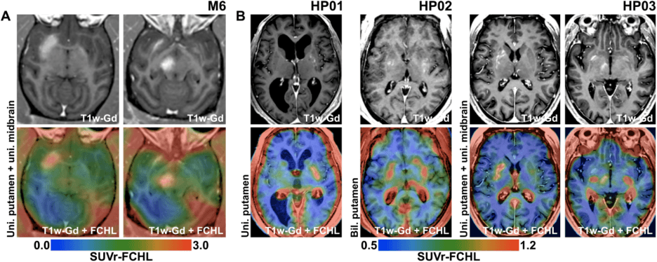**In vivo PET imaging of permeability BBB using** 18F**-holine at NHP and patients with PD**
*Source: Javier Blesa et al., Discovery BBB focused ultrasound in non-human primates and patients with Parkinson’s disease: targeted vector delivery AAV. Adv.9,eadf4888 (2023). DOI: 10.1126/sciadv.adf4888*
**Categories:** About the Treatment of Parkinson's Disease
---
### [PK-Merz (Amantadine) ](https://buzaevclinic.com/conditions/parkinsons-disease/treatment/pk-merz-amantadine-sulphate/)
**Published:** June 13, 2023
**Author:** Gulnara Akhmadeeva
**Excerpt:** The amantadine group is the only group of antiparkinsonian drugs that can reduce the manifestations of motor complications from long-term levodopa therapy in the form of choreiform dyskinesias. Most often, they are involuntary, dancing, chaotic movements in the limbs, swaying, and jerking movements in the torso, and most often occur at the peak dose of levodopa.
Amantadine has two representatives: PK-Merz (manufacturer - international company Merz), Midantan (produced in Belarus, this drug is included in the medication benefits for patients).
**Content:**
# PK-Merz (Amantadine)
**Amantadine group** – this is the only group of antiparkinsonian drugs that can reduce the manifestations of motor complications from long-term levodopa therapy in the form of choreiform dyskinesias. Most often, they are involuntary, dancing chaotic movements in the limbs, swaying and jerking movements in the torso, and most often occur at the peak dose of levodopa)
Amantadine has two representatives. PK-Merz (manufacturer – international company Merz), Midantan (produced in Belarus, this medication is included in the drug benefit for patients)
They have different chemical structures: PK-Merz is amantadine sulfate, and midantan is amantadine chloride. This affects differences in the duration of the effect (PK-Merz lasts slightly longer) and the rate of development of drug resistance (midantan is slightly faster).
Amantadines are effective due to several different **mechanisms of action**.
First, due to the blockade of specific glutamate NMDA-receptors. Glutamate is the main excitatory (and damaging) neurotransmitter, and when there is an excessive accumulation of sodium glutamate, neuron death occurs due to excitotoxicity. This provides an antidyskinetic effect and even possible neuroprotective action (convincing data is not yet available)
Secondly, due to the increased synthesis of dopamine and the decreased reuptake of it, which also increases its quantity. Therefore, the main antiparkinsonian effect manifests as a reduction in all three main symptoms of parkinsonism (hypokinesia/slowness, muscle rigidity/stiffness, resting tremor)
Thirdly, due to the mild anticholinergic effect resulting from decreased acetylcholine production. This may provide additional reduction of resting tremor.
Fourth, PC-Merz is the only antiparkinsonian drug available in infusion form (it can be administered intravenously via drip).
Therefore, **the main indications for the use of PC-Merz** are:
– early stages of PD (a rather small effect)
– advanced stages of Parkinson’s disease with the development of motor complications in the form of drug-induced dyskinesias
– atypical parkinsonism (together with levodopa medications)
– acute situations (akinetic crisis caused by withdrawal of antiparkinsonian drugs or introduction of neuroleptics, decompensation of PD)
The most common side effects may include skin discoloration in the form of white-blue star-shaped marbling (livedo reticularis) and a decrease in blood pressure. Taking the medication in the evening may cause difficulty falling asleep (therefore, PK-Merz is recommended to be taken in the morning and daytime, strictly before 16:00-18:00). The anticholinergic effect may also cause some confusion and visual hallucinations, exacerbate urinary retention symptoms in the presence of prostate adenoma, arrhythmia, and dry mouth.
**Contraindications include** renal and hepatic failure, epilepsy, thyrotoxicosis, closed-angle glaucoma, and prostate adenoma. The possibility of developing psychosis and hallucinations is assessed separately (based on existing hallucinations and age).
When prescribing amantadines, as with all other medications, a doctor’s consultation is necessary.
Bless you!
**Categories:** About the Treatment of Parkinson's Disease
---
### [How to organize medication intake for Parkinson's disease ](https://buzaevclinic.com/conditions/parkinsons-disease/treatment/how-to-take-parkinson-pills-properly/)
**Published:** January 15, 2024
**Author:** Gulnara Akhmadeeva
**Content:**
Taking antiparkinsonian medications is prescribed according to a specific regimen. Poor adherence to therapy leads to a decrease in the effectiveness of the prescribed treatment. To ensure that you or your loved one receives medication on schedule, it is necessary to maintain the regimen and prescriptions from your Parkinson’s specialist. In this article, we will provide you with tips on organizing the intake of antiparkinsonian medications.
1. Regularly keep a list of medications you are taking and bring it with you when visiting the doctor. Help your relative with Parkinson’s disease organize a convenient form for their records. The list must include the following items: medication name, dosage, frequency, and method of administration. If you are taking medications for another condition, they should also be noted in the list according to the above principle.
2. Store prescribed medications in a pill organizer with a secure lid. We recommend purchasing a pill container with a removable strip so that the daily dosage can be easily taken with you when leaving the house. Arrange all medications in the containers, including over-the-counter ones, not just those related to anti-parkinsonian treatment.
3. Set a reminder for taking your medication. You can ask for help from loved ones to set up a convenient timer on your mobile phone or purchase a watch with a vibrating alarm.
4. It should be remembered that a person with Parkinson’s disease may experience memory impairment, so close ones should carry the medication the person takes.
5. Consult a Parkinson’s specialist before independently adding new medications and over-the-counter supplements. If a person with Parkinson’s disease is taking MAO-B inhibitors (selegiline), some over-the-counter medications and supplements may be incompatible for combined use and may have a range of side effects.
6. Do not abruptly stop taking Parkinson’s disease medications for an extended period due to the risk of complications such as an akinetic crisis. If you need to adjust the dosage, our center’s neurologists specializing in Parkinson’s can assist you.
**Categories:** About the Treatment of Parkinson's Disease
---
### [Non-motor changes in Parkinson's disease ](https://buzaevclinic.com/conditions/parkinsons-disease/diagnostic/non-motor-symptoms/)
**Published:** January 9, 2020
**Author:** Aleksej Savelev
**Content:**
In Parkinson’s disease, in addition to movement disorders, there is a wide range of non-motor changes (not related to movements). Mood changes are quite common and are an important symptom. In some patients, they may reduce the quality of life more than motor symptoms. The most common symptoms that may occur in Parkinson’s disease include depression, anxiety, and apathy.
Depression is characterized by a low mood, sadness, a sense of hopelessness, feelings of loneliness or guilt.
Anxiety, i.e., a feeling of nervousness, worry, tension, excitement. Patients may experience fear or panic attacks.
Apathy – loss of interest, enthusiasm, motivation for daily activities or social interaction.
Symptoms of altered mood can appear at any time. In some patients, such symptoms may even precede movement disorders, while in others, they may occur several years after the first manifestations of the disease.
Non-motor changes in Parkinson’s disease occur due to alterations in brain regions that control mood. The disease reduces the levels of neurotransmitters such as dopamine, serotonin, and norepinephrine, which may underlie these symptoms. Your attitude towards the disease and certain psychosocial factors can also contribute to these symptoms.
There are several ways to treat mood disorder symptoms:
Antidepressants and anxiolytics can reduce the symptoms of depression and anxiety.
Dopaminergic medications that improve motor functions may also be effective for depression and anxiety, especially if you notice an improvement in your condition as a result of taking the medications.
Psychotherapy can be effective for these conditions, helping to reassess the attitude towards the illness.
Some physical activity programs, exercises, and an active social life can help in treating apathy.
Improving sleep quality and social support with stress limitation are also effective ways to combat non-motor symptoms of the disease.
**Categories:** About the Symptoms and Diagnosis of Parkinson's Disease
**Tags:** Parkinson's Disease
---
### [Instrumental Methods for Diagnosing Parkinson's Disease ](https://buzaevclinic.com/conditions/parkinsons-disease/diagnostic/instrumental/)
**Published:** November 23, 2022
**Author:** Aleksej Savelev
**Excerpt:** Diagnosis of Parkinson's disease in most cases remains purely clinical. However, sometimes certain instrumental methods can be used to slightly increase the confidence in the diagnosis.
**Content:**
# Instrumental Methods for Diagnosing Parkinson’s Disease
Diagnosis of Parkinson’s disease in most cases remains purely clinical. However, sometimes certain instrumental methods can be used to slightly increase the degree of confidence in the diagnosis.
This is about a simple and accessible method for visualizing the substantia nigra of the brain. Let us recall that this is the very area of the brain whose gradual atrophy causes the main motor symptoms of Parkinson’s disease—slowness of all movements (hypokinesia), muscle stiffness (rigidity), and trembling of the limbs (often resting tremor)
The technique is an ultrasound examination of brain structures conducted transcranially (through the open temporal window of the skull bones) and is called transcranial sonography (TCS).
In more than 90% of patients with Parkinson’s disease, a specific biomarker is identified using TCSG – an increased area of hyperechogenicity signal in the substantia nigra, or hyperechogenicity of the substantia nigra. The method is included in all expert recommendations for the diagnosis of PD – according to the recommendations of the International Parkinson and Movement Disorder Society (European Section) EFNS/MDS-ES MRS has an evidence level A in differentiating PD from atypical and secondary parkinsonism
For the hyperechogenicity of SN to be considered a sign of PD, the area must be more than 0.20 cm. The method has almost 90% specificity and almost 90% sensitivity. However, false-negative results may occur—most often observed in genetic forms of Parkinson’s disease caused by gene mutations PARK2 (parkin). Research has shown that the detection rate of this marker in parkin-associated genetic cases is no more than 50%
At the same time, the severity of SN hyperechogenicity is not a marker of the severity and progression of PD, nor does this indicator change depending on the stage. This means that TCS can be used for the diagnosis of PD at any stage—even in healthy individuals, to identify predisposition to the development of the disease.
The method is available at our Center (V.S. Buzaev International Medical Centre). We conduct this research together with the functional diagnostics physician Karina Fanilevna Valeeva.
Neurologist specializing in Parkinson’s disease at the V.S. Buzaev International Medical Centre, PhD, Gulnara Nailevna Akhmadeeva
**Categories:** About the Symptoms and Diagnosis of Parkinson's Disease
---
### [Parkinsonism-plus. Progressive Supranuclear Palsy ](https://buzaevclinic.com/conditions/parkinsons-disease/diagnostic/parkinsonism-plus-progressive-supranuclear-paralysis/)
**Published:** January 16, 2023
**Author:** Gulnara Akhmadeeva
**Content:**
# Parkinsonism-plus. Progressive supranuclear palsy
Progressive supranuclear palsy (PSP) is one of the most severe diseases in the parkinsonism-plus group.
I will remind you why these diseases are classified into a separate group and why they are called that. They belong to neurodegenerative diseases (like Parkinson’s disease and Alzheimer’s disease), meaning there is continuously progressive degeneration (atrophy) of brain cells in the brain. The cause is the accumulation of specific proteins, alpha-synuclein (as in the case of Parkinson’s disease, for example), or tau protein (as in the case of PSP).
«”Parkinsonism-plus” – because neurons die almost simultaneously in several different areas of the brain. This leads to the almost simultaneous appearance of several different symptoms and syndromes (in addition to the parkinsonism syndrome, as in the case of Parkinson’s disease). For example, speech disorders, swallowing difficulties and choking while eating, instability when walking and early falls, eye movement disorders, memory decline, and early development of dementia.
Compare – in Parkinson’s disease, for a long time, the only pronounced manifestation remains parkinsonism syndrome (slowness, muscle stiffness, resting tremor). Other symptoms (dementia, falls, swallowing and speech problems), if they appear, occur much later.
The problem is that often in the first 1-2 years of the development of any neurodegenerative disease, we can never be sure of the diagnosis, and frequently, over time, new symptoms appear that may lead to reconsidering the diagnosis towards another, more malignant and aggressively progressing disease from the parkinsonism-plus group.
The main, frequently occurring symptom of PSP is impaired eye movement. The patient finds it difficult to move their eyes, especially up and down. This becomes noticeable with the forced position of the head and neck (with the nose slightly raised upwards) and the patient’s particular facial expression (with eyebrows raised as if surprised, and infrequent blinking). To look to the sides or upwards, the patient has to turn their head, which is always difficult with PSP – because due to pronounced stiffness in the neck muscles, turning the head becomes very difficult
Unfortunately, treatment for progressive supranuclear palsy does not always bring clinical effect and improvement. This is because dopaminergic deficiency is not predominant in the pathogenesis of this disease, unlike Parkinson’s disease. Therefore, antiparkinsonian tablets, if they help, have a small effect and more often require large doses. Therefore, the general recommendation for treating PSP is to use the maximum tolerated doses of the most effective drug (levodopa)
Therefore, clinical rehabilitation and identification/treatment of all symptoms are very important in the management of a patient with Parkinson’s disease. This means preventing hypodynamia and stiffness, physical therapy (active and passive), massage, fall prevention, and the use of rehabilitation aids (walkers, canes, chairs, handrails), diet therapy, speech therapy, and much more.
Stay healthy!
Gulnara Nailevna Akhmadeeva,
PhD in Medicine,
neurologist specializing in Parkinson’s
V.S. Buzaev International Medical Centre
**Categories:** About the Symptoms and Diagnosis of Parkinson's Disease
---
### [Bicycle for Parkinson's Disease ](https://buzaevclinic.com/conditions/parkinsons-disease/lifestyle-lifehacks/bicycle/)
**Published:** January 9, 2020
**Author:** Aleksej Savelev
**Content:**
Bicycle for Parkinson’s Disease
Home physical exercises using a stationary bike and a remote monitoring app reduce the symptoms of movement disorders in patients with Parkinson’s disease \[1\].
High-intensity exercises have proven effective in reducing movement disorders in people with Parkinson’s disease, but prior to this study, the usefulness of home programs used by many patients was not clearly defined.
Scientists from the medical center Radboud University, Netherlands, developed the platform ” Park-in-Shape», which includes software with virtual reality and real-time video. They implemented the so-called approach ” exergaming» or fitness games, video games with physical activity, to allow patients to exercise on a stationary bike at home in more comfortable conditions
Centralized Randomized Study NTR4743) evaluated improvements from this type of aerobic exercise in the motor domain in patients with mild forms of Parkinson’s disease (stages 1 and 2 on the Hoehn and Yahr scale), who were either receiving continuous treatment with dopaminergic drugs or had not yet started it.
The study compared two groups of patients, all 130 of whom performed exercises three times a week for six months and either continuously took dopaminergic medications (stable dose for at least one month) or had not yet received pharmacological support. In the first group, 65 patients exercised for 30–45 minutes on a stationary bike at home (intervention group). Patients were advised to exercise within a comfortable heart rate zone, gradually increasing it as they became more trained. In the second group, 65 patients performed simple stretching, flexibility, and relaxation exercises for comparison (control group) during 30-minute sessions.
All participants had a motivational app with tips for optimal learning, support from loved ones, and information for tracking treatment progress. Everyone was monitored: once at home and remotely every two weeks.
Patients were evaluated at different times, both during dopaminergic treatment, which was still effective, and during periods when its effects had ceased.
The results showed that patients training on an exercise bike significantly improved motor function after six months. Specifically, during the period of medication action, their assessment scale was 4.2 points lower than the control group. Additionally, unlike the control group, patients engaging in aerobics improved their cardiovascular condition by the end of the study. However, no improvements were noted in non-motor symptoms such as fatigue, anxiety, depression, or cognitive functions, which researchers attribute to the short duration of the intervention.
This study once again proves the significant role of physical exercise in addition to medication therapy for improving the condition of patients with Parkinson’s disease. However, if you do not have a console with ” exergaming», nothing to worry about, – a regular stationary exercise bike, which many have gathering dust at home, is very suitable for achieving the first improvements
**Categories:** Lifestyle and Tips for Parkinson's Disease
---
### [Pathways to Improve Gait ](https://buzaevclinic.com/conditions/parkinsons-disease/lifestyle-lifehacks/tracks-for-gait-improvement/)
**Published:** January 9, 2020
**Author:** Aleksej Savelev
**Content:**
Pathways for Improving Gait
According to a new study, floor designs with large, clearly visible cross patterns improve motor activity in patients with Parkinson’s disease \[1\].
A characteristic feature of Parkinson’s disease is movement disorders, where patients temporarily feel as if their feet are glued to the floor, making it difficult to take a step forward. The use of visual stimulation to regulate or control movements has proven effective, helping patients improve their walking. This research showed that transverse and bright even lines are more effective than parallel stripes.
In many countries, when designing public spaces, accessibility and convenience for people in wheelchairs and with visual impairments are considered, but diseases like Parkinson’s disease are rarely taken into account. Researchers from Charles University and the Czech Technical University evaluated types of floor covering samples that can improve walking in this group of patients
The study included 32 patients (average age 67.5 years), whose disease was diagnosed on average 13 years ago.
Patients were asked to move forward and backward along an eight-meter path with six different floor patterns. Then, the scientists analyzed indicators such as timed walking, number of steps, number of steps in a turn, speed, and step length. They also analyzed the number of correct and incorrect backward counts, as well as the number and total duration of freezing episodes. Patients were asked to assess how the visual perception of the floor affected their walking.
The results showed that bright transverse lines on the floor improved time, speed, number of steps, and step length compared to diagonal lines. Other floor patterns contributed to improved walking and increased movement speed more than a plain floor, but to a lesser extent. Walking with virtual reality glasses also showed better results compared to a regular surface, but patients were often distracted by observing the surrounding reality.
Scientists recommend using research data when designing flooring in public places, medical facilities, and even at home to improve motor activity in patients with Parkinson’s disease.
In addition to a good floor, a favorite song can help you walk more steadily with Parkinson’s disease.
«”Road surface models should be developed to improve gait in patients with Parkinson’s disease” // Movement Disorders». 08.2019.
**Categories:** Lifestyle and Tips for Parkinson's Disease
---
### [Sex and Parkinson's Disease: Don't Be Afraid to Live Fully! ](https://buzaevclinic.com/conditions/parkinsons-disease/lifestyle-lifehacks/sex/)
**Published:** January 10, 2020
**Author:** Gulnara Akhmadeeva
**Content:**

Gulnara Akhmadeeva
[See Full Bio](https://buzaevclinic.com/author/gulnara-akhmadeeva/)
[YouTube](https://www.youtube.com/channel/UCLJMTPWarzceRoAglfcogKw)
# Sex with Parkinson’s Disease: Don’t Be Afraid to Live Fully!
Parkinson’s disease is a serious challenge that affects many aspects of a person’s life. However, research results PRIAMO, conducted by Italian scientists from the Center for Neurodegenerative Diseases CEMAND) University of Salerno provides an inspiring perspective on the importance of sexual activity for men with early stages of Parkinson’s disease. This data confirms: an active sex life can be an important tool in improving both motor and non-motor symptoms.
It turns out that sex can help improve the health of a person living with Parkinson’s disease. This conclusion was reached by the authors of a new two-year study that observed the sexual habits and disease progression among 355 patients at the Center for Neurodegenerative Diseases at the University of Salerno in Italy. Dr. Picillo and her colleagues concluded that sexually active male patients experience a “milder” progression of the disease and have less motor deficit, as well as report fewer other non-motor symptoms (compared to those who do not).
Parkinson’s disease can negatively affect a patient’s sex life in several ways.
1. Parkinson’s disease often causes tremor and stiffness throughout the body. This can complicate the technical aspects of sex and make it difficult, painful, or uncomfortable.
2. Men with Parkinson’s disease may experience erectile dysfunction (ED) due to nerve and muscle issues. ED can occur with impaired blood circulation in the genital area. This happens due to dysfunction of the autonomic nervous system in Parkinson’s disease (leading to arterial hypotension), as well as the use of antiparkinsonian medications (which in most cases can also lower blood pressure)
3. Women with PD due to the same autonomic dysfunction may have insufficient moisture in intimate areas and often need additional lubrication.
4. Both men and women may notice a decrease in libido (sexual desire) after being diagnosed. This may be related to a decrease in dopamine levels, the main causal neurotransmitter in Parkinson’s disease. Alternatively, it may be due to manifestations of anxiety-depressive disorder (this is also common in Parkinson’s disease)
5. Furthermore, some medications, including antidepressants, can themselves reduce sexual desire.
6. On the other hand, there is a group of drugs—dopamine receptor agonists (which include medications such as pramipexole, piribedil, ropinirole)—that can cause hypersexuality and excessive sexual desire, leading to deviant behavior. This is called impulsive-compulsive disorders, and if problems arise in the family, they need to be identified and controlled (primarily by changing the antiparkinsonian treatment regimen).
7. And finally, of course, the stress from the seriousness of the situation can also affect the patient and their partner—sometimes fatigue and lack of sleep can reduce (or temporarily postpone) the desire for sex.
## Why is sexual life important?
Sexual activity is not just an aspect of personal life but also an indicator of overall health. Research shows that men with Parkinson’s disease who maintain sexual activity experience slower disease progression, improved quality of life, and reduced symptoms of depression. This important finding highlights that intimate life can play a significant role in rehabilitation.
## What does research say?
As part of the research PRIAMO scientists analyzed data from 355 patients with early-stage Parkinson’s disease over two years. At the beginning of the study, 56.3% of participants reported their sexual activity, and after two years, this figure decreased to 50.8%. Interestingly, men were twice as sexually active as women. However, almost half of the sexually active men faced erectile dysfunction and other difficulties in their intimate lives.
## Positive Effects of Sexual Activity
The results of the study showed that men with an active sex life:
- less frequently experienced apathy and gastrointestinal issues
- had less pronounced movement disorders;
- reported better quality of life and lower levels of depression
- complained less about fatigue and attention problems;
- observed slow progression of the disease
The most important thing is that these positive changes were observed regardless of the severity of the disease.
## Why does it work?
Sexual activity can be considered a form of physical and emotional activity that positively affects the body. It helps maintain motivation, improves circulation, reduces stress levels, and promotes the production of happiness hormones. All of this can have a beneficial impact on the course of Parkinson’s disease.
## What to do if there are difficulties?
If you or your partner are experiencing difficulties in your sexual life, it is important not to ignore the problem. Discuss it with your healthcare provider. Modern treatment methods, including medication and psychological support, can help address issues such as erectile dysfunction or decreased sexual desire.
## The Importance of Open Dialogue
Scientists emphasize that doctors should actively inquire about the sexual life of their patients with Parkinson’s disease. This aspect should not be considered secondary—it can significantly affect overall health and quality of life.
## Interview with a Parkinsonologist: Sex and Sexual Activity in Parkinson’s Disease
Interview with Neurologist-Parkinsonologist, PhD Gulnara Akhmedeeva about Sex in Parkinson’s Disease ### Can sex improve the condition of patients with Parkinson’s disease?
**Gulnara Akhmadeeva:** Hello everyone! Today we are discussing a topic that might not be immediately addressed during a doctor’s appointment. It’s sex and sexual activity in Parkinson’s disease. It turns out that sex can improve motor activity in patients living with Parkinson’s disease. This conclusion was reached by Italian researchers who conducted a two-year study of 355 patients with the condition. They found that sexually active male patients experience milder disease progression, less motor deficit, and fewer non-motor symptoms compared to those who do not lead an active sexual life. Let’s explore how sexual activity can change depending on the course of Parkinson’s disease.
### What physiological problems in sex can arise due to Parkinson’s disease?
**Gulnara Akhmadeeva:** In fact, there can be several such influences. Parkinson’s disease often causes tremor and stiffness, which can complicate the technical aspect of sex, making it painful, difficult, or uncomfortable. Men with Parkinson’s disease often experience erectile dysfunction related to impaired blood circulation in the genital area. This occurs due to disruptions in the autonomic nervous system and decreased blood pressure. It is also worth considering that many antiparkinsonian medications can lower blood pressure, further affecting sexual function.
### What sexual problems do women with Parkinson’s disease face?
**Gulnara Akhmadeeva:** Women with Parkinson’s disease may also face issues such as insufficient moisture, requiring the use of additional lubrication. Both men and women may notice a decrease in libido after diagnosis. This is related to a decrease in dopamine levels, a key neurotransmitter, as well as serotonin, which often leads to depression and anxiety. Some medications, including antidepressants, may also reduce sexual desire. In such cases, considering a medication change or other treatments for anxiety and depression may be helpful.
### Which medications for Parkinson’s disease can affect sexual activity?
**Gulnara Akhmadeeva:** On the other hand, there is a group of medications, such as dopamine receptor agonists (pramipexole, ropinirole), which can enhance sexual desire. However, in rare cases, they may cause hypersexuality or impulse control disorders. If this leads to family issues, it is necessary to adjust the treatment plan by discontinuing or replacing the medication.
### How do stress and fatigue affect sexual life in Parkinson’s disease?
**Gulnara Akhmadeeva:** Moreover, the stress of a chronic illness, fatigue, and lack of sleep can reduce the desire for sex. In such cases, you can shift sexual activity to another time of day, such as in the morning or on weekends. To maintain an active sex life, it is important to openly discuss your issues with your partner and doctor. If you are concerned about depression, fatigue, erectile or mobility problems, be sure to talk to your healthcare provider, neurologist, or urologist. Modern treatment methods, including psychotherapy, meditation, and support groups, can significantly improve your condition.
### What specialists can help with addressing sexual activity issues in Parkinson’s disease?
**Gulnara Akhmadeeva:** If you have problems with erection or urination, such as incontinence or retention, a neurourologist can offer safe medications for treatment. If the disease progresses, with increased tremor, stiffness, or other symptoms, be sure to contact your healthcare provider. The specialists at our center are always ready to help you adjust your treatment and maintain your active sexual life. Wishing you health and goodbye!
## Conclusion
Sexual life is not only a possible way to improve health but also a source of joy and intimacy. If you are living with Parkinson’s disease, remember: an active sexual life can be an important element in your fight against the disease. Don’t be afraid to openly discuss this with your doctor and your partner. Life goes on, and you deserve to enjoy it to the fullest!
Problems with erection and other genitourinary disorders (frequent urges, difficult urination) can be corrected with medications that are quite safe for Parkinson’s disease. Information about this, as well as treatment recommendations, can be obtained from our team’s neurourologist – Ivan Alekseevich Kuvin.
And of course, with the progression of the disease and deterioration of the motor condition, you can always contact me and the neurologists at our center.
## Sources:
European Journal of Neurology. 2.07. 2019. URL: [https://onlinelibrary.wiley.com/doi/full/10.1111/ene.13983 ](https://onlinelibrary.wiley.com/doi/full/10.1111/ene.13983%20)
**Categories:** Lifestyle and Tips for Parkinson's Disease
**Tags:** Video
---
### [10 Tips for Caring for a Loved One with Parkinson's Disease ](https://buzaevclinic.com/conditions/parkinsons-disease/lifestyle-lifehacks/ten-tips-for-caregivers/)
**Published:** January 10, 2020
**Author:** Aleksej Savelev
**Content:**
### 10 tips for those caring for patients with Parkinson’s disease:
1. **Be organized**
Keep all medical records, notes, appointment schedules, and phone numbers of doctors and clinics in one folder. Take this folder with you to all appointments so that everything you need is always at hand.
2. **Take care of yourself**
To care for a loved one, you must be physically and emotionally healthy. Take breaks, engage in activities you enjoy, ask for help from other family members or friends, and consider the possibility of professional support.
3. **Study the disease**
Learn as much as possible about Parkinson’s disease, its stages, and possible treatment options. This will help you better understand your loved one’s condition and be prepared for changes.
4. **Join the support group**
Find support groups in your region or online for caregivers of patients with Parkinson’s disease. This will give you the opportunity to share experiences, receive advice, and emotional support.
5. **Expect changes in relationships**
Mood swings, depression, and irritability are common companions of Parkinson’s disease. Be prepared for changes and strive to maintain open and honest communication. This will help both of you adapt to new roles in the relationship.
6. **Monitor the symptoms**
As a caregiver, you are likely to be the first to notice changes in condition or behavior. Inform doctors of any new symptoms or deteriorations to receive timely assistance.
7. **Encourage independence**
Do not do too much for your loved one. Try to support their desire to be independent. Ask if they need help and respect their aspiration for independence.
8. **Know Your Rights**
Familiarize yourself with the rights of caregivers and persons with disabilities. Find out what benefits or allowances you may be eligible for to ease the financial burden.
9. **Discuss the future**
Despite the complexity of the topic, it is important to talk with a loved one about their future wishes. Discuss wills, treatment options, and end-of-life issues.
10. **Receive up-to-date information**
Stay updated on news about Parkinson’s disease treatment, explore new therapy methods, and consult with doctors. This will help you stay informed about all available options to improve your loved one’s quality of life.
Caring for a loved one diagnosed with Parkinson’s disease is very challenging both emotionally and physically. It is painful to watch your loved ones suffer, and it makes you want to do everything possible for them, but it’s important to remember that you must also take care of yourself. We have gathered some practical tips for those caring for patients with Parkinson’s disease, which we hope will ease your life and help relieve stress. Be as organized as possible: keep all medical notes, records, appointments, contacts of clinics and doctors of your loved one in a folder that is easily accessible at any time. Take this folder with you to all appointments so you always have everything at hand. Take care of yourself: to be able to care for your loved one, you must be physically and emotionally healthy. Take a break – it’s important to relax and relieve stress. Ask other family members and friends for help or seek professional assistance. Spend some time doing what you enjoy and take a few hours to distract yourself. Make sure you fully understand your loved one’s condition, the stage of the disease, and the medical care that can be received. Join a support group: find out if there are any active support groups in your area. If not, find an online support group for those caring for people with Parkinson’s disease. This will provide you with both practical and emotional support, and you can ask for advice and share your experiences with others going through the same thing. Learn more about Parkinson’s disease: stay informed about all the latest news on the condition, read about the symptoms, and talk to your loved one’s doctor about what to expect as the disease progresses. Expect changes in relationships: mood swings and depression are often observed in Parkinson’s disease, and your loved one may also become upset. It is difficult to cope with these changes in relationships, so open and honest communication is critically important. Both of you will need time to adjust to new roles in the relationship. Observe symptoms and report any changes. As a caregiver for your loved one, you will likely be the first to notice any changes in behavior or worsening symptoms. Report these to your loved one’s doctors as they occur so they can be addressed as soon as possible. Encourage independence: try not to do too much for your loved one, as they will want to maintain their independence as long as possible. Ask if they need help and respect their autonomy. Know your rights: familiarize yourself with the rights of caregivers and the rights of people with disabilities. Find out if you are entitled to any disability benefits or caregiving allowances. After accepting the disease, talk about the future. Although it is a difficult topic to discuss, it is important to understand what your loved one wants going forward. You need to discuss wills, treatment options, and end-of-life decisions.
**Categories:** Lifestyle and Tips for Parkinson's Disease
---
### [Breastfeeding with Parkinson's Disease ](https://buzaevclinic.com/conditions/parkinsons-disease/lifestyle-lifehacks/breastfeeding/)
**Published:** September 7, 2022
**Author:** Aleksej Savelev
**Content:**
Despite the fact that Parkinson’s disease is more common in the elderly, 4-10% of patients experience the onset of the disease before the age of 40. In the USA, about four hundred women under the age of 50 are diagnosed with Parkinson’s disease each year, while such statistics for Russia could not be found.
Young women start families, become pregnant, and give birth. Breastfeeding is important for the mother and baby, both as the best nutrition for the child and as a contribution to their future health, as well as the closest bond—love between the little one and their mother. Is it possible to achieve such natural feeding when you have Parkinson’s disease?
One of the most commonly used drugs for treating Parkinson’s disease is Levodopa. It is a natural amino acid, a direct precursor of dopamine, and has similar effects. This drug is considered low risk during breastfeeding. Levodopa is excreted in breast milk in clinically insignificant amounts, 100 times lower than the dose used for infants who need it ( Thulin, 1998). In infants whose mothers took Levodopa, no short-term or long-term problems were observed ( Thulin, 1998). The risk of taking this medication is that it reduces prolactin secretion ( WHO 2002, Petraglia 1987, Nattero 1986, Linkowski 1983, Kaulhausen 1982, Rao 1982), prolactin is the hormone responsible for the volume of milk produced
Two cases are described: in one case, the mother breastfed for 4.5 months while taking the medication, and stimulation through sucking was sufficient to maintain a good milk supply despite the therapy. In the second case, the mother, suffering from Parkinson’s disease, was very difficult to treat and received continuous intravenous infusion of Levodopa and duodenal gel of Carbidopa during pregnancy and after childbirth. This mother breastfed for 3 months, the child’s psychomotor development was normal, unlike the child’s weight, which was affected by intrauterine malnutrition (Zlotnik 2014)
To avoid the side effects of levodopa (nausea, vomiting, arrhythmias), levodopa is combined with carbidopa or benserazide, which block peripheral enzymes that metabolize levodopa, allowing more levodopa to reach the brain, thus enabling a lower dose of levodopa to be prescribed. Carbidopa, which is combined with levodopa, inhibits levodopa’s suppressive effect on prolactin and increases its level, which is beneficial for breastfeeding ( Camanni 1978). To date, there are no published data on the excretion of this drug in breast milk. The wide volume of distribution and short half-life make it unlikely for carbidopa to enter breast milk in significant amounts.
If we take a broader look at the group of anticholinergic and dopaminergic agents used to treat Parkinson’s disease, it can be noted that the drugs are categorized by their impact on breastfeeding into groups of low, high, and very high risk.
In the low-risk group: levodopa, carbidopa, amantadine hydrochloride, apomorphine hydrochloride, biperiden, orphenadrine. These drugs have a very low likelihood of entering breast milk, and most of them reduce prolactin levels. Biperiden is the only drug that does not affect prolactin. Caution is advised with apomorphine and orphenadrine when prescribing to mothers of premature infants and infants in the first 7 days of life, when intestinal permeability is increased.
High-risk group: bromocriptine mesylate, pramipexole, procyclidine, ropinirole hydrochloride. This group has a stronger effect on prolactin and suppresses lactation. Bromocriptine does not enter breast milk at all and poses no danger to the infant when taken by the mother. It is better to postpone the use of ropinirole hydrochloride by the mother for the first 20 days of the child’s life.
Rasagiline also suppresses prolactin production. The effect of this drug on breastfeeding has not yet been studied, so it is currently in the very high-risk group.
If you have Parkinson’s disease and are a nursing mother, consult a neurologist specializing in Parkinson’s to select the optimal and safe therapy for you and your baby.
If you have been prescribed medications that affect prolactin levels, to ensure everything goes well, it is necessary to monitor several things:
- purchase or rent a baby scale and monitor your baby’s morning weight once a week. In the first 3 months of life, a baby gains at least 150g per week, optimally around 200g. If the weight gain is less, you will need to weigh more often and seek assistance in organizing breastfeeding and calculating supplementary feeding
- ask for help from a professional in latching the baby to the breast, the baby must be latched very well to ensure good breast emptying and sufficient milk quantity;
- use asymmetric latch, carefully monitor the swallowing, enhance the milk ejection reflex with breast compression according to Newman, massage, warm drinks;
- supplement feeding with pumping if there is not enough milk. Depending on the amount of milk and how much energy you have, this may be a couple of pumping sessions or more. In this case, it is better to get a recommendation from a breastfeeding consultant
- Skin-to-skin contact with your baby during the day will help you relax and make feedings easier.
Thus, breastfeeding, even with Parkinson’s disease, is a great benefit for your baby. You should not give it up—just choose the right medications and treatment plan. Specialists at our medical center will gladly assist and support mothers with this condition.
Olga Vladimirovna Nodvikova,
Breastfeeding Consultant, Neonatologist
V.S. Buzaev International Medical Centre®
**Categories:** Lifestyle and Tips for Parkinson's Disease
**Tags:** Parkinson's Disease
---
### [How to maintain balance and avoid falling with Parkinson's disease? ](https://buzaevclinic.com/conditions/parkinsons-disease/lifestyle-lifehacks/fall-prevention/)
**Published:** January 10, 2023
**Author:** Gulnara Akhmadeeva
**Content:**

Gulnara Akhmadeeva
[See Full Bio](https://buzaevclinic.com/author/gulnara-akhmadeeva/)
[YouTube](https://www.youtube.com/channel/UCLJMTPWarzceRoAglfcogKw)
## Why do falls occur in Parkinson’s disease?
Falls in Parkinson’s disease are an almost inevitable symptom that may indicate the gradual progression of the disease. Several causes can lead to this:
- Balance disorder
- Stooping and Other Postural Changes
- Freezing and delayed muscle response
- Poor vision
- Muscle loss weakness
- Side effects of medications used for other conditions (depression, insomnia, hypertension)
- [Dangerous Surrounding Environment ](https://buzaevclinic.com/conditions/parkinsons-disease/lifestyle-lifehacks/safe-home-environment/)
## **What are the dangers of falls?**
The fact is that as a result of falls, patients may suffer injuries and sustain concussions, bruises, and fractures, up to a hip fracture, which can lead to reduced mobility and a “bedridden patient” state. Additionally, any injuries can lead to the decompensation of Parkinson’s disease itself and reduce the effectiveness of antiparkinsonian therapy.
## Environment around
[It is necessary to arrange the home to prevent falls in Parkinson’s disease, about which we have a separate article. ](https://buzaevclinic.com/conditions/parkinsons-disease/lifestyle-lifehacks/safe-home-environment/)
It is very important for reducing the risk of falls to engage in fall prevention through kinesitherapy and therapeutic exercise
## How to improve your balance and stability with Parkinson’s disease?
Exercises for improving balance and coordination are very useful and effective. Kinesiotherapists or physical therapy specialists help select the right exercises suitable for each specific patient. [We have an online school for training different patients with various stages of Parkinson’s disease](https://buzaevclinic.com/conditions/parkinsons-disease/lifestyle-lifehacks/parkinsons-disease-community/). How can you improve your stability to avoid falling?
- make sure the shoes are in good condition – laces tied, soles undamaged
- Footwear should be comfortable – fit well to the foot, not be loose, and must have a back part (heel)
- wearing flip-flops (without backs) and high heels is not recommended;
- when walking, avoid multitasking – when walking, ONLY walk (do not talk on the phone, do not watch TV, do not listen to music with headphones). Remember that in Parkinson’s disease, postural reflexes (these are automatic trunk reactions for naturally maintaining balance and a certain posture in space) are reduced. Therefore, there is a risk of losing balance when attempting to perform multiple tasks simultaneously
- always keep one hand free – this will allow you to quickly find support on surrounding furniture or walls
- do not carry items in both hands
- when standing still – keep your feet shoulder-width apart. If you keep your feet closer together, the risk of falling increases
- in winter – beware of ice
- the phone should always be nearby and within easy reach from the floor (if a fall does occur, it should be easy to reach)
- a bracelet with an alarm or the ability to call for help might be useful if you need to call for assistance
- sometimes a whistle on a cord around the neck can help for the same purpose
## What to do if you have fallen due to Parkinson’s disease?
If a fall occurs and there’s no one to help you get up, you can remember a specific algorithm of sequential actions to return to an upright position:
- lie on your back
- turn onto your side
- sit with the help of your hands
- slowly crawl to the nearest object (chair, sofa, armchair) that can support a person’s weight
- place your hands on this object and spread them wide apart
- raise your strong leg so that the knee is close to the chest
- push off with your hands and feet to stand up
Stay healthy!
[Do you know that tremor and stiffness can be reduced or even eliminated without increasing the dose of levodopa? ](https://buzaevclinic.com/services/tremor-mrgfus-treatment/)
**Categories:** Lifestyle and Tips for Parkinson's Disease
---
### [April 11 - World Parkinson's Disease Day ](https://buzaevclinic.com/conditions/parkinsons-disease/lifestyle-lifehacks/world-parkinsons-day/)
**Published:** April 12, 2023
**Author:** Aleksej Savelev
**Excerpt:** The first World Parkinson's Day was held in April 1997. It was established by the European Parkinson's Disease Association and co-sponsored by the World Health Organization (WHO). This date is dedicated to the birthday of Dr. James Parkinson.
**Content:**
# Why is World Parkinson’s Day important?
World Parkinson’s Disease Day is an opportunity:
1. Unite those affected by Parkinson’s disease around the world – to highlight the impact of Parkinson’s disease on individuals, families, and communities
2. Celebrate the tremendous work that people living with Parkinson’s disease and those working in the field do to treat and raise awareness about this disease
3. Celebrate the resilience and strength of people who live with this condition every day
4. World Parkinson’s Disease Day calls for:
5. World leaders should take the necessary actions to ensure that people with Parkinson’s disease receive the quality of life they deserve
6. National leaders and policymakers should recognize the impact of Parkinson’s disease on individuals, their families, and caregivers, and develop appropriate strategies and policies to mitigate them
7. Healthcare professionals should be aware of the latest research and treatment methods to improve care and services for people with this condition
## World Parkinson’s Day – A Brief History
First World Parkinson’s Disease Day ( World Parkinsin Day) was held in April 1997. It was established by the European Parkinson’s Disease Association and co-sponsored by the World Health Organization (WHO). This date is dedicated to the birthday of Dr. James Parkinson.
**Doctor James Parkinson (1755 – 1824)**
Born on April 11, 1755. James Parkinson is best known for his essay “An Essay on the Shaking Palsy” in 1817, in which Parkinson’s disease was first recognized as a medical condition.
The launched European Parkinson’s Charter has garnered worldwide attention and received support from many influential figures, including British royalty Her Royal Highness Princess Margaret and Princess Diana, UK Prime Ministers John Major and Tony Blair, and Pope John Paul II, Italian opera singer Luciano Pavarotti and American boxing legend Muhammad Ali
As a result of World Parkinson’s Day in 1997, the WHO formed a Working Group on Parkinson’s Disease, which led to the development of the first Global Declaration on Parkinson’s Disease, adopted in Mumbai (India) in December 2003, aimed at encouraging a change in attitudes towards Parkinson’s disease.

During the 9th conference WPD (Luxembourg, 2005), the red tulip was adopted as the official symbol of the disease.
**What is “Spark ” («The Spark»)?**
In 2022, efforts were combined to develop and launch a new logo for World Parkinson’s Day, named ‘Spark’.
Design Spark inspired by dopamine – the neurotransmitter that people with Parkinson’s disease strive to preserve. A group of organizations aims to engage the community living with Parkinson’s disease this year around a more coordinated annual event dedicated to World Parkinson’s Day, ultimately bringing organizations and individuals together under one recognized banner, leading to greater awareness, impact, and ultimately positive change
**Categories:** Lifestyle and Tips for Parkinson's Disease
---
### [What to give a person with Parkinson's disease? ](https://buzaevclinic.com/conditions/parkinsons-disease/lifestyle-lifehacks/best-present-for-ill-person/)
**Published:** January 15, 2024
**Author:** Dinara Nabiullina
**Content:**
In this article, we will share tips to help bring joy to your loved ones suffering from Parkinson’s disease.
1. Sports Equipment or Home Exercise Machine. Research shows that regular physical exercise helps alleviate the symptoms of Parkinson’s disease. You can organize a space for sports activities in the living room and set up a portable pedal exerciser. To maintain physical activity, we recommend exercising with your loved ones.
2. Maintaining dental health is important for people with Parkinson’s disease, as some symptoms can complicate daily dental care. An electric toothbrush or toothpaste dispenser makes these tasks easier. You can’t go wrong with a gift if you give an electric shaver.
3. Every person with Parkinson’s disease finds joy in different activities. Ask your loved one what they would like to do. It might be drawing or yoga classes, acupuncture sessions, or a massage course. We remind you that our team has created an entire Community for patients with Parkinson’s disease(hyperlink), where materials on various activities (boxing, tai chi, yoga) and cognitive training are posted daily.
4. People with Parkinson’s disease often experience weight fluctuations. An excellent addition to a gift can be ordering prepared meals from a local restaurant or purchasing ready-made groceries for a week. Share our nutrition guide with the person with Parkinson’s disease, which we have posted in the Community, or send our article dedicated to the [nutritional features of Parkinson’s disease](https://buzaevclinic.com/conditions/parkinsons-disease/digestion-nutrition/food-and-meals/)
5. Gift special drop-resistant dishes or choose a coffee set: research shows that consuming caffeine in moderate amounts can improve motor symptoms of Parkinson’s disease.
6. Foot or neck massager can help you relax. Handheld massagers and a heating pad help relieve muscle stiffness and soreness.
7. Parkinson’s disease can interfere with daily life, such as during dressing. Slip-on shoes and a long-handled shoehorn can simplify getting ready for daily walks.
8. Aromatherapy can help you relax before sleep: bath bombs and oils can promote relaxation before bedtime.
9. A photo book or a custom portrait can be a great reason for a warm conversation and memories. Choose a theme that holds special value for your loved one: family vacations, favorite places, hobbies. Memories help train memory and evoke positive emotions.
10. Audiobooks can make reading a pleasant and convenient activity during walks or long trips. Gift a subscription to any service with audio and e-books.
**Categories:** Lifestyle and Tips for Parkinson's Disease
---
### [How to Set Up a Home for a Patient with Parkinson's Disease ](https://buzaevclinic.com/conditions/parkinsons-disease/lifestyle-lifehacks/safe-home-environment/)
**Published:** February 16, 2024
**Author:** Gulnara Akhmadeeva
**Content:**

Gulnara Akhmadeeva
[See Full Bio](https://buzaevclinic.com/author/gulnara-akhmadeeva/)
[YouTube](https://www.youtube.com/channel/UCLJMTPWarzceRoAglfcogKw)
Living with Parkinson’s disease is truly a challenge. You need to follow many rules to maintain quality of life. The main thing you can and should do is consciously respond to emerging difficulties. Over time, you will certainly accept and incorporate all the changes in your life, and we will explain how to better adapt to Parkinson’s disease and properly organize your living space.
What problems do people with Parkinson’s disease most often face? The most common issues are unsteadiness when walking (stumbling, episodes of “freezing”) and instability when maintaining posture. This often leads to falls and serious injuries. To protect yourself from these problems and create a safer environment, we recommend adapting your home space.
## Checklist for Organizing Space in Your Home:
### Ensure good visibility and lighting
Create a high level of illumination throughout the house, adjust blinds and curtains to minimize glare.
There should be sufficient lighting in the apartment even at night. If the patient wakes up, small light sources (nightlights) should be on for safe movement to the bathroom or kitchen.
It’s convenient to mark the beginning and end of the stairs with paint
### Make the space familiar
Furniture should not be moved frequently—this way, the patient will be familiar with the home environment and can find support in case of an emergency.
### Clear space and remove obstacles to movement
Create the most spacious area for movement in the corridors.
There should be no loose objects on the floor that could cause tripping. Remove all household items that could lead to a fall.
- pets (cats or dogs lying on the floor)
- scattered things
- Mats
- wires
- Interior thresholds
Ensure that the floors are stable, free of gaps and cracks, and have a smooth, non-slip surface.
If there are unsecured rugs in the apartment, they should be smoothed out (and the ends/corners attached to the floor, for example, with double-sided tape), or removed entirely to prevent them from becoming obstacles and causing falls.
Remove all electrical wires from common walking areas to avoid tripping and falling while moving.
Ensure that all pathways and steps in the apartment/house are in good condition
### Eliminate the causes of slipping
If the floors are not covered with anything, it’s important to consider making them less slippery – for example, walking around the house not in slippers, but in socks with special non-slip coating.
Replace slip-on sandals with boat-style slippers (with backs) to prevent them from slipping off the foot
### Create stillness of what can be leaned on
Furniture must be sturdy enough to support a person’s weight. It can help in case of loss of balance
Ensure that all furniture is securely fastened
Furniture should not rotate.
Chairs should be stable, without a swivel base, have armrests, and a sufficient seat height to facilitate standing up.
If necessary, part of the space is recommended to be furnished with walkers or wheelchairs.
Handrails in the corridor or bathroom (toilet), as well as on both sides of the stairs, can be very useful.
### Reduce the risk in case of a fall
A heavy carpet with thick pile, securely attached to the floor, can serve as a cushioning pad if a fall does occur.
Eliminate sharp corners on furniture, use special covers.
Avoid furniture with glass tabletops – they can be dangerous.
### Create an option to call for help
If a house/apartment has two floors, there should be a phone on each floor (preferably cordless).
Carry a mobile phone or an emergency button with you.
## How to Arrange Rooms for Parkinson’s Disease?
### Bedroom for Parkinson’s Patients
1. Set the bed height so that your feet touch the floor when sitting on the bed.
2. Install side handles on the bed or a special support to facilitate turning and getting out of bed.
3. Ensure comfortable and quick access to lighting sources to avoid walking in the dark.
4. Use a bright light source (nightlight, flashlight) to have good visibility on the way to the bathroom.
5. Make it a habit to carry a flashlight.
6. Near the bed in case of a power outage.
7. Place clothing hangers at a height that is convenient to use, ensure the wardrobe is well-lit and accessible, and that clothes can be retrieved without additional effort.
8. Keep frequently used devices (watch, phone) on the bedside table for easy access without getting out of bed at night.
9. Avoid using sheets and nightwear made from slippery materials.
What should a bedroom be like for someone with Parkinson’s disease?
### Bathroom for Parkinson’s Disease
The risk of being in the bathroom and toilet is high. Most falls occur due to difficulties when getting up from the toilet, entering, and exiting the bath. You may experience difficulties due to poor lighting in the bathroom, and it may be hard for you to stand on a wet surface.
1. Install grab bars near the toilet, bathtub, and shower. Do not use towel racks, faucets, or soap dishes for support when standing up.
2. For safety, add a sturdy bench with a backrest to the bath or shower.
3. Provide a seating area for brushing teeth and shaving.
4. Do not walk when the bathroom floors are wet. Do not store anything unnecessary in the bathroom.
5. The bathroom can also be made safer with a non-slip mat on the floor (or on the bathtub floor itself), and a seat/bench across the bathtub.
6. A shower cabin can be safer than a bathtub.
### Cuisine for Parkinson’s Patients
Cooking often represents a labor-intensive process. You may experience difficulties operating many kitchen appliances. Due to balance disorders, opening the refrigerator door, oven, and kitchen shelves can be challenging.
1. Install convenient and durable handles for cabinets.
2. Store necessary items in drawers accessible for quick use
3. Use a single-handle faucet for the sink.
Take care of yourself, and we will help you with that!
[In addition to setting up your home, it may be helpful to work on stability and balance. ](https://buzaevclinic.com/conditions/parkinsons-disease/lifestyle-lifehacks/fall-prevention/)
[Did you know that tremor and stiffness can be reduced or even eliminated without increasing the dose of levodopa? ](https://buzaevclinic.com/services/tremor-mrgfus-treatment/)
**Categories:** Lifestyle and Tips for Parkinson's Disease
---
### [Vitamin D and Parkinson's Disease ](https://buzaevclinic.com/conditions/parkinsons-disease/digestion-nutrition/vitamin-d/)
**Published:** January 10, 2020
**Author:** Guzaliya Sakharova
**Content:**
## **Vitamin D and Parkinson’s disease**
Vitamin D — this is a fat-soluble vitamin that plays a key role in maintaining human health. It is primarily known for its influence on calcium and phosphorus metabolism, which ensures normal growth and development of bone tissue. However, its functions are not limited to this. Thus, vitamin D is involved in the regulation of numerous physiological processes. Recent studies indicate its involvement in dopaminergic neurotransmission, neurogenesis, and neuron development, which is particularly important for the functioning of the nervous system in Parkinson’s disease.
## Is there a vitamin D deficiency in Parkinson’s disease
Authors of the Russian study found that more than 65% of patients with Parkinson’s disease have a vitamin deficiency D \[1\]. According to data from foreign colleagues, vitamin deficiency D was found in 55% of patients with Parkinson’s disease \[2\]. Currently, great attention is being paid to the role of receptors (natural sensors) for vitamin D (VDR) in the pathogenesis of Parkinson’s disease, a genetic deviation of this receptor causes a more pronounced vitamin deficiency D in patients with Parkinson’s disease
## Low Vitamin D Levels and Severity of Parkinson’s Disease
Despite the theoretical justification of the neuroprotective properties of the vitamin D, clinical studies evaluating its effectiveness in treating Parkinson’s disease are still limited. In the article « A Review of theRelationship Between Vitamin D and Parkinson Disease Symptoms» the authors analyzed the relationship between vitamin levels D and symptoms of Parkinson’s disease. It was noted that low levels of vitamin D in blood plasma is associated with more severe movement disorders, such as tremor, rigidity, and gait disturbance. Taking vitamin D reduced the severity of these symptoms in some patients
## Slowing the Progression of Parkinson’s Disease with Vitamin D Intake
Research has also shown that maintaining vitamin levels D above 30 ng/ml helps slow the progression of movement disorders \[3\]. For example, one study found that men working outdoors had a lower risk of developing Parkinson’s disease compared to those working indoors, which may be related to increased vitamin production D under the influence of sunlight \[4\].
In addition, vitamin D has antioxidant activity, which allows it to reduce oxidative stress—one of the factors contributing neurodegeneration in Parkinson’s disease. It has been shown that vitamin D prevents excessive calcium accumulation in nervous cells, what slows down their apoptosis (programmed death). This is especially important for the protection of neurons in the substantia nigra (the location in the brain, where dopamine is produced) \[5\].
## Can vitamin D be taken for Parkinson’s?
Recommendations for Vitamin Use D in the treatment of Parkinson’s disease may be justified in the case of a detected deficiency of this vitamin, especially in the older age group, where vitamin deficiency D is a common problem
However, for accurate dosage determination and optimal treatment plans, a specialist consultation is necessary!
## Links to cited research:
1\. Novotny D. A. et al. The Influence of Vitamin D and other indicators of phosphorus-calcium metabolism on cognitive functions and quality of life in patients with Parkinson’s disease //Neurology neuropsychiatry, psychosomatics. – 2022. – Vol. 14. – No. 1. – pp. 38-44.
2\. Pignolo A. et al. Vitamin D and Parkinson’s disease //Nutrients. – 2022. – Vol. 14. – No. 6. – P. 1220.
3\. Fullard M. E., Duda J. E. A review of the relationship between vitamin D and Parkinson disease symptoms //Frontiers in neurology. – 2020. – T. 11. – S. 454
4\. Wang W., Li Y., Meng X. Vitamin D and neurodegenerative diseases //Heliyon. – 2023. – T. 9. – No. 1.
5\. Rimmelzwaan L. M. et al. Systematic review of the relationship between vitamin D and Parkinson’s disease //Journal of Parkinson’s disease. – 2016. – Vol. 6. – No. 1. – P. 29-37.
**Categories:** Nutrition for Parkinson's Disease
---
### [The Role of Vitamins in the Course of Parkinson's Disease ](https://buzaevclinic.com/conditions/parkinsons-disease/digestion-nutrition/vitamins/)
**Published:** September 7, 2022
**Author:** Dilara Kuznetsova
**Content:**
# The Role of Vitamins in the Course of Parkinson’s Disease
## Vitamins C and E (antioxidants)
One of the key mechanisms of neurodegeneration in Parkinson’s disease is lipid peroxidation, which leads to the formation of free radicals that are toxic to nerve cells. Vitamins E и C play an important antioxidant role as they can neutralize free radicals and protect neurons from oxidative stress \[[Scioli MG, 2020](https://pubmed.ncbi.nlm.nih.gov/32630452/)\]. The results of epidemiological studies demonstrate an inverse negative relationship between the consumption of antioxidant-rich foods and the risk of Parkinson’s disease \[[Erik Greb, 2021](https://www.medscape.com/viewarticle/943986?form=fpf)\]. Such a beneficial effect may be due to the ability of vitamin E to protect against oxidative damage and inhibit cell death (apoptosis) \[[Seidl](https://www.frontiersin.org/journals/aging-neuroscience/articles/10.3389/fnagi.2014.00036/full?pn=8559660636&GID=8559660636pinterest&alah=endometriosis), 2014; [Zhao](https://onlinelibrary.wiley.com/doi/full/10.1155/2019/9426867), 2019\]
In the study published by [Essi Hantikainen](https://www.neurology.org/doi/abs/10.1212/WNL.0000000000011373#con1) in 2021, from 1997 to 2016, the authors analyzed 41,000 questionnaires and medical histories. The 36-page questionnaire allowed for identifying the influence of various lifestyle factors on the onset of Parkinson’s disease. The average follow-up time was 17.6 years. In the examined group, 465 cases of Parkinson’s disease were identified. 74.6 years was the average age at which this disease was diagnosed. Here are some data from this study:
Study participants with the highest level of vitamin intake E и C had a 38% lower risk of developing Parkinson’s disease compared to participants with the lowest level of vitamin intake
In people with overweight or obesity, the effect of vitamin E is particularly noticeable. The effect when taking vitamins E и C may be altered with obesity and coffee consumption. Obesity is associated with increased oxidative stress, which in turn is linked to low serum antioxidant levels. In people with obesity, vitamin levels are sometimes insufficient, especially fat-soluble vitamins, folic acid, vitamin B12 and vitamin C. On one hand, this may be related to low consumption of foods rich in antioxidants. On the other hand, these associations may result from changes in the physiology of micronutrient metabolism, as greater fat mass can lead to increased sequestration of fat-soluble vitamins, such as vitamin E, in adipose tissue \[[Thomas-Valdes](https://www.tandfonline.com/doi/abs/10.1080/10408398.2015.1117413), 2017\].
Currently, there is no information on the specific amount of vitamins E and C that is most beneficial for reducing the risk of Parkinson’s disease.
It is important to focus on diet in young and early adulthood, as Parkinson’s disease has a very long preclinical stage, and pathological changes may occur many years before the first manifestation of Parkinson’s disease.
## Vitamin A in Parkinson’s disease
Vitamin A in Parkinson’s disease reduces neuroinflammation and oxidative stress, slows brain aging, and enhances neuronal plasticity \[[Pareek A, 2024](https://pubmed.ncbi.nlm.nih.gov/39181315/)\]. One study found no association between beta-carotene (vitamin A) and the risk of developing Parkinson’s disease \[[Essi Hantikainen,](https://www.neurology.org/doi/abs/10.1212/WNL.0000000000011373#con1) 2021\]. In a meta-analysis of 9 observational studies focused on vitamin A and carotenoids, in which a slight association was found between beta-carotene intake and the risk of Parkinson’s disease \[[Takeda](https://karger.com/ned/article-abstract/42/1/25/211019/Vitamin-A-and-Carotenoids-and-the-Risk-of), 2013\].
## Vitamins of group B in Parkinson’s disease
B vitamins play an important role in maintaining gut health and may be linked to the development of PD \[[Schaffner A, 2019](https://pubmed.ncbi.nlm.nih.gov/30858560/)\]. Research shows that deficiencies in vitamins B2 and B7 may contribute to the weakening of the intestinal mucosal barrier, allowing toxins to enter the bloodstream and cause inflammation of nerve cells \[[Alizadeh M 2023](https://pubmed.ncbi.nlm.nih.gov/37509049/), [Peterson CT 2020](https://pubmed.ncbi.nlm.nih.gov/33158037/), [Uebanso T 2020](https://pubmed.ncbi.nlm.nih.gov/32761878/)\]. K.S. Hossain and co-authors emphasize that vitamins B2 and B7 play a key role in metabolic processes, the production of short-chain fatty acids and polyamines, which help maintain the integrity of the intestinal barrier \[[Hossain K, 2022](https://pubmed.ncbi.nlm.nih.gov/35744686/)\].
### Vitamin B3
Vitamin B3 (Niacin) Has anti-inflammatory effects and may protect brain cells. Also plays a role in improving metabolism and energy exchange \[[Jacob E Earp, 2023](https://academic.oup.com/nutritionreviews/article/81/8/1063/7049590?login=true)\].
### Vitamins B6, B9, B12
Help reduce the level of toxic homocysteine, which can damage brain cells. These vitamins are especially important for maintaining the health of the nervous system and preventing cognitive impairments \[[Jacob E Earp, 2023](https://academic.oup.com/nutritionreviews/article/81/8/1063/7049590?login=true)\].
### Vitamin B12 in Parkinson’s Disease
The use of vitamin B12 inhibits the influence of lytic enzymes, preventing the development of neurotoxic effects of free radicals (laboratory study) on neurons.
Vitamin B12 plays an important role in the functioning of the nervous system and the overall condition of the body. It participates in energy production, supports the health of nerve cells, and helps synthesize DNA. In elderly people and patients with Parkinson’s disease (PD), vitamin deficiency B12 occurs more frequently than in other population groups. This is related to both age-related changes and treatment characteristics, such as taking levodopa medications, which can lower the level of this important vitamin in the body.
Vitamin deficiency B12 may manifest with various symptoms such as fatigue, memory decline, numbness of the limbs, and coordination disorders. In patients with Parkinson’s disease, these symptoms may overlap with the main manifestations of the disease, making diagnosis more difficult. However, early detection and treatment of vitamin deficiency B12 can significantly improve the quality of life by preventing irreversible changes in the nervous system
One of the causes of vitamin deficiency B12 is a disruption of its absorption in the gastrointestinal tract. In patients with Parkinson’s disease, this may be related to both age-related changes and infection Helicobacter pylori, which damages the stomach lining. In addition, long-term use of levodopa drugs increases homocysteine levels—a substance that negatively affects the health of nerve cells and is associated with vitamin deficiency B12 \[[Zalyalova, 2023 ](https://cyberleninka.ru/article/n/defitsit-vitamina-v12-i-bolezn-parkinsona/pdf)\].
Vitamin Deficiency Treatment B12 includes the intake of high-dose medications. Modern research shows that the oral form of vitamin B12 (for example, 1000 mcg per day) is as effective as injections, but more convenient to use and does not cause post-injection reactions. This is especially important for elderly patients and people with Parkinson’s disease who require long-term therapy.
For the prevention of vitamin deficiency B12 it is important to include foods rich in this substance, such as meat, fish, eggs, and dairy products, in the diet. Patients at risk, especially those taking levodopa or having gastrointestinal issues, should regularly check their vitamin levels B12. Early detection and timely treatment of deficiencies can help maintain nervous system health and improve overall well-being.
Currently, there are no recommendations for the prevention of vitamin B12 deficiency in patients with Parkinson’s disease. However, it is important to focus on consuming foods rich in cobalamin. For high-risk patients, such as those with rapid disease progression, neuropathy, or receiving intraduodenal administration of levodopa-intestinal gel, oral intake of cyanocobalamin at a dosage of 1000 mcg/day may be a reasonable option. This approach offers an optimal benefit-risk ratio and helps prevent serious disorders in the body. Considering the need for long-term treatment, some authors recommend preferring the oral form of the drug \[[Zalyalova, 2023 ](https://cyberleninka.ru/article/n/defitsit-vitamina-v12-i-bolezn-parkinsona/pdf)\].
## [Vitamin D in Parkinson’s Disease ](https://buzaevclinic.com/conditions/parkinsons-disease/digestion-nutrition/vitamin-d/)
Vitamin D in Parkinson’s disease is also important, as its deficiency is linked to neurological disorders, including PD. Read more about [vitamin D in Parkinson’s disease here](https://buzaevclinic.com/conditions/parkinsons-disease/digestion-nutrition/vitamin-d/)
## Vitamin K2 in Parkinson’s Disease
Serum level VK2 in patients in one of the studies, patients with Parkinson’s disease had lower levels than healthy individuals. Decreased levels VK2 may be associated with the onset and progression of the disease due to weakened regulation of inflammatory reactions and coagulation cascades \[[Yu](https://pmc.ncbi.nlm.nih.gov/articles/PMC7485738/), 2020\].
In 2019, a potential protective effect of vitamin K2 against the progression of Parkinson’s disease was discovered in animals \[[Ramazani](https://link.springer.com/article/10.1007/s11033-019-05011-2), 2019\]. In 2021, a study on the effect of vitamin K2 was initiated \[[Prasuhn](https://www.frontiersin.org/journals/neurology/articles/10.3389/fneur.2020.592104/full), 2021\] in 130 people with a genetic form of Parkinson’s disease (26 carriers of a biallelic gene mutation Parkin/PINK1, 52 patients with sporadic Parkinson’s disease and 52 healthy individuals from the control group)
## The Importance of the Right Doctor and the Right Treatment
It should be noted that there are still no clear dosages for the use of various vitamins in the treatment of Parkinson’s disease. A diet high in vitamins, the absence of harmful habits (smoking, alcohol abuse, gluttony) contributes to reducing the risks of developing Parkinson’s disease and effective drug therapy for this condition.
A person is a complex system. Sometimes vitamin deficiency exacerbates disease symptoms, and sometimes Parkinson’s disease itself or its treatment worsens vitamin deficiency. These general tips will help you understand the essence of the problem, but if you suspect a vitamin deficiency, discuss it with your doctor. Every person is unique, with different predispositions to vitamin deficiency related to genetics, dietary habits, and gut microflora. Only a good doctor can view you as a whole system and may recommend tests for deficiency of one or more vitamins.
If there is a significant deficiency of a vitamin, it is not always safe and effective to address it by consuming large amounts of foods containing that vitamin. For example, it may not be absorbed, or you might end up with an excess of something unnecessary. Treating vitamin deficiency usually cannot be replaced by diet and foods. A doctor can help you eliminate the cause, if there is one, and quickly and safely restore the necessary levels with the help of medications in correct, safe, and effective doses.
## Did you know that [tremor and rigidity in Parkinson’s can be significantly reduced with ultrasound](https://buzaevclinic.com/services/tremor-mrgfus-treatment/)?
In addition to a healthy lifestyle, [exercise](https://buzaevclinic.com/conditions/parkinsons-disease/lifestyle-lifehacks/parkinsons-disease-community/), [proper nutrition](https://buzaevclinic.com/conditions/parkinsons-disease/digestion-nutrition/food-and-meals/), and medication treatment such as Levodopa, a new treatment method has recently emerged that allows [significant reduction in hand tremors and stiffness in Parkinson’s disease](https://buzaevclinic.com/services/tremor-mrgfus-treatment/). The method is not experimental; it is approved worldwide, although very new. [Read more about Parkinson’s treatment with ultrasound here](https://buzaevclinic.com/services/tremor-mrgfus-treatment/)
## List of References
Oxidative stress and new pathogenetic mechanisms in endothelial dysfunction: potential diagnostic biomarkers and therapeutic targets. Scioli MG, Storti G, D’Amico F, et al. J Clin Med. 2020;9:1995. doi: 10.3390/jcm9061995.
Vitamins C and E Linked to Reduced Risk for Parkinson’s Disease. Medscape Medical News. Erik Greb, January 12, 2021
Pareek A, Singhal R, Pareek A, Ghazi T, Kapoor DU, Ratan Y, Singh AK, Jain V, Chuturgoon AA. Retinoic acid in Parkinson’s disease: Molecular insights, therapeutic advances, and future prospects. Life Sci. 2024 Oct 15;355:123010. doi: 10.1016/j.lfs.2024.123010. Epub 2024 Aug 23. PMID: 39181315.
Schaffner A., Li X., Gomez-Llorente Y. et al. (2019) Vitamin B12 modulates Parkinson’s disease LRRK2 kinase activity through allosteric regulation and confers neuroprotection. Cell Res., 29 (4): 313–329.
Alizadeh M, Kheirouri S, Keramati M. What Dietary Vitamins and Minerals Might Be Protective against Parkinson’s Disease? Brain Sci. 2023 Jul 24;13(7):1119. doi:3390/brainsci13071119. PMID: 37509049; PMCID: PMC10377174.
Peterson CT, Rodionov DA, Osterman AL, Peterson SN. B Vitamins and Their Role in Immune Regulation and Cancer. Nutrients. 2020 Nov 4;12(11):3380. doi: 10.3390/nu12113380. PMID: 33158037; PMCID: PMC7693142
Uebanso T, Shimohata T, Mawatari K, Takahashi A. Functional Roles of B-Vitamins in the Gut and Gut Microbiome. Mol Nutr Food Res. 2020 Sep;64(18):e2000426. doi: 10.1002/mnfr.202000426. Epub 2020 Aug 19. PMID: 32761878.
Hossain KS, Amarasena S, Mayengbam S. B Vitamins and Their Roles in Gut Health. Microorganisms. 2022 Jun 7;10(6):1168. doi: 10.3390/microorganisms10061168. PMID: 35744686; PMCID: PMC9227236.
Zalyalova Z. A., Ekusheva E. V. VITAMIN B12 DEFICIENCY AND PARKINSON’S DISEASE // Neurology, Neuropsychiatry, Psychosomatics. 2023. No.3 URL: https://cyberleninka.ru/article/n/defitsit-vitamina-v12-i-bolezn-parkinsona (date of visit: 02/03/2025)
Hantikainen, Essi, et al. “Dietary antioxidants and the risk of Parkinson disease: the Swedish National March Cohort.” Neurology 96.6 (2021): e895-e903.
Dietary Antioxidants and the Risk of Parkinson Disease
Seidl SE, Santiago JA, Bilyk H, Potashkin JA. The emerging role of nutrition in Parkinson’s disease. Front Aging Neurosci 2014;6:36.
Zhao X, Zhang M, Li C, Jiang X, Su Y, Zhang Y. Benefits of vitamins in the treatment of Parkinson’s disease. Oxidative Med Cell Longevity 2019;2019.
Dietary Antioxidants and the Risk of Parkinson Disease
Takeda A, Nyssen OP, Syed A, Jansen E, Bueno-de-Mesquita B, Gallo V. Vitamin A and carotenoids and the risk of Parkinson’s disease: a systematic review and meta- analysis. Neuroepidemiology 2014;42:25–38.
Dietary Antioxidants and the Risk of Parkinson Disease
Thomas-Valdes S, Tostes M, Anunciacao PC, da Silva BP, Sant’Ana HMP. Associa- tion between vitamin deficiency and metabolic disorders related to obesity. Crit Rev Food Sci Nutr 2017;57:3332–3343.
Yu, Yan-Xia, et al. “The association of serum vitamin K2 levels with Parkinson’s disease: from basic case-control study to big data mining analysis.” *Aging (Albany NY)* 12.16 (2020): 16410.
Jacob E Earp, Cristina Colon-Semenza, Dara L LoBuono, Considerations for developing a targeted amino acid supplement for people with Parkinson’s disease that promotes health while accounting for pathophysiology and medication interference, *Nutrition Reviews*, Volume 81, Issue 8, August 2023, Pages 1063–1076, <https://doi.org/10.1093/nutrit/nuad008>

Dilara Kuznetsova
Clinical resident doctor of the Department of Neurology at BSMU. Laureate of the V.S. Buzaev Award. [See Full Bio](https://buzaevclinic.com/author/dilara-kuznetzova/)
**Categories:** Nutrition for Parkinson's Disease
---
### [Difficulty swallowing in Parkinson's disease ](https://buzaevclinic.com/conditions/parkinsons-disease/digestion-nutrition/swallowing-disorders/)
**Published:** October 5, 2022
**Author:** Gulnara Akhmadeeva
**Content:**
# Swallowing Disorder in Parkinson’s Disease
## How can you modify your diet to make swallowing easier?
## 1. Choose moist and soft foods that are easier to eat:
· smooth texture soups
· Omelet
· well-cooked meat in sauces
· Casseroles
· macaroni (not spaghetti)
· Puree
· Custard creams
· Jelly
· yogurt
· ice cream
2\. Eat whole grain bread instead of white (it’s less likely to get stuck around your mouth)
3\. Try pasta or noodles instead of bread as carbohydrates during meals.
4\. Foods with an appetizing smell and taste are generally easier to swallow as they stimulate saliva production.
5\. Thick drinks (milkshakes and smoothies) may be easier to swallow than thinner liquids. Thickeners can be added.
6\. A semi-solid diet will ease swallowing, but it’s important to maintain overall calorie intake, so sometimes a doctor may recommend special drinks to provide nutrients (protein shakes)
### Avoid the following products:
· Dry, crumbly, or hard food that is more difficult to swallow and more likely to cause choking
· Soups and other products with mixed consistency (i.e., solid and liquid at the same time, such as watery soup with separate ingredients). Choose thick, creamy soups rather than thin, watery soups
· Foods that stick to the roof of the mouth or around the mouth (tomatoes with skin, cookies, bran flakes, and boiled eggs
· Nuts, grains, and seeds
· Tough, fibrous fruits and vegetables
### Drinking Recommendations
· Regularly sip water during meals
· Take a break between sips to completely empty your mouth
· Try to fill the glass when it is half empty (so you don’t have to tilt your head back too far when drinking)
· Use a special cup so you can drink without tilting your head.
### Dietary Recommendations
· Try eating small portions, but more frequently.
· Use a metal teaspoon instead of a plastic dessert spoon to feel the sensation of a teaspoon in your mouth.
· Relax and enjoy your food. If you are relaxed, the muscles in your throat will also be relaxed, making swallowing easier.
· You can relax the muscles of the oropharynx by yawning while eating.
· Try sitting upright for 10 minutes before eating, during the meal, and for 30 minutes afterward. Good posture aids in eating, swallowing, and digestion.
· Do not tilt your head back while eating, as it makes swallowing more difficult and less safe.
· Try to eat in a state of “engagement”
· Try not to eat when you are tired.
· Minimize distractions during meals – turn off the TV.
· Ensure that dental prostheses fit well and do not interfere with chewing.
· Brush your teeth at least three times a day, checking for food particles on the sides of your mouth.
· Engage your friends and family – make sure they are aware of the issues you are facing and the need to take your time.
These recommendations will help avoid choking and other swallowing disorders in Parkinson’s disease.
Akhmadeeva Gulnara Nailevna, neurologist specializing in Parkinson’s disease
V.S. Buzaev International Medical Centre
**Categories:** Nutrition for Parkinson's Disease
**Tags:** Parkinson's Disease
---
### [Constipation in Parkinson's Disease ](https://buzaevclinic.com/conditions/parkinsons-disease/digestion-nutrition/constipation/)
**Published:** January 11, 2023
**Author:** Gulnara Akhmadeeva
**Content:**
# Constipation in Parkinson’s Disease
Constipation, like other autonomic disturbances, often accompanies Parkinson’s disease. Research has shown that in many cases, constipation may precede the appearance of other movement symptoms of the disease. However, Parkinson’s disease may not be the only cause, and it is essential to identify or rule out additional, rarer factors contributing to difficult bowel movements. For example:
- Lack of physical activity and patient mobility
- Lack of Fiber in the Diet
- Insufficient fluid intake – taking certain medications
- Hypothyroidism
- Intestinal cancer
- Kidney failure
Why is constipation bad and what are its consequences?** Constipation can lead to many serious problems in a person:
- Nausea (combined with nausea due to the intake of dopaminergic drugs can be very unpleasant)
- Vomiting
- Poor and unpredictable absorption of medications (this can lead to worsening of the patient’s movement well-being)
- Bloating
- Abdominal pain
- enlargement of the colon
- Intestinal perforation
Constipation can also have an emotional impact on patients with Parkinson’s disease. Often, patients feel most comfortable if defecation occurs at the beginning of the day, bringing a sense of relief and a “good day” feeling. Conversely, on days without defecation, anxiety and stress may occur, along with related motor deterioration. How can the risk of constipation be reduced?** **Add physical exercise.**Exercises can be very simple—15-minute daily walks, possibly several times a day; seated torso twists and turns (based on the “remember-do” principle), regular joint exercises; workouts on home trainers with minimal load. **Follow the principles of a healthy diet.**
- Meal at the usual time
- Do not skip any meals
- Regular bowel movements at approximately the same time each day (ideally about thirty minutes after eating)
- Include fiber in your diet (whole grain bread, oatmeal, and bran flakes)
**Liquid.**
- Increase fluid intake – it is recommended to drink about 4-8 glasses of water every day. Senna tea and prune and beet juice are also beneficial
- Hot drinks can have a laxative effect, but they are most effective when consumed in the morning.
**If lifestyle changes are ineffective, psyllium-based laxatives may help (** contribute to increasing stool volume and bowel evacuation), lactulose syrup (like Duphalac), or glycerin suppositories. If constipation is prolonged and chronic, microenemas (like Microlax, they are fast-acting and usually cause multiple bowel evacuations – recommended for morning use) may be required. If necessary, various methods can be used simultaneously. However, for the safe use of laxatives, consultation with a doctor or pharmacist is often required. In particularly severe cases of constipation in Parkinson’s disease, botulinum toxin injections into the anal sphincter may be possible.

Gulnara Akhmadeeva
[See Full Bio](https://buzaevclinic.com/author/gulnara-akhmadeeva/)
[YouTube](https://www.youtube.com/channel/UCLJMTPWarzceRoAglfcogKw)
**Categories:** Nutrition for Parkinson's Disease
---
### [The first doctoral dissertation in Russia on the treatment of tremor with ultrasound has been defended ](https://buzaevclinic.com/research/completed/rezida-galimova-doctor-of-science-dissertation-focused-ultrasound/)
**Published:** June 6, 2024
**Author:** Aleksej Savelev
**Content:**
# The first doctoral dissertation in Russia on the treatment of tremor with ultrasound has been defended
## Congratulations to Rezida Galimova on her defense!
4 June 2024, at the meeting of the dissertation council at the Federal State Budgetary Scientific Institution “Research Center of Neurology,” [Rezida Maratovna Galimova](https://buzaevclinic.com/team/doctors/rezida-galimova/), a neurosurgeon and co-founder of the V.S. Buzaev International Medical Centre®, defended her dissertation for the degree “Doctor of Medical Sciences “.
Research work “MRI-guided focused ultrasound in the treatment of central nervous system diseases: patient management algorithms, short-term and long-term results, new technological solutions ” reflects the results of non-invasive transcranial treatment of patients with various central nervous system disorders (Parkinson’s disease, essential tremor, dystonia, neuropathic pain, and hypothalamic hamartoma)
In the dissertation, the MRgFUS method is considered an integral part of the system for providing highly specialized medical care. The role of qualified neurological support for the patient at all stages of treatment is shown as an important factor determining the treatment outcome.
The MRgFUS method has shown extremely high effectiveness in treating patients with essential tremor (a positive result was achieved in 98% of the study group) and safety (within 3 weeks, none of the patients experienced any neurological complications)
The long-term treatment outcomes were analyzed (the longest period being 627 days), and the strategy for repeated interventions was developed to maximize tremor control and maintain a high quality of life for patients.
A new method for bilateral treatment of patients with essential tremor using MRgFUS was substantiated and demonstrated for the first time in global practice ( VIM-thalamotomy) in a single operation.
The MRgFUS method in unilateral intervention has also shown high effectiveness in treating tremor forms of Parkinson’s disease. Even considering the degenerative nature of the disease and the high tendency for tremor recurrence, repeat procedures at other locations were successfully performed, significantly reducing disease manifestations and improving patients’ quality of life.
The MRgFUS technique was developed and improved thanks to the work carried out.
For the first time in Russia, successful non-invasive treatment was conducted for hypothalamic hamartoma and neuropathic pain, and a method for treating various forms of dystonia with focused ultrasound was developed.
The work also described the safe technology developed at the V.S. Buzaev International Medical Centre for launching a telemedicine remote treatment program using MRgFUS under online supervision. Foreign centers are already using our clinic’s example to launch MRgFUS technology.
Rezida Maratovna proposed new technological solutions to reduce the risk of ultrasound exposure: improved the WHO-recommended surgical checklist; developed protocols for patient selection and preparation, a safety protocol in the operating room; substantiated the concept of an “open operating room”; substantiated the minimally necessary protocol for neurological examination between sonications; developed a non-magnetic MRI-compatible device for assessing head tremor, fixed in a stereotactic helmet, allowing effective intraoperative monitoring of hyperkinesis.
New linear and neural network models have been proposed to predict temperature during MRI-guided focused ultrasound treatment at target points, and practical recommendations for physicians have been developed.
The dissertation work of Rezida Maratovna covered a large scope of collaborative work by the team at the V.S. Buzaev International Medical Centre, completed during the first three years of the clinic’s establishment, and is a source of pride for every member of the Centre.
Thanks to this work, global and domestic healthcare have expanded their arsenal with new approaches to treating central nervous system diseases, and people with severe central nervous system conditions worldwide have gained a new opportunity to improve their condition and quality of life with a new non-invasive, highly precise, safe, and highly effective method.
[Read more ](https://buzaevclinic.com/services/tremor-mrgfus-treatment/) about the technology of non-invasive treatment with focused ultrasound
[Download dissertation text for free ](https://buzaevclinic.com/downloads/Disser-Rezida-Maratovna-Galimova-2024.pdf)about the treatment of Parkinson’s disease, tremor, dystonia, hypothalamic hamartoma with MRI-guided focused ultrasound
[Download dissertation abstract for free ](https://buzaevclinic.com/downloads/Avtoreferat-Rezida-Maratovna-Galimova-2024.pdf) about the treatment of Parkinson’s disease, tremor, dystonia, and hypothalamic hamartoma with MRI-guided focused ultrasound
**Categories:** Completed Research
---
### [At the conference "Current Issues in Epileptology 2024" ](https://buzaevclinic.com/research/presentations/conference-epilepsy-2024/)
**Published:** March 21, 2024
**Author:** Aleksej Savelev
**Content:**
Ufa, Verkhnetorgovaya Square, Nesterov Plaza Hotel, 2
Interregional Conference “Current Issues in Epileptology 2024” Ufa, Verkhnaya Torgovaya Square, Nesterov Plaza. 15 March 2024 in Ufa, at the address: Verkhnetorgovaya Square, 2, Hotel ” Nesterov Plaza», Conference Hall “Gafuri” will host the Republican Scientific and Practical Conference with federal participation “Current Issues in Allergology and Clinical Immunology.” Scientific organizers of the conference: Bashkir State Medical University of the Ministry of Health of Russia, Russian Association of Allergologists and Clinical Immunologists, Federal State Budgetary Institution “State Research Center – Institute of Immunology” of the Federal Medical-Biological Agency of Russia, Ministry of Health of the Republic of Bashkortostan. Leading specialists from the “State Research Center – Institute of Immunology” together with our University’s faculty will discuss the most relevant practical issues of managing patients with allergic diseases and immunodeficiencies in light of the latest Russian and international recommendations. The event will be of interest to a wide range of specialists. The scientific program is accredited by the CME council with 4 credits in the specialties of allergology-immunology, therapy, pulmonology, general medical practice (family medicine), pediatrics, dermatovenereology, otorhinolaryngology.
**Program Committee:**
**Magzhanov R.V.** *– Honorary Professor of the Department of Neurology, FSBEI ВО «Bashkir State Medical University, Ministry of Health of Russia, Professor, MD, Ufa*
**Еникеева A.R.** *– Chief Freelance Pediatric Neurologist of the Ministry of Health of the Republic of Bashkortostan; Neurologist at the State Budgetary Healthcare Institution “Republican Children’s Clinical Hospital”, Ufa*
**Nurmukhametova S.R.** – *assistant professor of the Department of Neurology, FSBEI ВО «Bashkir State Medical University, neurologist epileptologist Groups of Companies “Mother and Child”, PhD, Ufa*
**08:30 – 09:00** Participant Registration
**09:00 – 09:10** Conference Opening
**Rakhmatullin A.R.,** *Minister of Health of the Republic of Bashkortostan, PhD, Ufa*
**Magzhanov R.V.,** *Honorary Professor of the Department of Neurology, FSBEI ВО «Bashkir State Medical University of the Ministry of Health of Russia, Professor, MD, Ufa*
**Kutlubaev M.A.** ., *Head of the Department of Neurology, FSBEI ВО «Bashkir State Medical University of the Ministry of Health of Russia, Chief Freelance Specialist – Neurologist of the Ministry of Health of the Republic of Bashkortostan, MD, Ufa*
**Еникеева A.R.** , *Chief Freelance Pediatric Neurologist of the Ministry of Health of the Republic of Bashkortostan; Neurologist at the State Budgetary Healthcare Institution “Republican Children’s Clinical Hospital,” Ufa.*
**General Questions Epileptology**
**Hall «Gafuri »**
**Moderators:** MD, PhD Magzhanov R.V., PhD Nurmukhametova S.R.
**09:10 – 09:30 The impact of third-generation antiepileptic drugs on cognitive functions in patients with epilepsy according to international and Russian research data** *(with the support of Intelligent neurosurgery clinic, Ltd Eisai », this report is not awarded CME points)*
**Mukhin Konstantin Yuryevich,** *scientific director of the Center for Pediatric Neurology and Epilepsy, head of the clinic of the Institute of Pediatric Neurology and Epilepsy named after St. Luke, full member and expert of the European Academy of Epilepsy EUREPA), professor, MD, Moscow*
Discussion: 5 minutes
**09:35 – 09:55 Rational Strategy polytherapy epilepsy in routine clinical practice. Review of international and Russian data .** *(п at the support of Intelligent neurosurgery clinic, Ltd Eisai », this report is not awarded CME points)*
**Vlasov Pavel Nikolaevich,** *professor of the Department of Neurology, Faculty of Medicine, N.A. Semashko Scientific and Educational Institute of Clinical Medicine, FSBEI ВО «Russian University of Medicine, Ministry of Health of Russia, Professor, MD, Moscow*
Discussion: 5 minutes
**10:00 – 10:20 Possibilities of Using Antioxidants in Patients with Structural Focal Epilepsy . *(п with the support of “PIK-PHARMA”, this report is not awarded CME points*
**Lebedeva Anna Valeryanovna** *professor of the department neurology, neurosurgery, and medical genetics of the Faculty of Medicine of FGAOU ВО «Russian National Research Medical University верситет named after N.I. Pirogov Ministry of Health Russia, Head of the Organizational and Methodological Department on neurology, State Budgetary Institution “Research Institute for Healthcare Organization and Medical Management of the Moscow City Health Department”, professor, MD, Moscow*
Discussion – 5 min.
**10:25 – 10:45 – Choice of Therapy for Focal Epilepsy in Everyday Clinical Practice** *(with the support of JSC “Firma EUROSERVICE”, this report does not provide CME credits)*
**Burd Sergey Georgievich** , *professor of the department neurology, neurosurgery, and medical genetics of the Faculty of Medicine of FGAOU ВО «Russian National Research Medical University named after N.I. Pirogov Ministry of Health Russia, Head of the Epilepsy and Paroxysmal Conditions Department, Federal Brain Center neurotechnology » Federal Medical-Biological Agency of Russia, Professor, MD, Moscow* .
Discussion – 5 min.
**10:50 – 11:10 – Features of Therapy for Epileptic Seizures Associated with Brain Tumors . *(п at the support of Intelligent neurosurgery clinic, Ltd Eisai », this report is not awarded CME points)*
**Zhidkova Irina Alexandrovna,** *professor of the Department of Neurology, Faculty of Medicine, N.A. Scientific and Educational Institute of Clinical Medicine Semashko FSBEI ВО «Russian University of Medicine, Ministry of Health Russia, Professor, MD, Moscow* .
Discussion – 5 min
***11:15-11:30 Coffee break***
**Children’s Questions Epileptology**
**Hall « Gafuri »**
**Moderators: MD, PhD Mukhin K.Yu., MD, PhD Belousova E.D.**
**11:30 – 11:50 Sleep Epilepsy: Diagnosis and Therapy** *(with the support of Intelligent neurosurgery clinic, Ltd Rinpharm », this report is not awarded CME credits)*
**Belousova E.D.** *chief research associate, head of the psychoneurology department and Epileptology Research clinical institute pediatrics named after Academician Y.E. Veltishcheva » ФГАОУ ВО «Russian National Research Medical University named after N.I. Pirogov, Ministry of Health of Russia, MD, Professor, Moscow.*
Discussion – 5 minutes
**11:55 – 12:15** – **Febrile-Associated Epilepsies: Clinical and Genetic Landscape**
**Sharkov Artem Alekseevich,** *research associate of the psychoneurology department and epileptology Research clinical institute pediatrics named after Academician Y.E. Veltishcheva » ФГАОУ ВО «Russian National Research Medical University named after N.I. Pirogov, Ministry of Health of Russia, research associate at the Institute of Bioorganic Chemistry RAS, head of the neurology department and Video-EEG laboratory at the Medical Genetic Center Genomed », Moscow.*
Discussion – 5 min
**12:20 – 12:40 – Sequencing Exome in neurologist practice or why Epileptology will never be the same again**
**Головтеев Alexander Leonidovich,** *neurologist epileptologist Clinic “Epilepsy Center,” Moscow*
Discussion – 5 minutes
**12:45 – 13:05 – Genetic Causes of Epilepsy in Children with Cerebral Palsy**
**Kovalskaya Valeria Alexandrovna** *Researcher at the Federal State Budgetary Scientific Institution “N.P. Bochkov Medical Genetic Research Center”, geneticist National Medical research center for obstetrics, gynecology, and Perinatology Named after Academician V.I. Kulakov, Ministry of Health of Russia, Moscow*
Discussion – 5 min
**13:10 – 13:30 – Therapy of Infantile Spasms** *(with the support of Intelligent neurosurgery clinic, Ltd Rinfarm », This report is not eligible for CME credits)*
**Bean goose Elena Valeryevna,** *neurologist epileptologist , Deputy Chief Medical Officer for Therapeutic Work at the St. Petersburg Pediatric Neurology Clinic and epileptology «EpiJay», PhD, Saint Petersburg*
Discussion – 5 minutes
**13:35 – 13:55 – Glucocorticosteroids – impact on a child’s body. Forewarned is forearmed**
**Shreder Ekaterina Vladimirovna,** *endocrinologist at the Federal State Budgetary Institution “Central Clinical Hospital with Polyclinic” of the Administrative Department of the President of the Russian Federation, endocrinologist at the Clinical Hospital Lapino , Moscow.*
Discussion – 5 min.
**14:00 – 14:20 – Ketogenic Diet – New Opportunities for Epilepsy Therapy**
**Tomenko Tatyana Rafailovna** *, Head of the Center for Brain Dysfunctions and Epilepsy at the “UGMK – Health” Clinic, Assistant Professor of the Department of Nervous Diseases, Neurosurgery, and Medical Genetics at FSBEI ВО «Ural State Medical University, Ministry of Health of Russia President of the Ural Association epileptologists ; PhD, Yekaterinburg*
Discussion – 5 min
**14:25 – 14:45 – Polymorphism of Clinical Manifestations and Features of EEG Patterns in Progressive myoclonic epilepsy**
**Yakupov Timur Zulfarovich ,** *neurologist, эпилептоло г , Neurophysiologist of the Clinical Hospital «”Mother and Child”, assistant professor of the Department of Neurology FGBOU ВО «Bashkir State Medical University of the Ministry of Health of Russia, Ufa*
**Psyanchin Ainur Albertovich,** *neurologist epileptologist , neurophysiologist of the Medical Center ” Pharmmed », Sterlitamak*
Discussion – 5 min
**14:50 – 15:05 – «Interdisciplinary approach to treating children with neurological disorders using the example of the “Health Geography” project**
**Piatnitsa Igor Olegovich,** *project manager of “Health Geography” at the “Korablik” Charitable Foundation for Helping Children with Severe Diseases and Their Families Moscow*
Discussion – 5 minutes
**15:10 – 15:30 – Epilepsies with Absence ми . Controlled and not very** *(with the support of Intelligent neurosurgery clinic, Ltd Torrent », this report is not eligible for CME credits ).*
**Yamin Maxim Anatolyevich** *chief freelance specialist epileptologist Ministry of Health of the Rostov Region, Head of the Neurology Center, epileptology и botulinum therapy MC “Hippocrates 21st Century”, assistant professor of the Department of Neurology and Neurosurgery with a course in manual therapy and reflexotherapy FSBEI ВО «Rostov State Medical University, Ministry of Health of Russia, PhD, Rostov-on-Don*
Discussion – 5 minutes
***15:35 – 16:15* *– Lunch***
**Surgery epilepsy**
**Hall Gafuri »**
**Moderators: MD, PhD Zuev A.A., PhD Галимова R.M.**
**16:15 – 16:35 – Features of MRI Diagnostics in Structural Epilepsy**
**Маринец Aleksei Anatolievich,** *radiologist of the Radiology Department at the N.I. Pirogov National Medical and Surgical Center, Moscow*
Discussion – 5 min
**16:40 – 17:00 – How to find FCD 2 b Type. Analysis of Clinical Cases**
**Vlasov Pavel Alexandrovich** *neurologist epileptologist FGAU “Research Medical Research Center named after Academician N.N. Burdenko, Ministry of Health of Russia, Moscow*
Discussion — 5 min
**17:05 – 17:25 – Pre-surgical diagnosis of focal epilepsy**
**Utyashev Nikita Pavlovich,** *head of the neurology department of the Federal State Budgetary Institution «”Pirogov National Medical and Surgical Center” Assistant departments Neurosurgery Institute for Advanced Medical Training FSBI “N.I. Pirogov National Medical and Surgical Center” of the Ministry of Health of Russia, Moscow*
Discussion – 5 min
**17:30 – 17:50 – Result ы surgical treatment of temporal epilepsy**
**Zuev Andrey Alexandrovich** *Head of the Neurosurgery Center of the Federal State Budgetary Institution “N.I. Pirogov National Medical and Surgical Center,” Head of the Department of Neurosurgery at the Institute for Advanced Medical Studies of the Federal State Budgetary Institution “N.I. Pirogov National Medical and Surgical Center” of the Ministry of Health of Russia, Professor, MD, Moscow*
Discussion – 5 minutes
**17:55- 18:15 – Modern Treatment Options pharmacoresistant epilepsy. Surgical treatment outcomes**
**Nurmukhametova Svetlana Rafailovna,** *assistant professor of the Department of Neurology, FSBEI ВО «Bashkir state medical university Ministry of Health of Russia , невроло г, epileptologist Clinical Hospital «”Mother and Child”, PhD, Ufa*
Discussion – 5 min
**18:20 – 18:40 – First Experience in Russia Dissections Hamartomas hypothalamus using MRI-guided focused ultrasound for treatment pharmacoresistant геластических attacks**
**Galimova Rezida Maratovna,** *CEO of the International Medical Centre им V.S. Buzaev , neurosurgeon, PhD, Ufa.*
Discussion – 5 min
**18:45 – Conference Closing**
**Conference Organizers:**
Ministry of Health of the Republic of Bashkortostan
Bashkir State Medical University
«”Mosneuro”
**Partners:** LLC “Eisai”, LLC “PIC-PHARMA”, LLC “Rinfarm”, LLC “Geropharm”, JSC “Firm EUROSERVICE”, LLC “Torrent”
Charitable Foundation for Helping Children with Severe Diseases and Their Families “Korablik” Clinical Hospital “Mother and Child”
**Categories:** Presentations and Reports
---
### [Nailya Mukhamadeeva at the VII Congress of Interventional Cardiologists ](https://buzaevclinic.com/research/presentations/mukhamadeeva-seventh-interv-card-congress/)
**Published:** January 25, 2024
**Author:** Nailya Mukhamadeeva
**Content:**
На VII At the Russian Congress of Interventional Cardiologists, cardiologist Nailya Rifatovna Mukhamadeeva from the V.S. Buzaev International Medical Centre spoke
Nailya Rifkhatovna Mukhamadeeva, Deputy Chief Physician for Medical Affairs, Cardiologist, Therapist at V.S. Buzaev International Medical Centre, at the podium VII Congress of the Russian Society of Interventional Cardiologists (January 24, 2024, Moscow) delivered a report “Ischemic Preconditioning as Preparation for Non-Invasive Neurosurgery with MRI-Guided Focused Ultrasound “

**Categories:** Presentations and Reports
---
### [Presentation about the V.S. Buzaev International Medical Centre ](https://buzaevclinic.com/compliance-documents/presentation-rus/)
**Published:** November 16, 2020
**Author:** Aleksej Savelev
**Content:**
[Click here to download pdf.](/downloads/vs-buzaev-international-medical-centre-presentation-rus.pdf)
**Categories:** Legal Information
---
### [The buzaevclinic team at the Eighth Ufa International Marathon ](https://buzaevclinic.com/prices/partners/charities/buzaevs-team-8th-ufa-marathon/)
**Published:** August 4, 2022
**Author:** Aleksej Savelev
**Content:**
4 In September 2022, the 8th Ufa International Marathon took place. The team of amateur athletes from the V.S. Buzaev International Medical Centre, united by a love for sports and an active lifestyle, demonstrated the necessity of physical activity in human life by personal example, thereby encouraging patients with tremor and spasticity to lead an active lifestyle. Running, dancing, therapeutic exercises, yoga, and fitness all have a positive impact on the condition of people suffering from movement disorders.


**Categories:** Charitable Foundations
---
### [Golden Scalpel for the First MRgFUS Operation in Russia ](https://buzaevclinic.com/awards/golden-scalpel-award-2020-neurosurgery-mrgfus/)
**Published:** May 4, 2021
**Author:** Aleksej Savelev
**Content:**
Award “Golden Scalpel ” for the first operation in Russia to treat tremor in Parkinson’s disease with focused ultrasound MRgFUS – the complex work of a large team. Undoubtedly, “First violin ” – this is neurosurgeon Rezida Maratovna Galimova
But the procedure is impossible without the coordinated work of administrators, radiologists and MRI-technicians, neurologists, nurses, and technical staff. Curators – both Russian and international – are ready to participate during challenging moments, always in touch and also involved in the process.
We are pleased that the Association of Surgeons of Bashkortostan highly appreciated this team effort, which helps to eliminate severe neurological pathology.
**Operation:** Non-invasive thalamotomy using MRI-guided focused ultrasound for a patient with Parkinson’s disease
**Team:**
**1. V.S. Buzaev International Medical Centre Intelligent Neurosurgery Clinic, Ufa**
Rezida Maratovna Galimova – operating neurosurgeon, PhD, director
Dmitry Konstantinovich Krekotin – radiologist, MRI doctor
Elvira Nailevna Zakirova – radiologist, MRI doctor, PhD
Olga Valeryevna Kachemaeva – neurologist, PhD
Agash Beymirza-ogly Alyshov – anesthesiologist
Igor Vyacheslavovich Buzaev – interventional cardiologist, MD, PhD
Tatyana Yuryevna Koveshnikova – MRI-technician
Ayvar Ramilevich Kireev – analyst, communication specialist
**2. Online**
**2.1. Bashkir State Medical University, Ufa** Professor Shamil Makhmutovich Safin, Head of the Department of Neurosurgery and Medical Rehabilitation at Bashkir State Medical University, Head of the Department of Neurosurgery at GBUZ GKB named after G.G. Kuvatov. Chief Freelance Neurosurgeon of the Ministry of Health of the Republic of Bashkortostan, Doctor of Medical Sciences, Professor
**2.2. Scientific Center of Neurology, Moscow:** Professor Illarioshkin S.N., Corresponding Member of the Russian Academy of Sciences, Deputy Director for Research and Head of the Brain Research Department at the Scientific Center of Neurology, President of the National Society for the Study of Parkinson’s Disease and Movement Disorders (Moscow)
**2.3. INSIGHTEC, clinical specialists in the implementation of the method:**
Paul Wragg (London, United Kingdom)
Tessa Case (Malaga, Spain)
Daniel Molchanov (St. Petersburg, Russia)
**3. Postoperative Management**
Ildus Minnulovich Zaitov, neurosurgeon, Kuvatov Republican Clinical Hospital
Igor Alexandrovich Timofeev, anesthesiologist, City Clinical Hospital No. 13, Ufa
Kochkina Rezeda Fanisovna, Head of Neurology Department, Neurologist
Gimaletdinova G.R., neurologist, City Clinical Hospital No. 13, Ufa
Aidar Askatovich Gafarov, Acting Head of ICU, City Clinical Hospital No. 13, Ufa
**4. Rehabilitation**
Vladimir Alexandrovich Voevodin, Head of the Rehabilitation Department at the Kuvatov Republican Clinical Hospital.
At the V.S. Buzaev International Medical Centre “Intelligent Neurosurgery Clinic” (Ufa), a multidisciplinary international team successfully performed the first neurosurgical operation in the Russian Federation using MRI-guided focused ultrasound. On May 5, 2020, a sixty-eight-year-old resident of the Republic of Bashkortostan with Parkinson’s disease was treated for tremor.
In addition, the team became the first center where the company Insightec implemented an online communication operation. Successful joint treatment under online proctorship paved the way for the launch of other centers in Australia and Belarus, which were awaiting the end of the epidemic Covid-19 for the personal visit of specialists for the implementation of MRI-guided focused ultrasound. The joint implementation of new methods in clinical practice using online technologies has made it possible to treat patients in situations when the virus epidemic COVID-19 does not allow specialists to work side by side
We were able to perform a surgical operation on the deep structures of the brain predictably, painlessly, non-invasively, and without anesthesia by heating with focused ultrasound waves, visualizing the impact on the MRI image. This is the first operation of its kind in Russia. The patient was a successful veterinarian for many years, even performing Caesarean sections on animals, and recently the disease significantly limited his ability to perform routine tasks due to severe hand tremor.
The treatment lasted several hours from the moment a special unique helmet was fixed on the patient until a stable effect from the impact of ultrasound waves was achieved. As a result, complete disappearance of the tremor was achieved, and a non-invasive thalamotomy was performed. During the operation and the postoperative period, the patient did not report any complaints. In the evening, the patient underwent a control MRI of the brain, and no complications were detected. The patient rated the improvement from the therapy at 100%. Postoperative management of the patient was conducted at City Clinical Hospital No. 13. Subsequently, the patient underwent rehabilitation at a sanatorium in Oktyabrsky and in the rehabilitation department of the Kuvatov Republican Clinical Hospital.
Congratulations to everyone involved in these operations on receiving the Golden Scalpel!
**Categories:** Awards
---
### [Alina Bikbulatova: Residency, Fears, and a Brilliant Performance ](https://buzaevclinic.com/research/presentations/alina-bikbulatova-speaker-neuro-conference-2024/)
**Published:** November 15, 2024
**Author:** Igor Buzaev
**Content:**
## Meet Alina Bikbulatova
Meet Alina Bikbulatova, a person who proved through her own example how important it is to believe in yourself and your abilities. Her story begins from the very start, when as a child she dreamed of being part of a team capable of performing miracles in medicine. However, she had to face the problem of public speaking. Fear of the audience, the fear of forgetting words or saying something wrong—all of this prevented her from expressing herself.
A turning point in Alina’s life was the [opportunity to intern at the **Buzaev Clinic**](https://buzaevclinic.com/education/healthcare-professionals/events/buzaevs-awards-2024-results/), an institution known for its [advanced techniques](https://buzaevclinic.com/services/tremor-mrgfus-treatment/) and unique approach to training young specialists. This internship was a real challenge for her, as it taught not only modern medical technologies but also nurtured true leaders in their fields. Alina’s mentors, [Rezida Maratovna](https://buzaevclinic.com/team/doctors/rezida-galimova/) and [Igor Vyacheslavovich](https://buzaevclinic.com/team/doctors/igor-buzaev/), helped her tackle this task. A tremendous amount of joint work was done, providing valuable advice and recommendations that helped her become more confident in herself and her abilities. She was ready to be anxious but knew she could overcome her fears thanks to them.
> «I won an [internship at the Buzaev Clinic](https://buzaevclinic.com/education/buzaevs-fellowship-award/), and in two months of training, I managed to start mastering not only [MRI diagnostics](https://buzaevclinic.com/services/expert-mri-imaging/) but also enhance my knowledge in neurosurgery and develop soft skills — public speaking. It was an incredible challenge, but the support of mentors helped me grow”
>
> — tells Alina
All this led her to participate in the **[Conference of Neurosurgeons of the Volga Federal District](http://scaf-spb.ru/spisok-konferencij/konferenciya-universitetskie-vstrechi-i-konferenciya-nejrohirurgov-privolzhskogo-federalnogo-okruga/)**, where she first presented a report on the [revolutionary technology for treating hypothalamic hamartoma without incisions](https://buzaevclinic.com/conditions/hypothalamic-hamartoma/)
> «I presented the experience of one of the first treatments in the world for hypothalamic hamartoma without incisions using the latest MRgFUS technology. It was an incredible experience that allowed me to grow as a professional. I am grateful to my educational base for the support and to all the mentors for the opportunity to touch the future of medicine”
>
> — a girl shares her experiences
Thanks to the support of the clinic team and her belief, she overcame all barriers, proving that even a novice doctor can become a true innovator in their field.
> «Let this post be a push for someone toward new achievements. Believe in yourself — and one day you too can stand on stage among legends!»
>
> — encourages Alina
Alina Bikbulatova runs her Telegram channel [“Resident’s Room”](https://t.me/+cEe3zlr1weI0MTEy)
## Participate in the Buzaev Award competition next year!
Aline’s story is not just a personal victory. It is an invitation for all young doctors to join her path and try their hand by participating in the [Buzaev **Fellowship Award**](https://buzaevclinic.com/education/buzaevs-fellowship-award/) competition.
Who knows, maybe you will become the next innovator changing the world of medicine!
[Conference of Neurosurgeons of the Volga Federal District ](http://scaf-spb.ru/spisok-konferencij/konferenciya-universitetskie-vstrechi-i-konferenciya-nejrohirurgov-privolzhskogo-federalnogo-okruga/), took place on November 15, 2024, at Bashkir State Medical University, Ufa, Zaki Validi St. 47/1, Building 8.
**Categories:** Presentations and Reports
**Tags:** V.S. Buzaev Internship , Video
---
### [Congratulations to anesthesiologist Valery Sakharov on the defense ](https://buzaevclinic.com/research/completed/valeriy-sakharov-finished-phd/)
**Published:** January 29, 2025
**Author:** Igor Buzaev
**Content:**
21 In January 2025, in the city of Yekaterinburg, our anesthesiologist-resuscitator Valery Igorevich Sakharov defended his **PhD** thesis on the topic “Features of the Clinical Course and Prognosis Assessment in Severe Community-Acquired Pneumonia Caused by Klebsiella pneumoniae”. The dissertation was written at Bashkir State Medical University, supervised by Doctor of Medical Sciences, Professor Mironov Petr Ivanovich
Anesthesiologist Valery Igorevich Sakharov at the defense of his PhD thesis
**Categories:** Completed Research
---
### [Deputy Mayor of Ufa visited the V.S. Buzaev Clinic ](https://buzaevclinic.com/collaborations/visit-ufa-mayor-deputy/)
**Published:** August 30, 2022
**Author:** Aleksej Savelev
**Content:**
30 In August 2022, our clinic was visited on a working visit by Deputy Head (Business Sheriff) of the Administration of the Urban District of Ufa, Republic of Bashkortostan, Damir Fayazovich Galiullin.
Igor Vyacheslavovich Buzaev introduced the guest to the clinic’s staff and the range of services provided. He talked about medical tourism to the clinic, as well as the challenges and prospects for the development of the medical center.
https://youtu.be/TxswNq_-aBU
**Categories:** Collaboration
**Tags:** Video
---
### [Cooperation between Ufa and Minsk for the development of medical tourism ](https://buzaevclinic.com/collaborations/belarus/)
**Published:** September 6, 2022
**Author:** Aleksej Savelev
**Excerpt:** During the meeting, Rezida Galimova discussed the center's capabilities and demonstrated clinical examples of using MRI-guided focused ultrasound to eliminate tremor and spasticity in patients with dystonia, essential tremor, and Parkinson's disease. The results of operations for treating tremor and various dystonias were presented, and complications and recurrences were analyzed. In turn, colleagues from the "Republican Scientific and Practical Center for Neurology and Neurosurgery" shared their experience in treating extrapyramidal disorders.
**Content:**
## Presentation of the V.S. Buzaev International Medical Centre
An online conference was held at the V.S. Buzaev International Medical Centre on the topic “Cooperation between Ufa and Minsk in the development of medical tourism. Presentation of the V.S. Buzaev International Medical Centre.” From Ufa, the video conference was attended by Deputy Head of the Ufa City Administration Damir Fayazovich Galiullin, Head of the Intelligent Neurosurgery Clinic Rezida Maratovna Galimova, and Chief Freelance Neurosurgeon of the Republic of Bashkortostan Shamil Makhmutovich Safin. Minsk was represented by the Deputy Minister of Health of the Republic of Belarus, the head, and leading staff of the “Republican Scientific and Practical Center for Neurology and Neurosurgery.” During the meeting, Rezida Galimova spoke about the center’s capabilities, demonstrating clinical examples of using MRI-guided focused ultrasound to eliminate tremor and spasticity in patients with dystonia, essential tremor, and Parkinson’s disease. The results of operations in treating tremor and various dystonias were announced, and complications and recurrences were analyzed. In turn, colleagues from the “Republican Scientific and Practical Center for Neurology and Neurosurgery” shared their experience in treating extrapyramidal disorders. The outcome of the meeting: a wish was expressed for the need for cooperation between specialists from Ufa and Minsk in treating patients with movement disorders.

**Categories:** Collaboration
---
### [Preparing a Child for Kindergarten ](https://buzaevclinic.com/conditions/peadiatric/podgotovka-rebenka-k-detskomu-sadu/)
**Published:** May 6, 2024
**Author:** Aleksej Savelev
**Content:**
Clinical psychologist, neuropsychologist at V.S. Buzaev International Medical Centre, together with the Center’s pediatrician, Yazgul Rakhimovna Ishkinina, discuss questions about preparing a child for kindergarten.
**Categories:** About Children's Health
**Tags:** Video
---
### [New Treatment for Dystonia: Ultrasound Surgery Without Incisions ](https://buzaevclinic.com/conditions/dystonia/new-review-ultrasound-treatment-published/)
**Published:** December 3, 2024
**Author:** Igor Buzaev
**Content:**
## В Neurological Science An article has been published, opening doors to a new non-invasive treatment for dystonia with focused ultrasound!
[Recent research ](https://pubmed.ncbi.nlm.nih.gov/39562491/), dedicated to MRI-guideded focused ultrasound (MRgFUS), has shown its promise in treating dystonia—a condition that causes involuntary movements and spasms. This method is non-invasive, meaning it does not require incisions. Instead, MRgFUS uses targeted ultrasound waves to heat specific areas of the brain, which can help alleviate symptoms of dystonia.
The global experience in treating dystonia with the MRgFUS method was until recently limited to a few publications, and pioneers in this field can be considered [Shiro Horisawa](https://doi.org/10.3171/2018.5.JNS173125) and [Takaomi Taira](https://doi.org/10.1002/mds.28613) (Japan)
The study discussed today analyzed 11 different works examining the results of using MRgFUS for the treatment of dystonia. Despite the limited amount of data, the analyzed studies show positive results, making this method promising for widespread practice.
Animation demonstrating the principle of treating tremor with MRI-guided focused ultrasound MRgFUS has several advantages over traditional surgical methods, including lower risks and more precise targeting of brain areas. This opens new horizons in the treatment of dystonia, and further research may lead to improved quality of life for patients suffering from this condition. [Guinal SM, Jamora RDG, Khu KJO, Aguilar JA. Magnetic resonance-guided focused ultrasound in dystonia: a scoping review. Neurol Sci. 2024 Nov 20. doi: 10.1007/s10072-024-07882-1. Epub ahead of print. PMID: 39562491.](https://pubmed.ncbi.nlm.nih.gov/39562491/)
## Why is this important?
The discovery related to the use of MRI-guided focused ultrasound (MRgFUS) for the treatment of dystonia is of great significance for patients suffering from this condition. [Dystonia manifests as involuntary muscle contractions](https://www.nhs.uk/conditions/dystonia/), which can cause painful spasms, abnormal postures, and movement difficulties. These symptoms can significantly impair quality of life, limiting people’s abilities in daily activities.
[MRgFUS is a non-invasive method that allows precise targeting of problem areas in the brain ](https://www.ajnr.org/content/early/2024/11/21/ajnr.A8405.abstract), causing dystonia. This means that patients can avoid traditional surgical interventions, which are associated with significant risks and prolonged recovery. [This research shows that the method already demonstrates positive results for patients with dystonia](https://pubmed.ncbi.nlm.nih.gov/39562491/), improving their condition and reducing symptoms.
Thus, MRgFUS opens new horizons in the treatment of dystonia, offering hope for more effective and safer ways to manage this complex condition. It is an important step towards improving the quality of life for patients, allowing them to return to active living and cope with symptoms that have long limited them.
Demonstration of MRI-guided focused ultrasound in the operating room for the treatment of tremor and Parkinson’s disease This publication not only inspires but also emphasizes the importance of further research in this field. We are on the brink of a new era in neurology, where technologies can radically change the approach to patient treatment. Each new study in this area brings us closer to more effective and accessible treatment methods, which undoubtedly inspires and gives hope to many people.
## Open the doors to tomorrow’s medical achievements by preserving health today
Patients can take steps today to maintain their health, even before the implementation of new technologies such as MRI-guided focused ultrasound (MRgFUS)
### The Importance of the Right Doctor, the Right Diagnosis, and the Right Explanation
It is important to follow your doctor’s recommendations, as they will help you better understand your condition and [select the most appropriate treatment methods](https://onlinelibrary.wiley.com/doi/full/10.1111/j.1468-1331.2010.03042.x). You should consult with an [knowledgeable and interested specialist experienced with dystonias](https://buzaevclinic.com/team/doctors/rezida-galimova/), because dystonias are often mistaken for various functional disorders and variants of Parkinson’s disease. Additionally, it is important to [conduct genetic testing.](https://onlinelibrary.wiley.com/doi/full/10.1111/j.1468-1331.2010.03042.x)
### An active lifestyle remains an important aspect in the treatment of dystonias
Maintaining an active lifestyle also plays a key role in health. Regular physical activities, such as walking, [swimming, or playing sports, can significantly improve your well-being](https://doi.org/10.1080/09593985.2018.1488319)
Don’t forget the importance of a balanced diet and [adequate sleep,](https://www.sciencedirect.com/research/article/abs/pii/S1087079215000702) which also contribute to overall health. Each of us can take steps to manage our health, and these simple actions will help you feel better today.
### When therapy does not help, surgery can be considered
The use of existing treatment methods, such as medication therapy or physiotherapy, can help manage symptoms and improve quality of life. If therapeutic agents do not help, there are surgical treatment methods:
- [radiofrequency ablation ](https://onlinelibrary.wiley.com/doi/full/10.1002/acn3.51170), when an electrode is inserted into the brain with microwave energy applied at its tip or
- [deep brain stimulation ](https://link.springer.com/article/10.1007/s004150170116), when a stimulator is implanted under the skin for 5-7 years, with an electrode running under the skin and through a hole in the skull into the brain
- the article we are discussing today suggests **MRgFUS treatment as a non-invasive surgical option**
## How are we involved in the transformation of dystonia treatment?
Our clinic is actively involved in the advancement of dystonia treatment using MRI-guided focused ultrasound (MRgFUS). We [are among the first in the world to implement this innovative technique](https://annaly-nevrologii.com/journal/files/journals/1/articles/1020/public/1020-34291-1-PB.pdf) and have already achieved significant success in its application.
In 2021, we conducted one of the world’s first treatments of dystonia using the MRgFUS method, marking an important milestone in our practice. In 2023, we continued to develop this technique, successfully applying it to treat various forms of dystonia, including cervical dystonia.
[Our research and treatment results also confirm the effectiveness of MRgFUS ](https://buzaevclinic.com/research/), what makes it a promising direction for patients suffering from this condition
Our clinic offers not only treatment but also [research in this field](https://buzaevclinic.com/research/). We actively collaborate with leading medical institutions and universities, which allows us to expand the possibilities of using MRgFUS and improve the quality of medical care.
We strive to make treatment as accessible and effective as possible, and are ready to support our patients at every stage of their recovery journey. MRI-guided focused ultrasound opens new horizons in the treatment of dystonia and other movement disorders, and we are proud to be part of this progress.
## How does our comprehensive approach help patients with dystonia?
Our approach to treating dystonia is comprehensive, individualized, and expert. It significantly increases our patients’ chances of improvement. We offer a rational selection of medications, allowing each patient to receive the drugs that suit them best.
Additionally, our clinic operates a [patient school](https://buzaevclinic.com/conditions/parkinsons-disease/lifestyle-lifehacks/parkinsons-disease-community/), where we teach you how to manage your condition, promoting more active participation in the treatment process and better outcomes. We also provide lifestyle and exercise recommendations, helping patients improve their quality of life and reduce disease symptoms.
### Modern surgical methods, such as MRgFUS (MRI-guideded focused ultrasound), are an important part of our approach.
These methods allow for minimally invasive procedures with high precision, significantly reducing risks and speeding up recovery. All these elements of our approach create synergy, giving patients a better chance of improving their condition and returning to a full life. We are confident that through our efforts and modern technologies, every patient can find hope for improvement and recovery.
## As research moves to new heights, let’s take a step together towards a better quality of life!
**If you or your loved one suffers from dystonia, pay attention to the new opportunity that can change your condition** . MRI-guided focused ultrasound (MRgFUS) is a non-invasive technique that has already shown promising results. [We invite you to schedule a consultation](https://buzaevclinic.com/telemedical-consultations/) to learn more about the benefits of this innovation. You can also seek diagnostics or ask questions to our specialists.
Your activity can be the first step toward a better quality of life.
Don’t miss the chance to participate in the transformation of medicine!
## History of Treating a Young Patient with Cervical Dystonia Using Focused Ultrasound
Today we will tell you the story of a 32-year-old patient from a small seaside town who, along with his wife Irina and their 8-year-old son, faced a challenging condition—cervical dystonia.
A few years ago, a young man noticed a strange tension in the neck muscles, reminiscent of a childhood sunburn. Gradually, his head began to turn to the left. Working as a truck driver, he initially attributed it to work-related strain—after all, he had to constantly look out the left window and mirror.
When willpower was no longer enough to keep his head straight, it began to seriously interfere with his professional activities. After consulting a neurologist, he was diagnosed with cervical dystonia. Despite undergoing [botulinum therapy courses](https://doi.org/10.1002/mds.21145), there were no significant improvements.
The pursuit of knowledge and faith in research led him to seek modern treatment. He discovered that there is a method of treatment using focused ultrasound. He found our [innovative V.S. Buzaev International Medical Centre](https://buzaevclinic.com/), we conducted an online consultation and prepared him for treatment.
Patient with cervical dystonia before MRI-guided focused ultrasound treatment. Video with the patient’s written consent. 29 October 2024, we conducted the treatment, and the result exceeded expectations. Before the treatment, the assessment on the scale Toronto Western Spasmodic Torticollis Rating Scale (TWSTRS) was 19 points. This is a true indicator of progress and the capabilities of modern medicine!
Patient with cervical dystonia after MRI-guided focused ultrasound treatment. Video with the patient’s written consent.
## Hope and Prospects
This is just one of many examples of how research is changing the understanding of treating complex diseases every day.
We lead in implementing the latest technologies and ideas to provide our patients with advanced solutions.
For those interested in research, the image below shows the results of our treatment of the first patients. One of the objective criteria for the severity of movement disorders is the scale TWSTRS (the more points, the more severe). You see in green on the left, the condition of patients (dots) before MRgFUS treatment of dystonia, on the right (dots) after treatment of dystonia with focused ultrasound. Lines connect the indicators of each patient, the rectangle with a line represents the median and quartiles. You see statistically significant improvements after treatment ( p=0.0025!). [We officially published the results of treating dystonia with focused ultrasound in a peer-reviewed research journal ](https://annaly-nevrologii.com/journal/files/journals/1/articles/1020/public/1020-34291-1-PB.pdf) (Galimova, R. M. et al. 2024: [MRI-guided focused ultrasound in the treatment of cervical dystonia. Annals of Clinical and Experimental Neurology](https://annaly-nevrologii.com/journal/files/journals/1/articles/1020/public/1020-34291-1-PB.pdf)), also our results are analyzed in a chapter of the monograph [“MRI-guided focused ultrasound in neurology” (Galimova R.M. et al., 2024)](https://buzaevclinic.com/kniga-fokusirovannyj-ultrazvuk-pod-kontrolem-mrt-v-nevrologii/) and [Rezida Maratovna Galimova’s doctoral dissertation](https://buzaevclinic.com/research/completed/rezida-galimova-doctor-of-science-dissertation-focused-ultrasound/)
Dynamics on the scale TWSTRS before and after applying the MRgFUS method in patients with cervical dystonia (experience of our center from the doctoral dissertation of Rezida Galimova) Today we offer [comprehensive treatment for movement disorders](https://buzaevclinic.com/services/tremor-mrgfus-treatment/), tremor, Parkinson’s disease, and dystonia. Our team selects rational medication therapy, and we have a school for patients where you will receive practical advice and life hacks. If necessary, we have advanced surgical methods for you.
We are moving forward in partnership with everyone who strives to make medicine more accessible and effective.
> Don’t miss the opportunity to move towards better health and well-being!
Using the current achievements of research and our center, we can offer hope for effective treatment today, providing patients with the highest standards of medical care.
## Debunking Myths: MRgFUS as a Hope for the Future of Dystonia Treatment
There are several myths about MRI-guided focused ultrasound (MRgFUS) that can be dispelled based on the presented information.
- The first myth is that MRgFUS is an experimental technology. In fact, the [MRgFUS method was approved by Roszdravnadzor in 2017](https://roszdravnadzor.gov.ru/services/misearch?downloadpdf=21623&table_name=med_products_new) for medical use in Russia. Additionally, it has been approved by regulators in other countries ([USA](https://www.fda.gov/news-events/press-announcements/fda-approves-first-mri-guided-focused-ultrasound-device-treat-essential-tremor), Europe, Israel, Japan), where the first conditions were [essential](https://www.fda.gov/news-events/press-announcements/fda-approves-first-mri-guided-focused-ultrasound-device-treat-essential-tremor) tremor and Parkinson’s disease. For reference, [the first MRgFUS operation in the world was performed for the treatment of neuropathic pain](https://onlinelibrary.wiley.com/doi/full/10.1002/ana.21801?casa_token=lTgpue65I3wAAAAA%3AAnML3EwkEMeJjn7TFsiAqVEpz9m2cWbsyBHMgeJP1KTg-4zKTq25gp0zyn9JMxo-yoIA-p-Zw39tAak)
- The second myth concerns the notion that research data on MRgFUS is insufficient. This article mentions that the available data is still limited. However, [the study discussed in this article already demonstrates the clinical benefit of the focused ultrasound method in treating dystonia](https://pubmed.ncbi.nlm.nih.gov/39562491/). This indicates that MRgFUS has potential and requires further research to confirm its effectiveness.
- The third myth is related to the notion that MRgFUS cannot be used on a wide scale. In reality, despite its challenges, MRgFUS is already considered a promising tool in the treatment of dystonia and other movement disorders, opening new horizons in medical practice.
Thus, MRI-guided focused ultrasound is not just a new technology, but an important step forward in the transformation of medicine, which can change the approach to treating dystonia and other diseases.

Igor Buzaev
Doctor of Medical Sciences, Professor of the Department of Hospital Surgery at Bashkir State Medical University, one of the creators of the first center in Russia for non-invasive neurosurgical treatment of tremor and Parkinson’s disease using MRI-guided focused ultrasound. [See Full Bio](https://buzaevclinic.com/author/igor-buzaev/)
[LinkedIn](https://linkedin.com/in/igor-buzaev)
**Categories:** About Dystonia
**Tags:** Video
---
### [Pituitary adenoma (prolactinoma) ](https://buzaevclinic.com/conditions/endocrinology-hormones/adenoma/)
**Published:** January 10, 2023
**Author:** Aleksej Savelev
**Content:**
In endocrinological practice, we encounter elevated prolactin levels, which can be caused by many factors, one of which is a **prolactinoma**.
**Prolactinoma** -it is a benign tumor (pituitary adenoma) that produces the hormone prolactin. This is the most common type of functioning pituitary tumor. Symptoms of prolactinoma occur due to too much prolactin in the blood (hyperprolactinemia) or due to the pressure of the tumor on surrounding tissues.
Clinical manifestations that an endocrinologist or any other therapeutic profile doctor may encounter with elevated prolactin are as follows:
- Thermoregulation disorder
- Emotional shifts such as tearfulness, depression, aggression, nervousness, lethargy
- Sleep disorder
- Pressing and bursting headache
- Nausea, vomiting
- Pressure sensation in the eyeballs, vision reduction
- Frigidity, decreased libido
- Infertility
- Nipple discharge outside the lactation period
- Weight gain, obesity
In men, the following may also be noted:
- Decreased Libido and Potency
- Infertility
- Gynecomastia (enlargement of the breast glands)
Diseases often associated with elevated prolactin:
- Polycystic Ovary Syndrome (PCOS)
- Cirrhosis of the liver
- Hypothyroidism
- Kidney failure
- Fibrocystic mastopathy
**Golden standard instrumentally й** **diagnostics adenomas pituitary is magnetic resonance imaging (MRI ,** which allows to identify even small adenomas sizes
up to 5 mm
To improve image quality and detect small adenomas, intravenous contrast enhancement is used during the examination.
When an elevated level of prolactin is detected and there is no pituitary adenoma, it is recommended to check the function of the kidneys, liver, thyroid gland, and mammary glands.
Endocrinologist-Dietitian at V.S. Buzaev International Medical Centre
Barsukova (Kuzina) Lilia Albertovna.
**Categories:** About Hormones
---
### [Kinesiology taping ](https://buzaevclinic.com/conditions/orthopaedics/taping/)
**Published:** December 10, 2023
**Author:** Ivan Osipov
**Content:**
**Kinesiology taping**
- Pain reduction
- Acceleration of fat tissue burning
- Protection from injuries and strains
- Elimination of swelling by normalizing lymph movement
- Reduction of cellulite appearance
- Increasing Endurance in Fat-Burning Exercises
Kinesiological Taping Taping at the V.S. Buzaev Clinic is performed by the orthopedic traumatologist [Ivan Aleksandrovich Osipov](https://buzaevclinic.com/team/doctors/ivan-osipov-orthopaedist/)
[Make an appointment. ](https://booking.medflex.ru/?user=a3519d7cde6091e1744f5ebaf0636db5&employeeId=984981)
**Categories:** About Bone and Joint Health
---
### [Intern from the Buzaev Clinic won at the international conference ](https://buzaevclinic.com/research/presentations/buzaevs-fellow-wins-international-conference-young-scientists/)
**Published:** December 6, 2024
**Author:** Igor Buzaev
**Content:**
## Amazing Success Story: Buzaev Clinic Intern Takes 2nd Place at International Conference!
Meeting within the walls of the First Saint Petersburg State Medical University named after Acad. I.P. Pavlov at the [Tenth International Youth Medical Congress](https://sovetsno1med.ru/mmmk) on December 5, 2024, **Alina Bikbulatova**, a laureate of the [Buzaev Fellowship](https://buzaevclinic.com/education/buzaevs-fellowship-award/), shared not only her unique experience but also warmly inspired colleagues with her dedication to helping patients. The confidence Alina exuded while presenting her report on “Dynamic Monitoring of the Brain after Treatment of Hypothalamic Hamartoma with Focused Ultrasound” was impossible to overlook.
Alina Bikbulatova at the 10th International Youth Medical Congress in St. Petersburg
> Today, I had the privilege of presenting at such a prestigious event. The questions from colleagues were very specific, and I was able to answer them with confidence. This is not just an exchange of knowledge; it’s an exchange of life stories about our patients and how our research can change their lives
>
> – recalls Alina
As part of her internship at the [Buzaevclinic](https://buzaevclinic.com/education/buzaevs-fellowship-award/), Alina not only studied advanced medical techniques but also made a significant contribution to the transformation of medicine through her presentation. She was not afraid to stand against the skepticism of colleagues, defending the MRgFUS method and proving its effectiveness and safety. Her persistence and professionalism led her to second place at such an important conference—the [Youth Medical Congress “St. Petersburg Scientific Readings – 2024”](https://sovetsno1med.ru/mmmk) ([conference program](https://buzaevclinic.com/downloads/programma_mmmk.pdf))
All this became possible thanks to the unique learning environment created at the Buzaev clinic, where young doctors not only become experts in modern medicine but also develop leadership skills, critical thinking, and openness to new ideas.
The Buzaev Clinic invites young doctors to [participate in the internship competition](https://buzaevclinic.com/education/buzaevs-fellowship-award/), where it is possible to go beyond the usual level of the profession and connect with the legends of neurosurgery. This experience will help any young specialist make a significant impact on patients’ lives step by step and transform medicine. Join the Buzaev Clinic and discover a world where the patient is at the center of attention thanks to advanced treatment methods and deep compassion.
Become part of this unique team, and perhaps the next success story will be yours!
**Categories:** Presentations and Reports
**Tags:** Awards , V.S. Buzaev Internship , Video
---
### [Launch of the International Operating Room for Tremor Treatment ](https://buzaevclinic.com/research/presentations/buzaev-5th-conference-questions-ultrasound-2021/)
**Published:** October 30, 2021
**Author:** Igor Buzaev
**Content:**
Presentation by Igor Vyacheslavovich Buzaev at the conference “Current Issues in Functional and Ultrasound Diagnostics ( V All-Russian Conference). Digital Transformation of the Healthcare System “. How remote medical technologies aid in the implementation of new complex minimally invasive interventions. October 30, 2021 13:10 — 13:25
**Categories:** Presentations and Reports
**Tags:** Video
---
### [Ultrasound in the Treatment of Movement Disorders. Neuroforum-2022 ](https://buzaevclinic.com/research/presentations/video-mrgfus-treatment-movement-disorders-neuroforum-2022/)
**Published:** February 16, 2022
**Author:** Aleksej Savelev
**Content:**
Neurosurgeon, General Director of the V.S. Buzaev International Medical Centre, Rezida Maratovna Galimova, presents a report “MRI-guideded focused ultrasound in the treatment of movement disorders “. Neuroforum 2022, Moscow
**Categories:** Presentations and Reports
**Tags:** Video
---
### [Valeeva Karina Fanilevna ](https://buzaevclinic.com/team/doctors/karina-valeeva-ultrasound-imaging/)
**Published:** May 20, 2022
**Author:** Guzel Ziyazetdinova
**Excerpt:** Ultrasound Diagnostic Doctor
**Content:**
## Ultrasound Diagnostics Doctor
Karina Fanilevna Valeeva ## Education
- 2022 Private Educational Institution of Additional Professional Education “Regional Academy of Business Education”, “Ultrasound Diagnostics”, Samara Region
- 2022 Private Educational Institution for Continuing Professional Education “Institute for Retraining and Advanced Training of Healthcare Specialists”, “Ultrasound Elastography and Elastometry in Modern Clinical Practice”, Novosibirsk Region
- 2018 Bashkir State Medical University, diploma of professional retraining in “Ultrasound Diagnostics”
- 2011-2012 Bashkir State Medical University, clinical residency in “Therapy”
- 2010 Bashkir State Medical University, internship in “Therapy”
- 2009 Bashkir State Medical University, diploma in “General Medicine”
## Work Experience
- 2019 – 2022 «”A-Clinic” on Bratyev Kadomtsevykh
- 2014 – 2019 All-Russian Center for Eye and Plastic Surgery
- 2010 – 2014 GBUZ City Clinical Hospital No. 21
## Publications
Co-author of 4 publications in research journals
## Membership in Professional Societies
- Member of the Russian Association of Specialists in Ultrasound Diagnostics in Medicine
- Member of the European Federation of Societies for Ultrasound in Medicine and Biology EFSUMB)
- Member of the World Federation for Ultrasound in Medicine and Biology ( WFUMB) since 2021
## Professional Interests
- Ultrasound of the abdominal organs — stomach with determination of EFJ, gallbladder with determination of PDF
- Ultrasound of the kidneys, adrenal glands, bladder
- Ultrasound of Superficially Located Organs (mammary glands, thyroid and parathyroid glands, csalivary glands, soft tissues, regional lymphatic collectors)
- Ultrasound of the pelvic organs (transabdominal and transvaginal)
- Ultrasound of the prostate (transabdominal and TRUS)
- Ultrasound in Children (including NSG, TBS)
- Ultrasound of the pleural cavities
- Ultrasound of joints
**Categories:** Doctors
**Tags:** Multidisciplinary team
---
### [Yuri Valeryevich Eremin ](https://buzaevclinic.com/team/doctors/iurii-eremin-mri-imaging/)
**Published:** March 21, 2023
**Author:** Guzel Ziyazetdinova
**Excerpt:** Radiologist
**Content:**
## Radiologist
MRI specialist, radiologist Yuri Valeryevich Eremin ## Education
Higher
2005 Ivanovo State Medical Academy, Ivanovo
Pediatric Faculty, Pediatrics
Internship
2006 Traumatology and Orthopedics
Ivanovo State Medical Academy, Ivanovo
2008 Radiology
Moscow State University of Medicine and Dentistry named after Evdokimov
## Professional Development, Courses
2022 NPCD DiT DZM “Magnetic Resonance Imaging of Prostate Diseases,” Moscow
2021 NPCD DiT DZM “CT and MRI of Spinal Diseases and Injuries,” Moscow
2021 NPCD DiT DZM “Basic Course on CT: Fundamentals of the Method and Head Studies,” Moscow
2020 NPCD DiT DZM “Neuroradiology in Outpatient Practice”, Moscow
2017 EMC Medical School “Head and Neck Diseases”, Moscow
2016 EMC Medical School “MRI of the Hip Joint and Spine”, Moscow
2016 EMC Medical School “Differential Diagnosis in Neuroradiology: Topical Approach”, Moscow
2015 EMC Medical School “CT and MRI of the Elbow Joint and Hand,” Moscow
2015 MIPMO “CT and MRI of the Ankle Joint and Foot”, Moscow
2015 MIPMO “CT and MRI of the Knee Joint”, Moscow
2014 MIPMO “CT and MRI of the Shoulder Joint”, Moscow
## Participation in Congresses, Forums, Presentations
Regional Scientific and Educational Conference of Traumatologists-Orthopedists “A General View on the Problem of Osteoarthritis from the Perspective of Modern Orthopedics and Rheumatology: Using Effective Methods!”, report
— 2022 – «Overview of Avascular Necrosis Classifications. Searching for a “Language” of Communication Between Radiologists and Orthopedic Surgeons in Determining the Strategy for Routing and Staging Treatment of Patients with Avascular Necrosis
## Membership in Professional Societies
Russian Society of Radiologists and Radiologists
Work experience — 17 years
December 2019 — Present
Regional Diagnostic Center, Ivanovo Radiologist
Description of MRI studies ( Siemens Symphony 1.5 T, Siemens Avanto 1,5 T)
December 2009 — July 2019 (9 years 5 months)
Intelligent Diagnostic Center MIBS Ivanovo, Ivanovo, Radiologist
Description of MRI studies ( Siemens Harmony 1.0 T, Siemens Symphony 1.5 T, Siemens Avanto 1,5 T)
June 2019 — August 2020 (1 year 3 months)
Intelligent neurosurgery clinic, Ltd “MRTshka” Volgodonsk, Sterlitamak, Volzhsky, Radiologist
September 2017 — December 2019 (2 years 4 months)
DC “BARSMED” Bugulma, Yelabuga, Chistopol, Izhevsk Radiologist
November 2015 — December 2017 (2 years 1 month)
OBUZ “Shuya Central District Hospital,” Shuya, orthopedic traumatologist
Emergency care and scheduled medical care.
September 2013 — November 2015 (2 years 2 months)
OBUZ “Regional Children’s Clinical Hospital”, Ivanovo, orthopedic traumatologist
Emergency care and scheduled medical care
July 2006 — September 2013 (7 years 2 months)
OBUZ “Children’s City Clinical Hospital No. 1”, Ivanovo, orthopedic traumatologist
Emergency care and scheduled medical care.
**Categories:** Doctors
**Tags:** Expert Magnetic Resonance Imaging Team , Radiology Team
---
### [Vitaly Nikolaevich Leushin ](https://buzaevclinic.com/team/doctors/vitalii-leushin-vitalii-mri-imaging/)
**Published:** September 12, 2024
**Author:** Guzel Ziyazetdinova
**Excerpt:** Radiologist
**Content:**
## Radiologist

**Categories:** Doctors
**Tags:** Expert Magnetic Resonance Imaging Team , Radiology Team
---
### [Liana Rafistovna Agzamova ](https://buzaevclinic.com/team/doctors/liana-agzamova-neuropsychologist/)
**Published:** August 6, 2021
**Author:** Guzel Ziyazetdinova
**Excerpt:** Clinical Psychologist/Neuropsychologist
**Content:**
## Clinical Psychologist/Neuropsychologist
Liana Rafistovna Agzamova ## Education
- 2015 Specialty: Special Psychologist, Bashkir State Pedagogical University
Named after M. Akmulla, Ufa
- 2016 Advanced Training: Psychological and Pedagogical Correction and Education of Children with Autism Spectrum Disorders (ASD), Moscow State University of Psychology and Education
- 2017 Specialty: Therapeutic Educator, Moscow State Pedagogical University
- 2017 Medical Rehabilitation for Medical Psychologists, Moscow State University
M.V. Lomonosov
- 2017 Training on the series “Early Assistance for Children with Autism Spectrum Disorders,” St. Petersburg Institute of Early Intervention
- 2020 PC ANO DPO “Clinical Institute of the Brain”, cycle: “Competencies of Multidisciplinary Team Members in Early Rehabilitation of Patients with Acute Cerebral Insufficiency”, Yekaterinburg
- 2021 Specialty: Clinical Psychologist, Bashkir State University, Ufa
## Work Experience
- 2015 – 2017 Psychologist, Federal Resource Center for the Organization of Comprehensive Support for Children with Autism Spectrum Disorders, Moscow State University of Psychology and Education, Moscow
- 2017 – to the present – Department of Adult Medical Rehabilitation, G.G. Kuvatov Republican Clinical Hospital,
medical psychologist
## Professional Interests
- Consequences of Stroke, TBI
- Disorders of higher mental functions (attention, memory, thinking, etc.)
- Preparation for School, Learning Difficulties in School
- Children with Autism Spectrum Disorder (ASD), Attention Deficit Hyperactivity Disorder (ADHD), early intervention
**Categories:** Doctors
**Tags:** Team "Nervous System"
---
### [Video Lecture for Patients on Dystonia ](https://buzaevclinic.com/conditions/dystonia/dystonia-video-lectures/)
**Published:** July 23, 2024
**Author:** Aleksej Savelev
**Content:**
# Dystonia. About the Disease. Causes of Development. Acquired Dystonia
Co-founder of the V.S. Buzaev International Medical Centre, neurosurgeon, Doctor of Medical Sciences Rezida Maratovna Galimova, talks about dystonias.
Dystonias
Causes of Dystonia
https://youtu.be/MN2JE9Pfaoc
Acquired dystonias
**Categories:** About Dystonia
**Tags:** Video
---
### [Dinara Teregulova trained at the EFIC Pain School (Austria) ](https://buzaevclinic.com/conditions/headache-facial-pain/dinara-teregulova-education-austria/)
**Published:** September 9, 2022
**Author:** Aleksej Savelev
**Content:**
From September 4 to 9, our center’s neurologist, Dinara Ravilievna Teregulova, attended training at the Pain School organized by the European Pain Federation ( EFIC), in Klagenfurt, Austria. Dinara Ravilievna received a training grant from the European Pain Federation and was the only representative from Russia in the training. The training consisted of 5 lecture days, each dedicated to a specific topic: general principles in diagnostics and treatment of pain, oncological pain, neuropathic pain, head and facial pain, pain in musculoskeletal disorders A lot of attention was given to the psychological aspects in the development of chronic pain and non-pharmacological methods of pain management. One day, a visit to the pain clinic was organized so that the doctors could familiarize themselves with its organization. We will apply new knowledge in our center!
Certificate Teregulova Dinara Ravilievna has completed training at the Pain School organized by the European Pain Federation EFIC), in Klagenfurt (Austria)
**Categories:** About Headache
---
### [We are hiring an administrator ](https://buzaevclinic.com/team/careers/open-vacancy-reception/)
**Published:** February 10, 2024
**Author:** Dinara Nabiullina
**Content:**
Clinic Administrator Vacancy Open (appointment scheduling for medical services, medical documentation processing). Shift schedule, “white ” salary. Payment for vacations, sick leave; corporate discounts on treatment and diagnostics (for the family)
Contact us at [+7 800 300-84-62](tel:+78003008462) (free within Russia).
**Categories:** Career
---
### [Veyn Readings 2023 ](https://buzaevclinic.com/research/presentations/vein-readings-2023/)
**Published:** February 8, 2023
**Author:** Aleksej Savelev
**Excerpt:** This year, the event was held in a hybrid format. The in-person part included plenary lectures, symposia, workshops, thematic training sessions, and informal discussions taking place simultaneously in six halls. In addition to the lecture part, an exhibition was available for in-person attendees. Furthermore, the entire research program was broadcast online on the conference platform for registered participants.
**Content:**
**8-10 In February 2023, the next Veyn Readings took place in Moscow—a large-scale educational event that has been held for over 20 years to unite and enhance the knowledge of specialists in various fields of neurology.**
Gulnara Nailyevna Akhmadeeva at the Veyn Readings 2023 Every year, the event is attended by more than 4,000 people. Doctors and researchers have an excellent opportunity to share their experiences, latest developments, and research results.
This year, the event was held in a hybrid format. The in-person part included plenary lectures, symposia, workshops, thematic training sessions, and informal discussions taking place simultaneously in six halls. In addition to the lecture part, an exhibition of medical equipment was available for in-person attendees. Furthermore, the entire research program was broadcast online on the conference platform for registered participants.
The themes of the conference were extensive and diverse, ranging from the fundamental basis of pathogenesis and the study of possibilities in personalized preventive neurology to the latest treatment methods, from rare genetic diseases to interdisciplinary neurology.
**Categories:** Presentations and Reports
---
### [Development of AI Technologies in Oncology and Radiology ](https://buzaevclinic.com/research/publications/creative-surgery-ai-developments-review-2018/)
**Published:** May 19, 2020
**Author:** Aleksej Savelev
**Content:**
The Industrial Revolution 4.0 occurred largely due to the implementation of artificial intelligence methods. In the past two to three years, artificial intelligence in medical decision-making practice has found real practical application. Tools for creating decision-making systems based on artificial intelligence methods have become commonplace and are accessible not only to mathematicians. [U.S. Food and Drug Administration ](https://www.fda.gov/) ([FDA](https://www.fda.gov/)) approved a number of applications for clinical practice, this change affected not only researchers but also practitioners. Most of these applications are used for analyzing medical images, including in oncology, and demonstrate comparable accuracy to a specialist.
The aim of our research was to demonstrate the **qualitative changes** that have occurred in the last 3 years in the use of artificial intelligence in healthcare by examining publication trends in the [PubMed](https://www.ncbi.nlm.nih.gov/pubmed/) database.
All article summaries with the keyword “ artificial intelligence” were uploaded into text files from the database <https://www.ncbi.nlm.nih.gov/pubmed/>. By summarizing key terms, we classified modern applications of artificial intelligence in medicine. 78,420 abstracts were extracted from the database [PubMed](https://www.ncbi.nlm.nih.gov/pubmed/), including 5558 reviews, 304 randomized studies, 247 multicenter studies. Then the typical areas of application were classified.
Interest in the topic of artificial intelligence in indexed in PubMed publications are growing according to the law of innovation development. The number of non-English publications increased until 2008 and was presented in Chinese, German, French, and Russian. After 2008, the number of non-English publications decreased in favor of English ones.
In collaboration with Professor Vladimir Vyacheslavovich Plechev, [Rezida Maratovna Galimova ](http://www.galimova.com/), Aivar Ramilievich Kireev, Lev Khadievich Yuldybaev, Published by Aigul Fazirovna Shaikhulova and Svetlana Herzenovna Akhmerova [the article “Development of Artificial Intelligence Technologies in Oncology and Radiology” in the peer-reviewed scientific and practical journal “Creative Surgery and Oncology” ](https://www.surgonco.ru/jour/article/view/334).
В [article ](https://www.surgonco.ru/jour/issue/download/21/19) a classification of the application of artificial intelligence technologies is presented
(download PDF: [the entire journal ](https://buzaev.ru/downloads/suronco20183full.pdf), [article ](https://buzaev.ru/downloads/suronco20183short.pdf))
#### For citation:
---
**Buzaev I.V., Plechev V.V., Galimova R.M., Kireev A.R., Yuldybaev L.Kh., Shaikhulova A.F., Akhmerova S.G.** Development of Artificial Intelligence Technologies in Oncology and Radiology *Creative Surgery and Oncology* . 2018;8(3):208-215. <https://doi.org/10.24060/2076-3093-2018-8-3-208-215>
[Source link ](https://www.surgonco.ru/jour/article/view/334?locale=ru_RU#)
**Categories:** Our Publications
---
### [MED TRAVEL LEADERS Laureates ](https://buzaevclinic.com/awards/med-travel-leaders-award/)
**Published:** December 12, 2023
**Author:** Aleksej Savelev
**Content:**
The V.S. Buzaev International Medical Centre has twice won the All-Russian Medical Tourism Competition MED TRAVEL LEADERS 2023 (Association of Medical Tourism and Export of Medical Services): in the nominations “Best promotional video ” и “Best Practices for Organizing an International Telemedicine Operating Room “.
Film “[Spiral ](https://buzaevclinic.com/values/spiral/)” Igor Buzaev, according to the ceremony host Evgeny Chernyshev, received very high marks from the jury. The film is described as “Art-house “, “The best medical video seen by the organizers “; “Masterpiece of Cinema ” и “Incredibly high standard “. We are very proud of such high ratings and especially thank Igor Vyacheslavovich for his excellent work!

**Categories:** Awards
---
### [Tremor can be treated! ](https://buzaevclinic.com/global/tremor-davolash-mumkin/)
**Published:** May 31, 2024
**Author:** Aleksej Savelev
**Content:**
If you or your loved ones are suffering from Parkinson’s disease and are looking for reliable, modern treatment methods, the V.S. Buzaev International Medical Centre offers you a unique opportunity — treating tremor and rigidity in Parkinson’s disease, essential tremor, and dystonia with MRI-guided focused ultrasound. This innovative method allows surgeries to be performed without incisions and anesthesia, ensuring maximum safety and comfort for the patient. Through our approach, you can not only reduce the symptoms of the disease but also decrease medication dosage, significantly improving quality of life.
Our international center is proud to be the first in Russia to implement this advanced technology and invites you to take advantage of the benefits of medical tourism. We offer a comprehensive service package that includes free follow-up for 12 months after treatment. Find out how we can help restore the joy of movement and confidence in the future. Learn more about medical tourism at the V.S. Buzaev International Medical Centre.
## Asosiy saifa
Essentsial tremor va Parkinson kasalligi bilan bog‘liq qo’l titrashini davolash mumkin! Bizning klinikamizda, biz, bemorlarni – zamonaviy, noinvaziv (jarrohsiz), FDA (U.S. Food and Drug Administration) va Roszdravnadzor (Federal Service for Surveillance in Healthcare of the Russian Federation tomonidan tasdiqlangan – MRT kontrolidagi fokuslangan ultratovush talamotomiya metodikasini ishlatib davolaymiz. Butun dunyoda bunday muolaja 2000 martadan ziyod muvofaqiyatli oshirilgan.
Siz ham O‘zbekistondan kelib bizning klinikamizda davolanishingiz mumkin.
[Boshlash](mailto:help@tremor.care)
## Biz
### Kesmalarsiz, implantlarsiz birgina muolaja – qollaringiz boshqa titramaydi
Titragan qo‘llarninq davolanishi odatda – har bir tomon uchun faqat bir seans talab etadi. Muolajaning hafvsiz va effektiv o‘tkazilishi biz uchun eng ustuvor vazifa. Shuning uchun biz, avval tremor va qo‘l titrashiga sabab bo‘lgan – bosh miyaning kichik bo‘limini bizning metodikamizni ishlatib, vaqtincha deaktivatsiya qilamiz. Bu muolaja – tekshiruv (test) muolajadir. Bu o‘zgarishlar faqat vaqtinchaliktir. Bizning test muolajamiz o‘zining samaradotligini va xavfsizligini isbotlaginligida – keyin asil muolaja qilinadi. Bizning usulimiz qo‘llanilganda – bemor bosh miyasiga hech qanday implant o‘rnatilmaydi, hech qanday kesmalar qilinmaydi, narkoz ishlatilmaydi va bizning usul bemor uchun mutloq og’riqsiz.
Biz Rossiyada, bu usulni neyrojarrohlikda yo‘lga qo‘ygan ilk klinika bo‘lganimizdan gururlanamiz. Bizning klinikamizda muolaja olgan bemorlarimizni, biz muolajadan keyin 12 oy ichida kuzatamiz. Bizning usulimiz yuqori samaradorligiga ishonganimiz uchun – muolajadan keyin 12 oy ichida agarda tremor takrorlanadigan bo‘lsa, biz muolajani takror – klinika hisobidan (bemor uchun mutloq bepul) amalga oshiramiz.
Bizning klinika yuqiri sifatli hizmatlar beradi. Bizga hozir telefon qiling va sizning hayotingizni qiyinlashtirayotgan qo‘l titrashidan halos bo’ling.
## Tarih
Yangi usullarni yo‘lga qo‘yish, ularni rivojlantirish juda qiyin va mashaqatli vazifa.
Ko‘p yillar oldin – inson badani ichida biron bir organning ishini yahshilash uchun – inson badani va to‘qimalari kesilardi. Bunga jarrohlik deb nom berildi. Bugungi kunlarda esa noinvaziv jarrohlik rivojlanib bormoqda.
Vacheslav Buzayev bizning o‘lkamizga noinvaziv kardiojarrohlikni olib kelganlardan biridir. U tufayli, hozirgi kunda bizning respublikamizda ko‘plab yurak arteriyalarining stentirovaniya va endovascular amaliyotlari qilinmoqda.
Biz uchun, noinvaziv jarrohlikni yangi cho‘qqilarga ko‘tarish katta sharaftir. Bugun organizm uchun stress bo‘lgan kesmalarsiz yo‘rdam berish juda muhim vazifamizdir. Shuning uchun klinikamizga Vacheslav Stepanovich nomini berdik va aholiga yangi darajadagi tibbiy yo‘rdam bermoqdamiz.
Rezida Maratovna Galimova, klinikamiz mudiri, neyrojarroh – har doim ochiq kesmalar orqali bosh miyaga kirib, unga zarar yetkazishni noto‘g‘ri deb ishongan. Hozirgi kunlarda uning orzusi – ro‘yobga chiqdi va endi miyani kesmalarsiz davolash mumkin.
## Jamoa
#### Rezida Galimova
Neyrojarroh
#### Igor Buzaev
Rentgenjarroh
#### Dmitry Krekotin
Rentgenolog/MRT mutaxassis
## Bizning xizmatlar
Jamoamiz, sizning tashrifingiz sizga qulay va samarali bo‘lishi uchun qo‘limizdan kelganin qilamiz.
### Birlamchi konsultatsiya
Siz bizga tibbiy yozuvlaringizni yuborasiz. Biz onlayn video qo’ng’iroqda uchrashamiz, keyin birgalikda davolanish imkoniyati haqida dastlabki qarorlarni qabul qilamiz.
### TAYYORGARLIK
Siz, sizga qulay bo‘lgan davolanish vaqtini tanlaysiz. Biz sizga, aviachiptalarni bron qilishda yordam beramiz. Safaringiz bilan bog‘liq butun kerakli ma‘lumotlarni beramiz. Ufa shahrining eng yahshi mehmonxonalaridan birida sizga xona bron qilamiz va klinikamiz hisobidan qoplaymiz.
### SAFAR
Safaringiz davomida siz bizga 24/7 telefon orqali ulashishingiz mumkin. Biz sizni Ufa aeroportida kutib olamiz va siz uchun bron qilingan mehmonxonaga olib boramiz. Registraciya kerakli muddatga mehmonxona hodimlari tarafidan qilinadi.
### MUOLAJA va DAVOLASH
Safardan dam olganingizdan so‘ng, biz sizni klinikamizga olib kelamiz. Ilk kun, tahminan 1.5-2 soat davom etadigan neyroxirurg va kardiolog ishtorokida uchrashuv o‘tkaziladi. Bu uchrashuvda kelajak muolajaning barcha muhim bosqichlari muhokama qilinadi. Odatda ikkinchi kun MRGFUS moulajasi amalga oshiriladi.
### QAYTISH
MRGFUS moulajasidan ertasi kun, klinikamizda takroriy tekshirish amalga oshiriladi va odatda uchunchi – kun siz vatanga qaytib ketishingiz mumkin. Biz sizni aerportga eltib qo‘yamiz. Agar istasangiz, Ufa shahrini ko‘rishingiz uchun – ekskursiya tashkil qilishimiz mumkin (qo‘shimcha muhokama qilinishi kerak).
### MUOLAJADAN KEYINGI 12 oy
Bu vaqt ichida biz siz bilan aloqada bo‘lamiz. Qo‘l titrashi (tremor) qaytish ehtimoli juda kichik bo‘lsada, agar u takrorlansa – biz sizni takror bizning klinika hisobidan davolaymiz.
## Narxlash
### Bizning narxlar
#### **Boshlash**
492 507 UZS
- **Onlayn maslahat**
- Chiptalar
- Turar joy
- Davolash
- 4 marta shifokor tashrifi
- Kuzatuv 12 oy
- Diqqatga sazovor joylarni tomosha qilish
#### **Standart**
So’rov bo’yicha (maxsus taklif)
- **Onlayn maslahat**
- Chiptalar
- **Turar joy**
- **Davolash**
- **4 marta shifokor tashrifi**
- **Kuzatuv 12 oy**
- Diqqatga sazovor joylarni tomosha qilish
#### **Elita**
Talab bo’yicha
- **Onlayn maslahat**
- **Chiptalar**
- **Turar joy**
- **Davolash**
- **4 marta shifokor tashrifi**
- **Kuzatuv 12 oy**
- **Diqqatga sazovor joylarni tomosha qilish**
## Videolar
[](https://youtu.be/wjSNWX_gh2Q?si=N-yQKXa43A6A6GLc)**Bizning direktorimiz hikoyasi:**
Parkinson rassomining sakkiz yoshli qizi ilmiy-fantastik filmdan ilhomlanib, orzusini amalga oshirdi.
[](https://youtu.be/O3XEFzstU1c)**Tibbiyotning kelajagi shu erda boshlanadi**
INSIGHTEC MRGFUS texnologiyasi haqida biz foydalanamiz.
[](https://youtu.be/onTE_z50ylg?si=nusV1NMIudC_pBnh)**Biz shunday boshladik**
Biz birinchi MRGFUS talamotomiyasini amalga oshirganimizdan beri biz kundan-kunga yaxshilandik va protsedura bo’yicha yaxshilanishda davom etamiz.
## Aloqa
Kompaniyamiz va davolanish tartibi haqida savollar uchun.
### Aloqa
Telefon: [+7 (905) 307 08 46](tel:+79677406099)
Email: <help@tremor.care>
### Manzil
17/4, Richarda Zorge street
Ufa, Republic of Bashkortostan
O‘zbekistonning istalgan shahridan kelgan bemorlarni davolaymiz.
Biz allaqachon Samarqand yoki Toshkent aholisini davolaganmiz.
[Pochta yuborish](mailto:help@tremor.care)
[](http://essentialtremor.ru/)**[Rus versiyasi](https://buzaevclinic.com)**
Qo’llash mumkin bo’lmagan holatlar mavjud. Mutaxassis bilan maslahatlashing [bizning markaz](https://buzaevclinic.com/).
[Yuridik ma’lumotlar.](https://buzaevclinic.com/legal-compliance/)
**Categories:** Global medical tourism
---
### [Presentation at the Interregional Neurosummit ](https://buzaevclinic.com/research/presentations/neurosummit-2021-rezida-galimova/)
**Published:** November 25, 2021
**Author:** Aleksej Savelev
**Content:**
GALIMOVA Rezida Maratovna, PhD, neurosurgeon, director of the V.S. Buzaev International Medical Centre, UFA, presented a report on “Focused ultrasound in the therapy of movement disorders. Analysis of the work of the first medical center in the Russian Federation for performing MRgFUS operations” at III Interregional Neurosummit on Parkinson’s Disease, November 25-26, 2021
III Interregional Neurosummit on Parkinson’s Disease, November 25-26, 2021
**Categories:** Presentations and Reports
**Tags:** Parkinson's Disease
---
### [Daily pediatrician appointment ](https://buzaevclinic.com/prices/promo/paediatrician-everyday/)
**Published:** January 11, 2024
**Author:** Aleksej Savelev
**Content:**
We are pleased to announce that our wonderful [pediatrician Alesia Raisovna Zakirova](https://buzaevclinic.com/team/doctors/alesia-zakirova-paediatrician/) has available slots for daily appointments starting in 2024. We look forward to seeing you and your little ones!
**Categories:** Special Offers
---
### [Neuropathic Pain. "Nervous Diseases" No. 1, 2024 ](https://buzaevclinic.com/research/presentations/neuropathic-pain-first-case/)
**Published:** May 28, 2024
**Author:** Aleksej Savelev
**Content:**
*Research article authored by neurologists from the V.S. Buzaev International Medical Centre published in a Russian medical journal “Nervous Diseases “. The research paper is dedicated to the first experience in the Russian Federation of treating neuropathic pain using bilateral thalamotomy with MRI-guided focused ultrasound (MRgFUS) at the V.S. Buzaev International Medical Centre. ®. Valuable clinical experience was detailed and formatted as a research article, receiving high praise from reviewers in the medical journal “Nervous Diseases”*
### Neuropathic Pain. Clinical Case
**Neuropathic (or neurogenic) pain – a type of pain,
caused by dysfunction of certain structures of the nervous system**
The main question that arises for the patient is whether it is possible to get rid of such pain? The difficulty in eliminating neuropathic pain lies in the ineffectiveness of medication in most cases. Analgesics provide a temporary effect, eventually causing other unwanted side effects In such situations, the possibility of directly affecting the brain structures involved in the development of neuropathic pain is considered.
Modern neurosurgery has two approaches:
1. Neuromodulation (electrical or pharmacological stimulation of the “target area” using implanted devices)
2. Local destruction (targeted destruction of areas of the nervous system responsible for the formation of pain sensations)
In the V.S. Buzaev International Medical Centre ® a 39-year-old patient N. presented with complaints of aching pain, burning sensation, and lack of sensitivity below the level of the nipples, absence of movement in the legs, and impaired pelvic organ function. Several years ago, he suffered from severe spinal cord inflammation, and life since then has become a real nightmare. The patient described his pain as so pronounced and intense that the use of strong medications, including narcotic analgesics, did not provide significant relief. Additionally, he noted the development of side effects due to high doses of these medications.
In 2017, a spinal cord stimulation electrode was implanted in the patient, but unfortunately, there were no changes in health status and quality of life.
Having studied the global experience in treating neuropathic pain, doctors at the V.S. Buzaev International Medical Centre ® decided to use MRI-guided focused ultrasound for pain relief – for the first time in Russia
A medical consultation was organized by a group of specialists from the V.S. Buzaev International Medical Centre to decide on the possibility of treating the patient, and on 14.07.2022, a non-invasive operation was performed—bilateral thalamotomy using the MRgFUS method, lasting 3 hours and 15 minutes. During the intervention, the patient was conscious, allowing for clinical effect monitoring directly during the operation. Postoperative monitoring on the second day revealed no complications, and the intensity of the pain significantly decreased!
Throughout the year, specialists at the Buzaev Clinic regularly monitored the patient’s condition and adjusted the treatment plan. The quality of life for patient N. significantly improved, pain decreased, and the need for regular use of painkillers almost completely disappeared, with narcotic drugs being completely discontinued.
Thus, the V.S. Buzaev International Medical Centre became the first center in the Russian Federation to apply thalamotomy using MRI-guided focused ultrasound (MRgFUS) in the treatment of neuropathic pain.
The advantages of MRI-guided focused ultrasound treatment are its non-invasive nature, no need for incisions, craniotomy, or anesthesia. There is no need for implants, and there are no cosmetic defects resulting from the procedure. Surrounding body tissues are not affected, which helps reduce the risk of serious complications.
In the V.S. Buzaev International Medical Centre, focused ultrasound ultrasound (MRgFUS, MRgFUS, Neuravive treatment) since 2020, it has been successfully used in the treatment of essential tremor, Parkinson’s disease, dystonias, for the dissection of small hypothalamic hamartomas, and the elimination of neuropathic pain
This clinical case demonstrates a significant breakthrough in the treatment of neuropathic pain.
Our priority is you and your health. We monitor the condition of our patients at all stages of treatment, including the long-term postoperative period, and are always ready to help.
Bless you!
V.S. Buzaev International Medical Centre®. Contact information:
+7 800 300-84-62 (Free within Russia ( GMT +5)). Email: help@tremor.care.
[Link ](https://neurology.ru/o-centre/news/novyy-nomer-zhurnala-nervnye-bolezni-1-2024.html) to the source
[Read ](https://buzaevclinic.com/wp-content/uploads/2024/06/opyt-lecheniya-nevropaticheskoy-boli-metodom-bilateralnoy-talamotomii-s-ispolzovaniem-fokusirovannogo-ultrazvuka-pod-kontrolem-magnitno-rezonansnoy-tomografii.pdf) article PDF).
**Categories:** Our Publications
---
### [Myths and Truths About Headaches ](https://buzaevclinic.com/conditions/headache-facial-pain/myths/)
**Published:** January 20, 2022
**Author:** Dinara Nabiullina
**Content:**
Headache is a widespread problem that each of us encounters at least once in our lives. There are many misconceptions about this topic among patients, and today we will address the most common ones.
1. Headaches are caused by osteochondrosis and/or intracranial pressure. No! Most headaches—90-95%—are primary headaches, independent conditions not associated with any other pathology. The most common types of primary headaches are migraines and tension headaches. In the remaining 5-10% of cases, headaches are secondary, meaning they occur against the background of other diseases—such as infections, injuries, tumors, and others.
2. If you have a headache, you need to undergo an MRI of the brain. For diagnosing primary headaches (such as migraines or tension headaches), a thorough patient interview about their headache and a neurological examination are sufficient, and in most cases, no additional tests are needed. If the doctor suspects a secondary headache due to the presence of so-called “red flags”—specific complaints, medical history, and examination findings indicating a possible secondary headache—then the patient will be referred for further examination. Undergoing an MRI for patients with headaches in most cases does not provide the doctor with any information about the causes of the headache.
3. Headaches cannot be cured. If we treat a patient with headaches correctly, we can significantly reduce the frequency and severity of headaches and improve the quality of life for patients. Yes, headaches will periodically occur, but a patient trained by a doctor will know what to do about them.
4. To prevent headaches, you need to have IV drips twice a year. No! No one in the world treats headaches this way; it does not align with effective headache treatment methods. For headaches, patients are prescribed preventive treatment—medications that the patient takes for a long time, about a year, to reduce the frequency and severity of headaches. For example, for migraine prevention, we have a choice of 8 different groups of medications.
5. Headaches due to high blood pressure. Many patients note that headaches are accompanied by increased pressure. Most often, the pressure rises by 10-30 mmHg from the usual level against the background of a headache, as a physiological reaction to pain, usually observed in migraines. The head really hurts due to blood pressure at levels of 180/100 mmHg and above.
6. Headache is associated with a previous traumatic brain injury, such as a concussion. Often, patients associate their headache with a head injury that occurred long before the headache appeared. According to the International Classification of Headache Disorders, 3rd edition (ICHD-3 beta, 2018), a headache is considered related to head trauma if it occurs within 7 days after the injury.
In other situations, we cannot associate the presence of a headache with a traumatic brain injury. It should be noted that a head injury is a stressful situation, against which any primary headache (migraine, tension headache) may manifest in the patient.
[At the V.S. Buzaev International Medical Centre, our neurologists specializing in headaches can assist you with the diagnosis and treatment of headaches ](https://buzaevclinic.com/services/head-and-facial-pain/).
Call us: [ +7 800 300-84-62](tel:+78003008462) Free in Russia ( GMT+5)
**Categories:** About Headache
---
### [Holter Monitoring at the Buzaev Clinic ](https://buzaevclinic.com/prices/promo/holter-monitor-ecg-offer/)
**Published:** May 20, 2022
**Author:** Aleksej Savelev
**Excerpt:** Research is important for diagnosing conduction and rhythm disorders of the heart, ischemic heart disease, and for monitoring therapy.
**Content:**
### In our clinic, you can undergo continuous Holter ECG monitoring for up to 7 days
Within a week, three days, or one day (your choice), we can monitor changes in heart function during physical and emotional stress, and during sleep. This study is important for diagnosing conduction and rhythm disorders, coronary heart disease, and for monitoring therapy.
**Categories:** Special Offers
---
### [Minister of Family, Labor and Social Protection of the Republic of Bashkortostan at the V.S. Buzaev International Medical Centre ](https://buzaevclinic.com/collaborations/visit-minister-lenara-ivanova/)
**Published:** December 11, 2022
**Author:** Aleksej Savelev
**Content:**
Visit to the Intelligent Neurosurgery Clinic
Today, we were visited by the Minister of Family, Labor, and Social Protection of our Republic, Lenara Khakimovna Ivanova. We showed Lenara Khakimovna the clinic and discussed joint projects to assist patients with movement disorders.
**Categories:** Collaboration
---
### [Get an MRI in Ufa on Sunday ](https://buzaevclinic.com/prices/promo/mri-ufa-on-sunday/)
**Published:** July 16, 2024
**Author:** Aleksej Savelev
**Content:**
Starting July 21, 2024, MRI diagnostics will also be available on **Sunday**. According to the updated schedule, you can have an MRI on Sunday from 9 AM to 5 PM. Examinations, as before, are conducted by prior [appointment](tel:+78003008462)
[Schedule an MRI ](tel:+78003008462)
**Categories:** Special Offers
---
### [Seminar with the Medical Ultrasound Laboratory of MSU ](https://buzaevclinic.com/collaborations/seminar-msu-limu-2024-sept-16/)
**Published:** September 23, 2024
**Author:** Aleksej Savelev
**Content:**
Seminar LIMU about non-contact treatment of movement disorders with powerful focused ultrasound
16 a Zoom seminar was held in September LIMU based on the [summer visit](https://t.me/limu_msu/118) of Lomonosov Moscow State University staff as part of research collaboration.
[Алиса Крохмаль ](https://istina.msu.ru/workers-beta/22800495/), [Ekaterina Ponomarchuk ](https://istina.msu.ru/profile/PonomarchukEkaterina/), and PhD student [Daria Chupova](https://istina.msu.ru/profile/DChupova/) shared:
▪️about the mechanisms of transcranial (= through the skull) [destruction of brain tissues by powerful focused ultrasound](https://t.me/limu_msu/36) ( HIFU)
▪️about movement disorders, [which are already being treated with the help of](https://t.me/limu_msu/104) HIFU
▪️about the stages of such an operation and the results achieved at the V.S. Buzaev International Medical Centre
▪️new challenges LIMU in the field of transcranial ultrasound, including in collaboration with the V.S. Buzaev International Medical Centre
🎬 You can watch the recorded seminar in the attached video, as well as in our group on [VK](https://vk.com/video-226969104_456239212) and on the [Lomonosov Moscow State University website](https://limu.msu.ru/content/lekcii-video-i-prezentacii)
**Categories:** Collaboration
---
### [Professor Naufal Zagidullin was awarded the title "Mentor of the Year" ](https://buzaevclinic.com/awards/naufal-zagidullin-mentor-of-the-2024-year/)
**Published:** October 6, 2024
**Author:** Aleksej Savelev
**Content:**
Diploma of Naufal Zaghidullin “Mentor of the Year “Our head of the cardiology department was awarded the “Mentor of the Year” diploma for being a “borderless” scientist and for an active research and life stance at the 61st Russian National Congress of Cardiologists.
Read more on the BSMU website (<https://bashgmu.ru/news/85176/>)
**Categories:** Awards
---
### [Модель нейронной сети как член команды в поддержке принятия решений ](https://buzaevclinic.com/research/publications/jacc-buzaev-alynx-concept/)
**Published:** June 5, 2020
**Author:** Aleksej Savelev
**Content:**
Buzaev, I. V., Plechev, V. V., Nikolaeva, I. E., & Galimova, R. (2017). TCTAP A-048 Neural Network Model as the Multidisciplinary Team Member in Clinical Decision Support to Avoid Medical Mistakes (aLYNX concept) // Journal of the American College of Cardiology, 69(16 Supplement), S25-S26.”
1Republican Centre of Cardiovascular Diseases, Bashkir State Medical University, Russian Federation
Magazine cover JACCBACKGROUND The feedback is the essential part of any system. aLYNX concept is an idea to use some fuzzy logic algorithm, for example, a neural network model in decision-making system to avoid possible mistakes in a choice between PCI and CABG.
METHODS aLYNX system contains:
first – a registry with parameters, decisions, and late results;
second – machine learning process based on successful cases registry data;
third – the use of the machine learning results as the adviser.
Objective: To show a possibility to build a mathematic model as an adviser for making a decision between CABG and PCI based on the experience of 5107 patients.
RESULTS The neural network was trained by 4,679 patients who achieved 5-year survival. Among them, 2,390 patients underwent
PCI and 2289 CABG. After training, the correlation coefficient (r) of the network was 0.74 for training, 0.67 for validation, 0.71 for test and
0.73 for a total. Simulation of the neural network function has been performed after training in the two groups of patients with a known 5- year outcome. The disagreement rate was significantly higher in the dead patient group than that in the survivor group between a neural network model and heart team \[16.8% (787/4679) vs. 20.3% (87/428).
**Categories:** Our Publications
---
### [The case when MRI is not for you. ](https://buzaevclinic.com/services/expert-mri-imaging/mri-contraindications/)
**Published:** December 14, 2022
**Author:** Kseniya Meshchanova
**Excerpt:** MRI is absolutely contraindicated for people with an implanted pacemaker and other implanted electronic devices.
The presence of iron-containing and electrical prostheses of the middle ear stapes (after reconstructive ENT surgeries), hemostatic clips (if they are not titanium) after surgeries on the brain, abdominal cavity, or lungs, metallic fragments in the eye socket area, large fragments, pellets, or bullets near neurovascular bundles and vital organs is unacceptable
**Content:**
# Contraindications for MRI
Currently, products made from various metals are widely used in surgery. These include clips, staples, prostheses, and more. The vast majority of these products are made from **non-ferromagnetic** metals, such as titanium and tantalum *,* which do not react to the magnetic field. Therefore, **most people after surgeries with the implantation of these metals can safely undergo magnetic resonance imaging.**
On the other hand, **MRI is absolutely contraindicated for people with an implanted MRI-incompatible pacemaker** and other **implanted electronic devices** (many are now compatible).
**Not allowed** **availability iron-containing and electrical prostheses of the stapes of the middle ear** (after reconstructive ENT surgeries), **hemostatic clips** (in cases where they are not titanium) after surgeries on the blood vessels of the brain, abdominal cavity, or lungs, **metallic fragments** in the orbital area, **large fragments, shot, or bullets** near neurovascular bundles and vital organs
> Usually, our MRI-technicians check the MRI compatibility of your devices using a special database.
**Relative contraindications** related to: **claustrophobia** (fear of enclosed spaces), **presence of massive non-ferromagnetic metal structures and prostheses in the patient’s body**, **presence of an IUD (intrauterine device),** as well as **pregnancy up to three months.**
> Can we perform an MRI with claustrophobia? We have a range of secrets and psychological tricks. We love helping such people, we dedicate time and effort to this, so we often manage to make the procedure tolerable for patients.
We will help with MRI for claustrophobia
Moreover, **all patients with MRI-compatible (non-ferromagnetic) metal structures** can only be examined **one month after the surgical intervention**.
If you have had a similar procedure, provide all the information you have to the technician or doctor when scheduling the examination and before the examination – the name of the device, model, and when and where it was installed.
Also, if you may have metal fragments left in your body after an injury or working with metal (grinding, cutting), this may interfere with the examination.
All this is important to know in advance to determine the safety of the MRI examination for you.
Consider these tips not as medical advice, but rather as general information. In any case, a specialist consultation is required before an MRI. The benefits and risks of the examination need to be weighed in each specific case.
**Categories:** MRI diagnostics of diseases
---
### [Why is an MRI scan not harmful? Or is it harmful after all? ](https://buzaevclinic.com/services/expert-mri-imaging/why-mri-is-not-harmful/)
**Published:** December 4, 2024
**Author:** Igor Buzaev
**Content:**
# Why is an MRI scan not harmful? Or is it harmful?
Today we will talk about the most common **misconception** related to MRI:
> «You can’t have an MRI often, you’ll get a high dose of radiation and cancer
## Let’s figure out if having an MRI is harmful?
Magnetic Resonance Imaging (MRI) is a method of radiological diagnostics, as well as computed tomography (CT). Both methods allow obtaining images of internal organs and soft tissues for the purpose of diagnosing diseases or injuries. However, the physical mechanisms for obtaining these images are completely different!
In this article, we discuss CT and MRI without contrast. The safety of contrast is a separate topic.
## Let’s compare the physics of X-ray and MRI
### X-ray
During a **radiological** examination (which is a CT scan), X-ray radiation is used – electromagnetic radiation with a high frequency **(3⋅1016 Hz — 6⋅1019 Hz).** Let’s simplify the explanation as much as possible, although everything in the body is much more complex.
- Penetrating the human body, part of the radiation is **absorbed**. It’s similar to projecting a film in a cinema. We see on the image the places where the X-ray passed through and where it was absorbed.
- When absorbed by tissues, X-ray radiation causes **ionization**, which is why the radiation is called ionizing.
- Ionization **chemically** activates atoms, such as oxygen.
- Ionization of oxygen atoms leads to activated oxygen atoms eagerly **combining** with molecules in the cell, including, for example, with **DNA**.
- DNA altered by attached oxygen **is copied incorrectly**. All its copies become altered. This is called a mutation.
- Reading altered (mutated) DNA provides **incorrect information**. It is similar to a choir of body cells starting to sing off-key.
- Incorrect information causes **incorrect processes in the body**. The degree of disorders depends on the mutation, just as music depends on a mistake in the notes. Sometimes the mistake destroys the entire harmony, sometimes we may not catch it, and sometimes, very rarely, it can make “музыку ” even better
CT scans can cause changes in DNA molecules, which can lead to two types of consequences: direct and stochastic.
- With **direct** exposure, a certain dose will always cause specific damage. Cells that divide frequently will suffer significant DNA damage. They will begin to die en masse. For example, someone who receives a lethal dose at once will experience life-incompatible damage. Direct damage is caused by doses much larger than those in CT. Even doctors working daily in interventional radiology and operating under X-ray do not receive such doses.
- But in everyday life, we experience **stochastic** effects. These are probabilistic events—receiving such a dose does not necessarily lead to specific damage. It is incorrect to talk about stochastic effects: “such a dose – cancer will definitely appear “, correct to say so: “the more you gain, the higher the probability “. It might be lucky if the body copes, or it might not be. It’s similar to a situation where you slightly ruin “Notes “, that the choir sings. A good musician can still sing correctly because they know the music even without the notes, and they can also correct these notes. This happens in the body. Radiation can lead to cancer or premature aging, or it may not.
Unlike CT, MRI machines **[do not use ionizing radiation](https://journals.lww.com/health-physics/fulltext/2017/03000/icnirp_statement_on_diagnostic_devices_using.10.aspx).** A recent research article in 2024 states that [field strength below 2.0 Tesla does not have a significant biological impact on humans.](https://www.ncbi.nlm.nih.gov/books/NBK604477/)
### MRI tomography
Did you know that initially, MRI was called nuclear magnetic resonance imaging (NMR)? This is because the method is based on the resonance of the magnetic field of small (at the quantum level) magnets in atomic nuclei – protons. The word “Nuclear was removed from the name, partly because nuclear reactions, which cause dangerous radiation, have nothing to do with MRI, and people are very afraid of this word, especially after Chernobyl.
**So how is an MRI image obtained?**
Let’s simplify again. In MRI diagnostics, [data is obtained through a magnetic field](https://pmc.ncbi.nlm.nih.gov/articles/PMC3078983/) [and the mechanism is not absorption, but **echo**](https://pmc.ncbi.nlm.nih.gov/articles/PMC3078983/)!
- In hydrogen protons, the positive charge does not have a specific location; it flies around randomly like a fly in a jar. But our fly is magical—it is charged. A moving charge **creates** an **electromagnetic field** and **interacts** with it
- When we **apply a constant magnetic field**, imagine a fly being attracted to the edge of a jar but continuing to fly. **The stronger we attract, the more it vibrates**, because there is less and less space for flying.
- Now let’s move on to another analogy: **different frequency** **resonance** for producing the sound of a guitar string **depends** **on the tension** of the strings. Similarly, the frequency of an imaginary fly in a jar depends on the strength of the magnetic field.
- Do you remember when you make a sound, the resonance of the guitar strings makes them sing back to us? The same is true with our hypothetical fly, **when we** **apply** **a variable electromagnetic field of a certain frequency**. Our “fly ” **sings in response to a variable electromagnetic field,** like a guitar hanging on the wall
- In [MRI, time-varying aperiodic electromagnetic pulses (approximately in the range of 100–1000 Hz) and radiofrequency electromagnetic waves (in the range of 20–300 MHz)](https://journals.lww.com/health-physics/fulltext/2017/03000/icnirp_statement_on_diagnostic_devices_using.10.aspx) are used. These frequencies are [billions of times lower](https://www.ncbi.nlm.nih.gov/books/NBK604477/) than those of X-rays. In the case of MRI, the photon energy, which is proportional to frequency, is insufficient for ionization.
- The magnetic field closer to the center of the MRI machine is significantly stronger. Let’s place the jars with these magical flies at different distances from the center of the MRI. Closer to the center “flies ” будут “sing ” **higher notes** , **further from the center lower** .
- Now, we will start producing sounds of different pitches (frequencies) and listen to which flies **respond to us**. All the flies sang in response to each of their notes – this means there were flies in all the jars.
- If you remove a fly from one jar, you won’t hear its notes at its frequency, but the other flies will sing their notes.
- Now imagine these jars are points on your MRI scan. If we color the jars differently where the flies responded and did not respond.
- MRI can be tuned to the protons of hydrogen atoms and create a map showing where there is more water. “Watery ” areas will be whiter “Sukhikh “.
The essence of MRI is the echo of magnetized protons. The magnetic field used in MRI has a relatively low frequency. Just like the field used in a radio or television, it does not cause ionization and does not affect chemical reactions. Therefore, MRI is no more dangerous than a television broadcast flying through the air to a turned-off TV.
## MRI is safe, but there are always nuances!
Despite the absence of physical and chemical prerequisites (ionization), there are several aspects that not everyone considers.
### The impact of MRI on the fetus in the first trimester
There is a 2016 study involving [one and a half million newborns](https://jamanetwork.com/journals/jama/fullarticle/2547756), which indicates no effect of MRI in the first trimester of pregnancy). However, there is still no consensus that there is enough research, especially [for the first trimester](https://www.thieme-connect.com/products/ejournals/abstract/10.3233/PNR-2012-025), so the decision is made individually based on risk and benefit.
### [MRI has contraindications and can be dangerous ](https://buzaevclinic.com/services/expert-mri-imaging/mri-contraindications/) in some cases.
[Article: When MRI Can Be Dangerous, for Example, MRI-Incompatible Metallic Foreign Bodies ](https://buzaevclinic.com/services/expert-mri-imaging/mri-contraindications/)(MRI-incompatible clips, MRI-incompatible pacemakers, or shrapnel fragments). The danger is related to the impact of the magnetic field on these foreign bodies.
### Magnetization
There is an [exotic theory that microgranules of iron can become magnetized in brain cells](https://www.sciencedirect.com/research/article/abs/pii/S0306987715001966). This hypothesis has not yet been confirmed by any real manifestations of this influence on health.
### Noise-Induced Hearing Loss
There are [publications](https://www.sciencedirect.com/research/article/abs/pii/S0385814610002555) suggesting that [MRI noise can cause hearing loss](https://www.sciencedirect.com/research/article/abs/pii/S0385814610002555). A case is described where a patient developed hearing loss and tinnitus after undergoing an MRI scan at 3 Tesla for the diagnosis of chronic back pain. Hearing tests revealed unilateral sensorineural hearing loss, which resolved after 3 days, although tinnitus persisted. This is likely related to a temporary increase in hearing threshold due to noise exposure, although the possibility of sudden sensorineural hearing loss cannot be ruled out.
### Medicine stickers
Some patches [contain foil, can heat up and burn.](https://ajronline.org/doi/full/10.2214/AJR.06.1616)
### Peripheral nerve stimulation during MRI
A changing magnetic field will generate current in a conductive medium and in the human body. Such currents can sometimes occur in nerve and muscle tissues, leading to a phenomenon known as peripheral nerve stimulation. [Peripheral nerve stimulation can cause temporary sensations in the patient – tingling, twitching, or tapping](https://www.jospt.org/doi/full/10.2519/jospt.2011.3906), but current safety regulations limit the parameters of magnetic field changes to levels well below those required to cause pain or heart stimulation. We provide patients with a handheld emergency signal if they become uncomfortable during the scan.
### The Human Factor as a Danger in the MRI Room
A properly conducted study, taking into account the indications and [MRI contraindications](https://buzaevclinic.com/services/expert-mri-imaging/mri-contraindications/), will be safe for you unless there’s human error from the staff! For example, someone might bring a heavy metal object into the MRI room in the hustle. There’s a tragic story where a six-year-old boy was [killed by a magnetized oxygen tank in an MRI](https://link.springer.com/chapter/10.1007/978-0-387-48896-7_11). If staff forget about this, inattention can end in tragedy. At our center, we have a [special philosophy](https://buzaevclinic.com/values/mission-vision/). We strive to eliminate the human factor as much as possible. Look at the photos below.
Objects near the MRI are secured with straps so that safety is ensured independently and does not rely on staff attention!
Nurse Yulia Mukhina secures the medical aspirator to prevent it from being brought into the MRI room at a dangerous distance if needed in an emergency situation
## Conclusion
[It is possible to perform an MRI without contrast multiple times if there are no contraindications, ](https://journals.lww.com/health-physics/fulltext/2017/03000/icnirp_statement_on_diagnostic_devices_using.10.aspx) without fearing the development of oncology. The question about contrast is a separate issue because contrast is a foreign medicinal substance.
> MRI is a compromise between quality and examination time. MRI examinations with us take longer. We do not believe in saving time at the expense of quality. We consider all, even non-obvious nuances, and our team provides you not just “MRI images “, and answers to questions and solutions to riddles!
Our Whatsapp
Bless you!

Igor Buzaev
Doctor of Medical Sciences, Professor of the Department of Hospital Surgery at Bashkir State Medical University, one of the creators of the first center in Russia for non-invasive neurosurgical treatment of tremor and Parkinson’s disease using MRI-guided focused ultrasound. [See Full Bio](https://buzaevclinic.com/author/igor-buzaev/)
[LinkedIn](https://linkedin.com/in/igor-buzaev)
**Categories:** MRI diagnostics of diseases
---
### [Искусственный интеллект – Развитие в медицине за последние два года ](https://buzaevclinic.com/research/publications/cdtm-galimova-ai-developments-2018/)
**Published:** May 19, 2020
**Author:** Aleksej Savelev
**Content:**
AI development timeline from 1993 to 2013 Finally released our article on development artificial intelligence in medicine in the past 2 years
Galimova RM, Buzaev IV et al. Artificial intelligence – Developments in medicine in the last two years // Chronic Diseases and Translational Medicine, https://doi.org/10.1016/j.cdtm.2018.11.004
In the article, you can read about what qualitatively new developments have occurred in the field of artificial intelligence in medicine recently.
[(download PDF)](https://buzaev.ru/downloads/cdtm_buzaev_ai_2018_12.pdf)
Quote: Galimova RM, Buzaev IV et al. Artificial intelligence-Developments in medicine in the last two years, Chronic Diseases and Translational Medicine, https://doi.org/10.1016/j.cdtm.2018.11.004
**Categories:** Our Publications
---
### [Early Signs of Alzheimer's Disease ](https://buzaevclinic.com/conditions/mental-health/alzheimers-disease-early-signs/)
**Published:** June 19, 2023
**Author:** Aleksej Savelev
**Content:**
# Early Signs of Alzheimer’s Disease
When the phrase is heard “Alzheimer’s disease ” regarding a close relative – it’s always frightening. Even if the signs or symptoms have been quite obvious for some time. It’s much easier (and psychologically more comfortable) to explain strange behavior with phrases like: “Oh, mom is just getting older ” or: “Well, we all forget things sometimes “. The following **seven symptoms** are **closely related to Alzheimer’s disease**. If your loved one exhibits these symptoms, it is essential to seek medical help and consult a neurologist.
1. Memory gaps
Does the person ask the same questions or repeat their stories and remarks after some time?
Are memory gaps getting worse and starting to affect information that was previously well-known?
Do the gaps occur more frequently – several times a day or even more often?
Is this forgetfulness unusual for a person – for example, someone who never needed to make lists or use a notebook?
We all forget things sometimes. But these more significant symptoms (short-term memory impairment) are the most common and earliest signs of the disease. There is a paradox: a person with Alzheimer’s disease easily forgets recently acquired information (for example, information in response to a just-asked question), but can remember information from the distant past well (such as events or people from childhood, often with vivid details)
2. Word confusion
Does the person have difficulty finding the right word in speech?
Does a person forget “Forgotten ” words for descriptive characteristics (for example, “the round thing in which soup is cooked”) ” instead of a pot or “what you comb your hair with” instead of a comb)?
Of course, it’s normal for anyone to occasionally forget words, especially those that are not often used. However, this symptom is considered a red flag for Alzheimer’s disease—especially if the problem with word retrieval occurs more frequently, and if “lost ” The words are quite simple and common.
The situation when words are forgotten during a conversation can be unpleasant and very embarrassing for the speaker. After all, a person may stumble during a conversation, get stuck searching for a specific word, or replace the needed word with another. Sometimes this replacement can be very close in meaning (for example, “hairdryer” instead of “hair salon”). At later stages of the disease, even the meaning of the newly chosen word may be completely different (for example, “Bank ” instead of “hair salon “) or meaningless ( “Hairstyling salon “).
3. Mood changes
Has a previously active person become more calm or apathetic? Has a reserved person become more active?
Does the patient avoid communication even with family and friends?
Has the person experienced mood swings and anxiety, especially related to awkward memory situations?
Has the patient developed unreasonable fears of something new? Distrust of other people?
Are there signs of depression (including insomnia, poor appetite, low mood)?
Mood swings are not a 100% sign of Alzheimer’s disease, because old age and any health condition can naturally cause changes in someone’s mood, personality, or behavior. However, when combined with other symptoms of Alzheimer’s disease, such mood changes can be very suspicious.
Remember that in the later stages of Alzheimer’s disease, a patient may exhibit aggressive behavior.
4. Problems with Abstract Thinking
How well does the patient handle relatively simple mathematical tasks, such as getting change in a store?
Does the patient have problems paying utility bills (assuming the patient used to handle this and there were no issues before)?
Does he or she have problems with discussing, understanding explanations, or following instructions?
5. Complex tasks
Does the person have problems with cooking?
Does the person engage less in a hobby they used to really enjoy?
Does a person stop in the middle of an action and not complete it (for example, forget that they started making soup)?
Has the person stopped engaging in activities they were previously interested in (singing, playing the piano)?
Organizing a process with several sequential actions is usually very difficult for a person with Alzheimer’s disease. The patient may become distracted, feel confused, or simply lose interest and leave the project unfinished.
6. Disorientation in space
Having trouble orienting yourself in new or unfamiliar environments (such as a hospital or airport)?
Have you experienced difficulty orienting yourself in a well-known and familiar area (for example, the way to the store)?
Have there been episodes of wandering in public, crowded places (for example, while driving, or in a parking lot, supermarket)?
Has the person started losing track of time, confusing days, months, or even years? For example, right after being reminded of a doctor’s visit, the patient might start getting ready to leave?
These vivid examples of disorientation are very typical symptoms of Alzheimer’s disease—more so in the later stages of the disease, but sometimes in the early stages as well.
7. Improper storage of items
Person often “loses ” things?
Are these items later found in unusual places (for example, in the refrigerator instead of the bread box)?
Loss of glasses, keys, documents sometimes happens to most people due to age or simply due to inattention. However, this may be a symptom of Alzheimer’s disease, especially if items are stored in inappropriate or unusual places, and the person does not remember putting them there.
Many of these symptoms of Alzheimer’s disease go unnoticed for a long time. If you observe at least 1-2 of the above signs, watch more closely. It is very likely that a visit to the doctor would be advisable.
**Categories:** About Dementia and Mental Health
---
### [Recommendations for Neurologists on Selection for MRgFUS ](https://buzaevclinic.com/research/publications/bulletin-mrgfus-therapy-movement-disorders/)
**Published:** May 8, 2020
**Author:** Aleksej Savelev
**Content:**
## Recommendations for Neurologists on Selection for MRgFUS Therapy for Movement Disorders Using MRI-guided Focused Ultrasound. Recommendations for Neurologists on Patient Selection
R.M. Galimova 1, S.N. Illarioshkin 2, I.V. Buzaev 3, O.V. Kachemaeva 4
1 Intelligent neurosurgery clinic, Ltd
V.S. Buzaev International Medical Centre (Ufa)
2 FSBSI “Research Center of Neurology” (Moscow)
3 State Budgetary Healthcare Institution “Republican Cardiology Center” (Ufa)
4 FSBEI HE “Bashkir State Medical University” Ministry of Health of the Russian Federation (Ufa)
DOI: 10.24411/2226-079X-2020-12168
The article was published in the [Bulletin of the National Society for the Study of Parkinson’s Disease and Movement Disorders](https://buzaevclinic.com/wp-content/uploads/2020/05/Maket-bulletin1_20.pdf) No.1 2020.
For patients who have exhausted pharmacological treatment options for tremor, there are currently several important alternative methods in our country – deep brain stimulation ( deep brain stimulation, DBS), radiofrequency
and radiosurgical thalamotomy, MRI-guided focused ultrasound (MRgFUS) \[1–7\].
Deep brain stimulation and radiofrequency thalamotomy represent
long surgical procedures with
all their difficulties, complications, and
subsequent selection of stimulation parameters, replacement of the generator if DBS и
etc. \[1, 8\]. Radiosurgical thalamotomy involves a lengthy preparatory
stage, the need for multiple exposures, manifestation of results only
in a few months, as well as radiation exposure to the patient’s body \[1\].
At the same time, therapy using MRgFUS technology is non-invasive
a single procedure performed on a conscious patient without anesthesia, ensuring continuous feedback with the registration of any discomfort or sensation during the procedure, with timely correction of the location and size of the target brain area \[2, 9, 10\].
The MRgFUS method is an alternative to neurosurgical interventions for therapy
movement disorders, it is based on two proven effective technologies: focused ultrasound and magnetic resonance imaging (MRI) with
real-time thermometry function
The central nervous system (CNS) has always been the most interesting area for
applications of focused ultrasound with overcoming the bone barrier and the possibility of implementing non-invasive therapy for various pathological conditions. When performing MRgFUS using the company’s neurosurgical system INSIGHTEC controlled non-invasive tissue destruction is performed with high precision (with an average error of 0.50–0.75 mm) without the use of ionizing radiation \[11\].
## Advantages of MRgFUS Therapy
for movement disorders
An important feature of the system is the ability for reversible impact due to
application of sub-therapeutic levels of ultrasound intensity. This allows for the adjustment of parameters in the area of impact, taking into account the patient’s physiological response even before the start of the treatment procedure
The impact on brain tissue is performed without surgical intervention, in an outpatient setting. The procedure does not require general anesthesia, allowing for constant contact with the patient during the treatment session and, if necessary, adjusting the focus of the impact based on the response to therapy to achieve the most pronounced therapeutic effect \[1, 12\].
Performing the procedure without surgical intervention means minimal risk of complications and a short recovery period. The result is achieved in just one session, and its intensity is monitored during the manipulation. An additional advantage of the method is that most side effects occur directly during the procedure.
Side effects that persist immediately after treatment usually resolve on their own within a few days \[2, 13\].
**Advantages of this method** from the perspective of clinical practice are:
1\) non-invasiveness of the method (without trepanation of the skull bones)
2\) absence of radiation;
3\) determination of the area of impact based on real-time MRI data
4\) real-time brain tissue thermometry, which prevents brain damage
5\) analysis of the patient’s physiological response during the procedure to minimize neurological complications;
6\) precise formation of clear destruction foci from 1 mm in size
7\) no need for hospitalization;
8\) attracting new categories of patients to the treatment group;
9\) achieving competitive advantage in the high-tech medical care market
**Advantages of Using MRgFUS for Patients** are:
1\) therapy without the implantation of any devices
2\) instant results
3\) minimal risk of infectious complications
4\) only 1 treatment session without general anesthesia
5\) quick recovery
6\) absence of hospitalization in the neurosurgery department
7\) personalized treatment – precise targeting of the specific area and affecting only it
## Indications and Contraindications for Therapy
using MRgFUS
Currently, the indications for using this method in neurology and neurosurgery are the treatment of essential tremor and Parkinson’s disease with predominance
tremor, neuropathic pain \[10, 14\]. Clinical research is being conducted on
destructive impact on primary and metastatic brain tumors, and
also on targeted drug delivery by opening the permeability of the blood-brain barrier, sonothrombolysis, treatment of mental disorders, epilepsy, Alzheimer’s disease \[9, 15–17\].
Review of clinical outcomes of MRgFUS in patients with medication-resistant tremor has allowed for the identification of clear indications and contraindications, with the development of corresponding clinical guidelines for neurologists, neurosurgeons, and other specialists (Table 1, 2)
**Table 1.** Indications for the use of MRgFUS in the treatment of patients with essential tremor and
Parkinson’s disease with predominant tremor \[11\].
| **Essential tremor** | **Parkinson’s Disease** |
|---|---|
| Age over 22 years | Age over 30 years |
| Confirmed diagnosis of essential tremor and refractoriness to drug therapy (propranolol or primidone) | Confirmed diagnosis of tremor (Tremor-rigid form of Parkinson’s disease. The tremor persists despite medication therapy. Tremor is the main factor of disability. |
| Absence of contraindications for performing a CT scan of the skull before therapy to determine bone density |
|---|
| Absence of contraindications for MRI. No pathological changes in the projection of the thalamus and subthalamic nucleus on brain MRI. |
| The patient’s ability to undergo a therapy session without sedation or with minimal sedation. |
| The patient’s ability to understand and perform movement tests during the procedure and activate the button to stop sonication ( Stop Sonication). |
Designations here and in Table 2: CT – computed tomography. **Table 2.** Contraindications for the Use of MRgFUS in the Treatment of Patients with Essential Tremor and Parkinson’s Disease with Tremor Dominance \[11\]
| **Contraindications** | **Explanations and clarifications** |
|---|---|
| Inability to perform MRI | Implanted metal devices, incompatible with MRI (see below); weight limitations; allergy contrast agent; severe claustrophobia that cannot be overcome |
| Presence of implants in the brain or on the skull bones | Shunting systems, electrodes, plates, artificial dura mater, clips |
| Presence of areas in the brain brain, bones, and skin that have increased ультразвуковой абсорбционной ability in projection proposed routes ultrasound waves | Residual changes in the projection of the removed shunt systems, scars on the skin surface, etc. |
| Presence of other diseases from the CNS | Brain tumors, aneurysms, etc. |
| Presence in medical history for previous year defined diseases | Acute Ischemic Cerebrovascular Accidents or hemorrhagic nature; seizure episodes |
| Taking certain medications | Anticoagulants in the previous 2 weeks or medications, increasing the risk of bleeding, within 1 month after the procedure |
| Unstable diseases cardiovascular system and severe arterial hypertension | Unstable angina, regardless of medication intake documented myocardial infarction in the previous 6 months congestive heart failure with ejection fraction left ventricle <40%; unstable ventricular arrhythmia; uncontrolled atrial arrhythmia; severe arterial hypertension (diastolic BP >100 mmHg during intake лекарств) |
| Chronic kidney Kidney failure and undergoing dialysis treatment | Severe renal impairment with an estimated glomerular filtration rate <30 mL/min/1.73 m²; patient on dialysis |
| Infectious disease | Acute period, remission of chronic disease |
| Presence of certain conditions in medical history pathological conditions | Abnormal bleeding; hemorrhages; coagulopathies |
| Pregnancy | At any stage |
| Alcohol abuse and other psychoactive substances | Taking medications that affect the CNS in the previous 6 months |
| Contrast agent administration in the previous 24 hours | Diagnostic studies (MRI, CT, ultrasound, radiological examination) using contrast agents |
| Critical values skull density ratio | Value of the coefficient ≤0.45 (±0.05) according to CT data |
| Personal Characteristics patient | The patient cannot or does not wish to undergo the required procedure stationary position during treatment (approximately 2 hours), cannot independently communicate with staff during therapy |
Designations: BP – blood pressure, US – ultrasound examination. **Table 3** . Differential Diagnostic Features of Essential Tremor and Disease
Parkinson’s with predominant tremor.
| Essential tremor | Tremor in Parkinson’s Disease |
|---|---|
| Tremor – primary, main symptom Diseases. Possible gait issues (tandem walking) and maintaining body balance, but these impairments are usually minimal | Symptoms include bradykinesia, muscle rigidity, resting tremor, and (in a more advanced stage) postural control disorders, freezing, falls, propulsion and retropulsion, etc. |
| High frequency of tremor (8–10 Hz), can decrease as the disease progresses | Low tremor frequency (4–6 Hz) |
| Tremor is postural-kinetic in nature. Resting tremor may be observed, more often in more severe patients with a long course of the disease | Rest tremor. In some patients it may postural tremor observed |
| Handwriting is large and shaky | Handwriting is small, the writing process becomes slow |
| The amplitude of kinetic tremor is greater than the amplitude of postural tremor | Kinetic tremor is rare |
| Family history of the disease in more than 50% patients | Family history of the disease is rare |
| Usually, both sides of the body are involved in the tremor (bilateral tremor), with the shaking possibly being unequal in identical parts of the body | Usually, tremor and other manifestations Parkinsonism symptoms appear on one side of the body and only later spread to the other side |
| The disease can begin at any age (most often – on average) | The disease usually begins at the 6th–7th Decade of life |
| Tremor involves the hands, head, and voice, less often the legs | The tremor involves the hands, legs, chin, and lower jaw, rarely the head and voice |
## Preparing Patients for Therapy
using MRgFUS
Patient selection should be conducted by an interdisciplinary team experienced in tremor therapy, including physicians
who have undergone special training in movement disorders. At the preparatory stage, it is necessary to accurately establish the diagnosis and cause of the tremor development.
Characteristic differential diagnostic features of the two most common diseases accompanied by tremor hyperkinesis are presented in Table 3.
Patients and caregivers must understand that currently
the duration of the MRgFUS procedure for treating tremor is initially conducted on one (dominant) side of the body, with a possible subsequent intervention on the contralateral side after 3 months or more (as needed). Before performing MRgFUS, it is mandatory to conduct a computed tomography (CT) scan of the skull bones to determine the overall bone density and identify areas of calcification in the brain. Results
CT scans conducted during the preparatory stage are uploaded to the workstation to determine bone thickness and density for calculating the necessary phasing of the ultrasound wave and ensuring precise focus through the cranial vault bones. Patients with an overall bone density coefficient ≤0.45 (±0.05) according to CT data cannot be admitted for therapy \[11\].
The results of previous brain MRIs can also help in treatment planning. MRI neuroimaging data is uploaded to the workstation and together with
CT results are used to identify areas to avoid
(for example, areas of calcification)
## **Stages of Therapy Using the Method
MRgFUS next**
1. Planning. Standard stereotactic planning using MRI data.
2. Anatomical verification. Confirmation of the area of impact using real-time thermometry.
3. Physiological verification. Checking the patient’s physiological reactions during
procedures for the final determination of the target area. During the procedure, the staff is located in the MRI room where the scanner is situated, and after each sonication, they assess the patient’s neurological status.
Control at each stage of the procedure means that doctors know exactly which
the area is affected, what are the thermal effects, is it necessary to make changes to the procedure parameters, and what will be the result immediately after the session
4. Correction of parameters. Procedure parameters can be adjusted if necessary
to ensure the safety and effectiveness of treatment.
5. Final therapy – impact on a part of the brain with ultrasound
waves
6. Evaluation of therapy results through neurological examination and
MRI of the brain.
## Economic feasibility of using therapy
using MRgFUS
Based on the analysis of the economic component, a statistically
reliable effectiveness of MRgFUS technology compared to DBS
(p < 0,001) and stereotactic radiosurgery ( p < 0,001) \[18\]. Projected costs
for therapy using MRgFUS is significantly less than for the procedure DBS
(p < 0,001), what is achieved by the absence of the need for hospitalization, the use of anesthesia, the absence of significant complications, the implantation of a device and its subsequent replacement, the need for repeated visits to adjust the generator parameters. It is precisely due to economic feasibility combined with high efficiency and safety that this method has become the standard of treatment in many countries \[5, 19\].
## Conclusion
MRI-guided focused ultrasound is an effective non-invasive personalized method for treating movement disorders
violations and meets all modern requirements for patient-centered medical care: it is performed quickly, safely, without surgery, without hospitalization, and without the need for general anesthesia.
Patients who have experienced tremor for years and were completely dependent on caregivers for simple daily routines, after a single procedure, easily perform them and become socially adapted individuals.
Today, there are more than 60 medical centers worldwide where
assistance is provided using MRgFUS. In the Russian Federation, the first
the device will be launched in March 2020 at the Intelligent Neurosurgery Clinic, Ltd, V.S. Buzaev International Medical Centre in Ufa. The next step
on the path to expanding the use of MRgFUS in neurology beyond tremor therapy will become
providing assistance to patients with neuropathic pain, depression, obsessive-compulsive disorders, epilepsy, brain tumors, hydrocephalus
and others \[9, 15–17\]. Given the current data on ongoing research and developments worldwide, it can be concluded that the application of MRgFUS in neurology holds tremendous potential.
## References
1. Campbell A, Glover J, Chiang V, Gerrard J, Yu JB. Gamma knife stereotactic radiosurgical thalamotomy for intractable tremor: a systematic review of the literature. Radiotherapy and Oncology 2015 Mar;114(3):296-301.
2. Chang W, Jung H, Zadicario E, Rachmilevitch I, Tlusty T, Vitek S, Chang JW. Factors associated with successful magnetic resonance-guided focused ultrasound treatment: efficiency of acoustic energy delivery through the skull. Journal of Neurosurgery 2016 Feb;124(2):411-6.
3. Elble RJ. Diagnostic criteria for essential tremor and differential diagnosis. Neurology 2000;54(11 Suppl 4):S2-6.
4. Fishman PS. Thalamotomy for essential tremor: FDA approval brings brain treatment with FUS
to the clinic. Journal of Therapeutic Ultrasound 2017;5:19.
5. Flora E, Perera C, Cameron A, Maddern G. Deep brain stimulation for essential tremor: a
systematic review. Movement Disorders 2010 Aug;25(11):1550-9.
6. Harary M, Segar D, Hayes MT, Cosgrove GR. Unilateral thalamic deep brain stimulation versus focused ultrasound thalamotomy for essential tremor. World Neurosurgery 2019 Jun;126:e144-52.
7. Iacopino DG, Gagiardo C, Giugno A, Giammalva GR, Napoli A, Maugeri R, Graziano F, Valentino F, Cosentino G, D’Amelio M, Bartolotta TV, Catalano C, Fierro B, Midiri M, Lagalla R. Preliminary experience with a transcranial magnetic resonance-guided focused ultrasound surgery system integrated with a 1.5-T MRI unit in a series of patients with essential tremor and Parkinson’s
disease. Neurosurgical Focus 2018 Feb;44(2):E7.
8. Frighetto L, Bizzi J, Annes R, Silva Rdos S, Oppitz P. Stereotactic radiosurgery for movement disorders. Surgical Neurology International 2012;3(Suppl 1):S10-6.
9. Fasano A, Llinas M, Munhoz RP, Hlasny E, Kucharczyk W, Lozano AM. MRI-guided focused ultrasound thalamotomy in non-ET tremor syndromes. Neurology 2017 Aug;89(8):771-5.
10. Ghanouni P, Pauly K, Elias W, Henderson J, Sheehan J, Monteith S, Wintermark M.
Transcranial MRI-guided focused ultrasound: a review of the technologic and neurologic applications. American Journal of Roentgenology 2015 Jul;205(1):150-9.
11. INSIGHTEC. Insightec for neurosurgery. Tirat Carmel (Israel): Insightec, 2017. Available from:
http://www.insightec.com/clinical/neurosurgery Accessed 2020 Jan 25.
12. Schlesinger I, Sinai A, Zaaroor M. MRI-guided focused ultrasound in Parkinson disease: a review.
Parkinson’s Disease 2017;2017:8124624.
13. Gallay M, Moser D, Rossi F, Magara AE, Strasser M, Bühler R, Kowalski M, Pourtehrani P, Dragalina C, Federau C, Jeanmonod D. MRgFUS pallidothalamic tractotomy for chronic therapy-resistant Parkinson’s disease in 51 consecutive patients: single center experience. Frontiers in Surgery 2020;6:76.
14. Health Quality Ontario. Magnetic resonance-guided focused ultrasound neurosurgery for essential tremor: a health technology assessment. Ontario Health Technology Assessment Series 2018 May;18(4):1-141. eCollection 2018.
15. Alkins R, Huang Y, Pajek D, Hynynen K. Cavitation-based third ventriculostomy using MRI-guided focused ultrasound. Journal of Neurosurgery 2013 Dec;119(6):1520-9.
16. Mainprize T, Huang Y, Alkins R, Chapman M, Perry J, Lipsman N, Bethune A, Sahgal A, Trudeau M, Hynynen K. Initial experience in a pilot study of blood-brain barrier opening for chemo-drug delivery to brain tumors by MR-guided focused ultrasound. Journal of Therapeutic Ultrasound 2016;4(Suppl 1):17-8.
17. Monteith S, Newell D, Vermeulen S, Cobbs C. Clinical trial update: treatment of metastatic brain tumors using MRgFUS. In: Current and future application of focused ultrasound: 4th International Symposium, abstracts. Bethesda, MD: 2014: 34.
18. Botzel K, Tronnier V, Gasser T. The differential diagnosis and treatment of tremor. Deutsches ÄrzteblattInternational 2014Mar;111(13):25-36.
19. Ravikumar V, Parker J, Hornbeck T, Santini VE, Pauly KB, Wintermark M, Ghanouni P, Stein SC, Halpern CH. Cost-effectiveness of focused ultrasound, radiosurgery, and DBS for essential tremor.Movement Disorders 2017Aug;32(8):1165-73.
**Categories:** Our Publications
**Tags:** Video
---
### [First in History Remote MRgFUS Program Start ](https://buzaevclinic.com/research/presentations/first-in-history-remote-start-mrgfus/)
**Published:** November 9, 2020
**Author:** Aleksej Savelev
**Content:**
## First in history remote start mrgfus treatment due to covid 19 epidemic Rezida Galimova. 7th International Symposium on Focused Ultrasound. November 9–13, 2020
https://youtu.be/4ZSoTaPXyDY
[7th International Symposium on Focused Ultrasound](https://www.fusfoundation.org/content/images/Focused_Ultrasound_Foundation_Symposium_2020_Proceedings-web.pdf). November 9–13, 2020. Virtual meeting. Rezida Galimova, MD, PhD, Neurosurgeon, CEO of Intelligent Neurosurgery Clinic (V.S. Buzaev Memorial International Medical Center® in Ufa, Russia).
First in history MRgFUS treatment procedures remote start and successful treatment of PD patients due to COVID-19 epidemic – MDS Abstracts
G*alimova, O. Kachemaeva, G. Akhmadeeva, YU. Sidorova, D. Krekotin, I. Buzaev (Ufa, Russian Federation)*
**Meeting:** MDS Virtual Congress 2021
**ABSTRACT NUMBER:** 1245
**Keywords:** Magnetic resonance imaging(MRI), Parkinson’s, Thalamotomy
**Category: Surgical Therapy: Parkinson’s Disease**
**Objective:** The aim of our study is to present the results of 20 PD patients treated with magnetic resonance guided focused ultrasound thalamotomy (MRgFUS) with the use of modern digital technologies under online supervision.
**Background:** MRgFUS is a FDA-approved treatment for refractory essential tremor and tremor dominant form of Parkinson’s disease (PD). Intelligent Neurosurgery Clinic in Ufa (Russia) is the first medical center adopting MRgFUS to cure movement disorders in Russia. Due to the new coronavirus epidemic, the company specialists (Insightec) were not able to be on the site to start the first treatment procedures. The decision to treat all patients remotely with the help of modern digital technology came during the collaborative work.
**Method:** MRgFUS therapies require difficult installation with the help of the company specialists. The new coronavirus epidemic put the limitations to start the treatment procedures. The medical center requested to start the therapies by inline supervision. One week online training was conducted. Secured VNC access was organized to the equipment, Remote Desktop Sharing, and Port Address Translation allowed the use of the working station by all participants, four videoconference cameras were installed in MRI rooms, Microsoft Teams program was used for the online communication. All 20 PD patients (13 male, 7 female), mean age 61,25±11,82 years (range, 38-81) with a mean PD duration of 7,55±6,07 years (range, 2–30) and mean stage Hean-Yar 2,45±0,96 were treated under remote collaborative work. The procedures were performed in a 1.5T MR imaging system (GE Optima 450, GE Healthcare, USA) using the ExAblate 4000 (InSightec, Israel). PD symptoms were estimated using of MDS-UPDRS scale (parts III).
**Results:** Patients’ data according to MDS-UPDRS changed from 41,15±15,69 to 29,53±13,69 (reduction is 28,24%) with tremor and rigidity decrease immediately after the final sonication. These effects were sustained for at least 6 months. The side effects were headache (n=7), gait disturbancy (n=2), numbness (n=4).
**Conclusion:** Despite the COVID infection limitations innovative digital technologies allow patients’ to have high-quality medical care. Our experience shows that even very difficult neurosurgical procedures can be performed with a good result after proper preparation and education under online supervision. Supportive teamwork from both sides is achievable.
First in history MRgFUS treatment procedures remote start and successful treatment of PD patients due to COVID-19 epidemic – MDS Abstracts
**To cite this abstract in AMA style:**
R. Galimova, O. Kachemaeva, G. Akhmadeeva, YU. Sidorova, D. Krekotin, I. Buzaev. First in history MRgFUS treatment procedures remote start and successful treatment of PD patients due to COVID-19 epidemic \[abstract\]. *Mov Disord.* 2021; 36 (suppl 1). https://www.mdsabstracts.org/abstract/first-in- history-mrgfus-treatment-procedures-remote-start-and-successful-treatment-of-pd-patients-due-to- covid-19-epidemic/. Accessed January 11, 2023.
**Categories:** Presentations and Reports
**Tags:** Video
---
### [Thyroid Hormones. What are T3, T4, and TSH? ](https://buzaevclinic.com/conditions/endocrinology-hormones/t3-t4-ttg/)
**Published:** March 1, 2023
**Author:** Aleksej Savelev
**Content:**
## Thyroid hormones
What are T3, T4, and TSH?
Thyroid diseases are complex, variable, and highly individual, meaning thyroid problems will look very different for each person. In any case, it is important to have a general understanding of how the thyroid works and what hormones it produces.
This understanding can help you advocate for your interests and ask the right questions when visiting a doctor. It can also give you insight into some of those mysterious symptoms you may be experiencing.
First of all: the thyroid gland is an integral part of the endocrine system, which is a collection of glands that produce all the important hormones responsible for metabolism, growth, sexual function, sleep, and your mood.
The gland, tiny and butterfly-shaped, is located in the lower front part of the neck. It produces two main thyroid hormones, triiodothyronine (T3) and thyroxine (T4), both of which play an important role in your energy levels, internal temperature, hair, skin, weight, and much more.
When the level of thyroid hormones decreases, several problems may arise.
You may have an overactive thyroid gland, which produces too much thyroid hormone (hyperthyroidism). An example of this is Graves’ disease.
You may also have reduced thyroid activity, where too few thyroid hormones are produced (hypothyroidism). An example of this is Hashimoto’s thyroiditis.
Understanding the Two Main Thyroid Hormones: T3 и T4
The thyroid gland is regulated by both the hypothalamus (located in your brain) and the pituitary gland, a pea-sized gland at the base of your skull.
The hypothalamus releases thyrotropin-releasing hormone, which then causes the pituitary gland to produce thyroid-stimulating hormone (TSH). It is TSH that helps the thyroid gland release T4 and T3. Without TSH, the system would fail.
Thyroxine (T4) is responsible, among other things, for your metabolism, mood, and body temperature. T3 is also produced in the thyroid gland and can also be produced in other tissues of the body by converting T4 (in a process called deiodination) into T3. This hormone is central to your digestive and metabolic function and also monitors bone health.
So, if your T3 and T4 levels are too low, the pituitary gland will secrete more TSH. If they are too high, the gland will secrete less TSH, but this feedback system only works if everything is functioning properly.
When you have too much T3 or T4, you may experience:
- Anxiety
- Feeling of irritation
- Hyperactivity
- Hair loss
- Irregular menstruation
- Tremor and Shaking
- Excessive sweating
When you have too much T3, you may experience thyrotoxicosis, a condition that arises from an overactive thyroid gland or hyperthyroidism.
It is also important to note that hormone levels are very complex. For example, an elevated level of free T4 along with a low level of TSH may indicate hyperthyroidism.
When you have too little T3 or T4, you may experience:
Weight gain
Memory issues
Fatigue
Constipation
Different Degrees of Consciousness Impairment
Dry skin
## Understanding Thyroid-Stimulating Hormone (TSH)
As mentioned above, thyroid-stimulating hormone (also known as thyrotropin or thyrotropin) is produced by the pituitary gland. It acts as the “master of hormones” and controls the production of T3 and T4 from its control center.
If you have too much TSH, it may mean that your thyroid gland is not producing enough T3 or T4. Remember, TSH should stimulate the thyroid gland, but if the gland does not respond, it means there is too much TSH in your body.
If your TSH level is too low, it may mean that your thyroid gland is producing too much thyroid hormone. This excessive thyroid production can actually suppress TSH.
Attention to pregnant women: It is incredibly important for your hormones to be balanced during pregnancy, as the thyroid-stimulating hormone plays a significant role in the development of a healthy fetus
## Diagnosis of Thyroid Condition
There is no single test for every situation; there are several types of thyroid examinations. If you are concerned about thyroid issues or if your hormones are unbalanced, your doctor may order an initial blood test for TSH levels. This is because TSH levels can signal other, more specific problems.
## What is the normal TSH level?
If your results are somewhere between 0.4-4.0 mIU/L, you are in the “normal” range. However, there are studies suggesting that the reference range for TSH should be narrowed to 0.4–2.5 during pregnancy.
As a rule, a healthy TSH level indicates that the entire system is functioning well, but this is at best a simplification. A normal T3 level can range from 100 to 200 nanograms per deciliter (ng/dL), while a normal T4 level ranges from 5.0 to 12.0 micrograms per deciliter (µg/dL)
Free T4, which determines the amount of T4 available in the body, should be in the range of 0.8 to 1.8 nanograms per deciliter (ng/dL)
However, it is important to note that different laboratories and doctors may have different “normal ranges.” There is also no single laboratory test that can accurately tell you what might go wrong or be absolutely precise in diagnosis, so you may need several different tests. Additionally, you may need a thyroid ultrasound or biopsy to determine the exact cause of your symptoms or hormone level imbalance. There are several different thyroid diseases, each with its own root causes. Your endocrinologist or thyroid specialist will know what to check for, as there are many different tests depending on your specific condition and level.
## Questions to Ask an Endocrinologist
What tests do I need to check my thyroid levels?
What are the normal ranges for each hormone?
Are my T3, T4, and TSH levels normal?
Do my symptoms indicate a thyroid disease?
How can we fix any issues with levels using medication or lifestyle changes?
Are there any side effects of the medications?
How long should it take before the medication starts to work?
## Support for a Healthy Thyroid
Now that you have a general understanding of thyroid function, you might be wondering if there are ways to support thyroid health beyond medication. This is important because sometimes it may take a while for medications to correct hormonal imbalances. In this case, the best thing you can do is change your lifestyle.
## Eat well
There are no diets or protocols for the thyroid gland.
But it is important to eat properly and in a balanced way.
Avoid non-nutritious foods or products full of empty calories; this will only worsen any feelings of fatigue or sluggishness caused by thyroid issues. Give up processed foods (anything in bags or boxes), candies, sodas, and junk food, and reduce their consumption.
## Movements
Exercise is crucial, but it’s important to coordinate any exercise with a doctor. This is because patients with hyperthyroidism (who already have an increased metabolism) may experience heart problems if they overtrain.
Lilia Albertovna Barsukova (Kuzina)
Endocrinologist-Dietitian
**Categories:** About Hormones
---
### [Rusfond - Helping Children with Genetic Diseases ](https://buzaevclinic.com/prices/partners/charities/rusfond-genetic-tests-and-solving-puzzles/)
**Published:** September 21, 2024
**Author:** Aleksej Savelev
**Content:**
Rusfond – Helping Children with Genetic Diseases or When No One Can Diagnose
Children from 0 to 18 years old with an undiagnosed condition and suspected genetic etiology, identified during the initial examination by a neurologist, are recommended to consult a neurogeneticist with subsequent confirmation through genetic analysis at the Medical Genetic Center “Genomed,” Moscow.
If your child needs clarification of the neurological diagnosis using genetic tests, the work of a neurogeneticist and the necessary volume of MRI studies will be funded with the support of RusFund
**Categories:** Charitable Foundations
---
### [How do I conduct a consultation for suspected dementia? ](https://buzaevclinic.com/conditions/mental-health/how-i-consult-dementia-patient/)
**Published:** February 28, 2023
**Author:** Gulnara Akhmadeeva
**Content:**
## How is consultative observation conducted in cases of memory decline and suspected dementia?
If there are **complaints about memory decline and suspicion of dementia**
First, you need to schedule an initial appointment with the neurologist specializing in cognitive impairments, G.N. Akhmadeeva.
It is preferable for a relative who lives with the patient to be present at the appointment.
1️⃣ First – a survey and thorough medical history collection (including medication history)
What was the first symptom and how long ago?
How everything developed so quickly
Have there been any visits to doctors
What were the treatment attempts (names of medications, dosages, duration of treatment, was there an effect, and how was the medication tolerated)
What comorbidities are there
What medications have you taken previously (for other conditions)
Is there a family history (have any close relatives experienced something similar)
2️⃣ **Neurological examination** (to identify neurological symptoms – this can aid in diagnosis, for example, early development of any neurological signs is not characteristic of Alzheimer’s disease. In contrast, early development of parkinsonism syndrome is typical for dementia with Lewy bodies)
3️⃣ **Neuropsychological Testing** – identify the predominant profile of cognitive decline, this will also help in differential diagnosis. For example, the amnestic type is characteristic of Alzheimer’s disease
4️⃣ We establish a **preliminary diagnosis** and discuss the **strategy for further examination**. Often, these are routine laboratory tests and neuroimaging. The method of choice is MRI of the brain in neuroregeneration mode using special programs to detect local atrophy or specific neuroimaging markers.
5️⃣ We recommend preliminary treatment and schedule a follow-up visit with a neurologist. This is usually 3-4 months, as it is within this period that it makes sense to assess the effectiveness of the prescribed medications.
In the presence of hallucinations or delusional/paranoid ideas (delusions of harm, fear of loss, etc.), a follow-up visit is scheduled with the gerontopsychiatrist of our Center – F.F. Kashapov. In this case, the prescription of antipsychotic drugs and mood stabilizers is required – but this is the “heavy artillery” (treatment almost never starts with them). Most often, such a follow-up examination is conducted jointly with a neurologist.
During the follow-up visit, the results of additional examinations are evaluated, based on which both the diagnosis and further treatment may be adjusted.
Further observation – if necessary, more often once every six months or less frequently
We want to remind you – **dementia CAN and SHOULD be treated!**
**Symptoms of dementia CAN and SHOULD be controlled** – to improve the quality of life for the patient and caregiver relatives
We will be happy to assist you during your visit to our Center for Extrapyramidal Pathology and Cognitive Disorders.
**Categories:** About Dementia and Mental Health
---
### [Presentation "Neurological Readings in Perm" – 2020 ](https://buzaevclinic.com/research/presentations/news-kravtzov-perm-2024/)
**Published:** November 26, 2020
**Author:** Aleksej Savelev
**Content:**
Presentation at the Interregional Annual Scientific and Practical Conference with International Participation “Neurological Readings in Perm,” dedicated to the memory of Professor Yu.I. Kravtsov
Presentation at the [Interregional Annual Scientific and Practical Conference with International Participation “Neurological Readings in Perm](https://www.psma.ru/files/other/conference/2020/%D0%9F%D1%80%D0%BE%D0%B3%D1%80%D0%B0%D0%BC%D0%BC%D0%B0_%D0%9D%D0%B5%D0%B2%D1%80%D0%BE%D0%BB%D0%BE%D0%B3%D0%B8%D1%87%D0%B5%D1%81%D0%BA%D0%B8%D0%B5_%D1%87%D1%82%D0%B5%D0%BD%D0%B8%D1%8F_2020.pdf)“, dedicated to the memory of Professor Yu.I. Kravtsov
26, 27, 28 November 2020, Perm
“New Method for Treating Tremor-Dominant Parkinson’s Disease “
Golchenko Ekaterina Alexandrovna, neurologist specializing in Parkinson’s disease, clinical-
diagnostic polyclinic of the State Healthcare Institution “Orenburg Regional Clinical
Psychoneurological Hospital for War Veterans (Orenburg)
Research Supervisor: Y.V. Karakulova
**Organizers:**
«”Professional Medical Community of the Perm Region”
FSBEI HE “Perm State Medical University” of the Ministry of Health of the Russian Federation
FSBEI HE “Perm State Medical University” Ministry of Health of the Russian Federation
At the sectional meeting “EXTRAPYRAMIDAL DISORDERS,” a report was presented: **Treatment of Essential Tremor Using MRI-guideded Focused Ultrasound.**
**Categories:** Presentations and Reports
---
### [Continuous Glucose Monitoring Systems for Diabetes ](https://buzaevclinic.com/conditions/endocrinology-hormones/glucose-monitoring-systems/)
**Published:** September 12, 2023
**Author:** Gulnaz Khakamova
**Content:**
Imagine yourself in an unfamiliar city. You need to get to a certain point, but the route is new to you. It seems someone verbally told you how to get there, but the streets are very narrow and winding, and you got lost. It’s a different story if you have a navigator that will open the entire city map and explain in detail how to reach your destination by the shortest route, avoiding traffic jams.
This often happens with glycemia as well, when those single blood glucose measurements with a glucometer, in terms of time in a day, cannot show the true picture of glycemia. There are situations related to different life periods, acute illnesses, physical activities—so-called “sugar spikes”—where it’s difficult to navigate even for patients with long-standing diabetes and experienced doctors. Confusion arises, and it’s unclear what to do—increase medication dosage or decrease it.
The incidence of diabetes is steadily increasing. However, over the past 50 years, there has been a breakthrough in diabetes treatment, with many highly effective medications becoming available. Despite modern advances, a significant portion of people with diabetes (DM) do not achieve target glycemic levels, leading to an increase in acute and chronic complications, significantly reducing quality of life and leading to disability. Chronic hyperglycemia, i.e., high blood sugar, as well as pronounced fluctuations in glycemic levels throughout the day, “sugar spikes,” play a leading role in their development – i.e., glycemic variability
According to the largest global studies, glycemic control is of paramount importance in the treatment of diabetes.
Continuous glucose monitoring has revolutionized the lives of patients and doctors. It’s as if our eyes have been opened to situations that were previously beyond understanding. Today, providing personalized therapy selection, especially for patients on insulin therapy, is very difficult without this data. This is a completely different level of glycemic control and correction, providing not only a complete picture of glycemia but also allowing remote transmission of information to the doctor and addressing therapy adjustments.
Look at this picture, where seemingly perfect blood glucose levels taken from a finger are recorded. And compare it with what actually happens a couple of minutes later according to continuous glucose monitoring data.
**Continuous Glucose Monitoring Application**
- helps identify hidden abnormalities, such as frequent cases of hypoglycemia (low blood glucose levels)
- will allow for a clear understanding of the nature of the glycemic curve throughout the day and over an extended period
- identify all issues on the path to diabetes compensation (insulin resistance, chronic insulin overdose – the so-called Somogyi syndrome, the “dawn phenomenon” and “dusk phenomenon,” “early breakfast phenomenon,” unclear hypoglycemia, unclear hyperglycemia)
- will help adjust glucose-lowering therapy (both insulin therapy and oral medication) considering individual characteristics;
- select and program the necessary insulin administration program;
- considering the duration of use (6-14 days), the system allows for the establishment of trends in glycemic changes and the identification of errors in treatment and insulin administration;
- allows understanding how certain products affect glycemic levels, which will help in selecting the right diet
- will allow you to understand how different types of physical activity affect glucose levels in the body, which will help in sports activities
Thus, for the first time, both the patient and the doctor have the opportunity to obtain a complete picture of blood glucose level fluctuations.
**Who can particularly benefit from the use of 24-hour monitoring**
- patients with type 1 diabetes or type 2 diabetes on insulin therapy;
- pregnant women;
- patients who have not reached target values HbA1 or another individual target indicator;
- in the presence of severe hypoglycemia (≥1 time in the past year)
- with frequent episodes of mild hypoglycemia (≥1 time per day)
- in case of impaired hypoglycemia recognition
- with high glycemic variability, regardless of the level of H bA1c;
- when the time in the target glucose range is less than 50% according to CGM data in “blind” mode
- to any patient, on any therapy, if they wish to conduct regular self-monitoring of glycemia and follow the recommendations of the attending physician
The continuous glucose monitoring system consists of devices that measure sugar in the interstitial fluid at regular short intervals (1–10 minutes) over several days (6-14 days) Interstitial fluid is a thin layer of fluid surrounding cells under the skin, where glucose freely diffuses from the capillaries. Glucose levels in interstitial fluid are similar to those in capillary blood, allowing the use of widely accepted standards for diabetes treatment.
Any continuous glucose monitoring system consists of three main parts: *sensitive sensor, monitor, and data transmission device* .
The principle of operation for most devices used is similar. A platinum sensor in a silicone sheath, impregnated with the enzyme glucose oxidase necessary for the enzymatic breakdown of glucose in the interstitial fluid of subcutaneous fat, is installed in the patient’s subcutaneous tissue. Measurement occurs through a two-step chemical reaction, during which a glucose molecule donates 2 electrons, creating an electric current, and the device for continuous glucose monitoring measures the current strength, but the result is displayed in millimoles per liter. The higher the glucose content in the interstitial fluid, the more electrons are produced, and consequently, the higher the electrical potential. This method of measuring glucose levels is called electrochemical. This same method is used in most modern glucometers.
Depending on the manufacturing technology, sensors may vary in sensitivity and accuracy, lifespan, selectivity, etc. An analyzer is connected to the sensor, which can either record and store information or transmit it via radio to a reading device for real-time display.
Devices for CGM measure glucose levels in interstitial fluid, which may lag behind changes in blood glucose levels measured by a glucometer by 5-15 minutes, especially in cases of rapid rise or fall in blood glucose concentration. This delay is due to three factors:
– physiological delay time and is associated with the time of blood flow to the skin
– sensor response time to glucose intake;
– signal processing time by the sensor
This necessitates calibration with blood glucose level measurement twice a day by most CGM devices. It is better to perform calibration when the glucose level is relatively stable (i.e., before meals, before physical activity, before administering bolus insulin).
**Currently available NMG systems can be divided into three categories:**
1\) Continuous Glucose Monitoring (CGM) in “blind” mode or so-called professional CGM. Used in medical facilities.
2\) Real-time HMG reflecting current glucose levels
3\) Periodically scanned/intermittently viewed continuous glucose monitoring (CGM) or flash glucose monitoring (FGM).
The greatest interest lies in real-time continuous glucose monitoring and flash glucose monitoring. The main difference between flash monitoring and real-time continuous monitoring is in the way the sensor processes and transmits information.
- Continuous monitoring transmits readings from the sensor to the receiver (smartphone, pump) in real-time via BlueTooth or NFC. This happens automatically, no actions are required from the patient.
- Flash monitoring (Freestyle Libre 1 and 2 generations) cannot do this. To get current readings, the user needs to take action: apply the scanner to the sensor. Although modern technologies allow flash monitoring to be turned into continuous monitoring. But more on that in another article.
There are many devices for self-monitoring of glycemia and glucose monitoring in the world. In Russia, devices for continuous glucose monitoring (CGM) by the company are certified MEDTRONIC and flash monitoring FreeStyle Libre Abbott.
Read about the features of each system in the following articles.
Endocrinologist Dr. Hakimova Gulnaz Albertovna
**Categories:** About Hormones
---
### [How telemedicine helps implement complex interventions ](https://buzaevclinic.com/research/presentations/how-telemedicine-helps-implement-innovation/)
**Published:** October 29, 2021
**Author:** Igor Buzaev
**Content:**
How telemedicine technologies assist in the implementation of new complex minimally invasive interventions “Current Issues in Functional and Ultrasound Diagnostics ” 2021 г.
https://youtu.be/HymBaDFqzss
Presentation by MD, PhD Igor Vyacheslavovich Buzaev at the online conference “Current Issues in Functional and Ultrasound Diagnostics ” 29 – 30 October 2021 ( V All-Russian Conference)
**Categories:** Presentations and Reports
**Tags:** Video
---
### [Hypothyroidism. Causes, risk factors, symptoms. ](https://buzaevclinic.com/conditions/endocrinology-hormones/hypothyroidism/)
**Published:** March 3, 2023
**Author:** Aleksej Savelev
**Content:**
# Hypothyroidism. Causes, Risk Factors, Symptoms
**Hypothyroidism is a condition in which the thyroid gland is unable to produce enough hormones. Since the main purpose of thyroid hormones is to “regulate the body’s metabolism,” it is clear that people with this condition will have symptoms related to a slowed metabolism.**
**Hypothyroidism. Causes**
- Inflammation of the thyroid gland (thyroiditis). Hypothyroidism often occurs as a result of previous or ongoing inflammation of the thyroid gland, which leaves a large percentage of thyroid cells damaged (or dead) and unable to produce sufficient amounts of hormone
- The most common cause of thyroid insufficiency is autoimmune thyroiditis (also known as Hashimoto’s thyroiditis) – a form of thyroid inflammation caused by the body’s own immune system attack.
- Procedures or medications that affect the thyroid gland’s ability to produce enough hormones to meet the body’s needs. For example, the treatment of many thyroid diseases, including thyroid cancer, requires surgical removal of part or all of the thyroid gland. If the total mass of thyroid cells responsible for hormone production is insufficient to meet the body’s needs, the patient will develop hypothyroidism.
- Thyroid neoplasms. They often require surgical removal. The operation involves removing the nodule, leaving half of the thyroid gland intact. In these cases, the remaining lobe of the thyroid and the isthmus (the thin section connecting the two lobes) produce enough hormone to meet the body’s needs. However, in some patients, it is discovered years later that the remaining part of the thyroid was unable to meet the hormone demand, resulting in hypothyroidism.
- Goiter and some other thyroid diseases can be treated with radioactive iodine therapy. The goal of radioactive iodine therapy for benign conditions is to destroy part of the thyroid gland to prevent goiter growth or excessive hormone production (hyperthyroidism). Sometimes, too many cells are damaged as a result of radioactive iodine treatment, leading to hypothyroidism within a year or two. This may not seem like a very good outcome, but it is usually preferable to the original problem.
- There are several other, less common causes of hypothyroidism, one of which is a completely “normal” thyroid gland that does not produce enough hormones due to a problem in the pituitary gland (in the brain). If the pituitary gland does not produce enough thyroid-stimulating hormone (TSH), then the thyroid gland simply does not receive the “signal” to produce the hormone.
- In addition, hypothyroidism can cause iodine deficiency. Despite recommendations to use iodized salt, this cause of hypothyroidism remains a global issue in the Russian Federation
- Some children are born with abnormally low thyroid function, known as congenital hypothyroidism. About half of the children, as they grow, transition to the healthy group, while others require lifelong treatment with thyroid hormone replacement medications.
**Risk Factors for Hypothyroidism**
Anyone can develop hypothyroidism, but you are at increased risk if you:
- woman
- over 50 years old
- have a family history of thyroid disease or any other autoimmune disease
- have an autoimmune disease, such as type 1 diabetes or rheumatoid arthritis
- took antithyroid medications (treatment for hyperthyroidism) or were treated with radioactive iodine
- postponed thyroid surgery (partial or total thyroidectomy)
- were exposed to radiation on the neck or upper chest
- were pregnant or gave birth within the last six months
**Symptoms of Hypothyroidism**
A person with hypothyroidism may experience any number of the symptoms listed below, and they will vary depending on the severity of the thyroid hormone deficiency and the duration of time the body has been deprived of the proper amount of hormones. Symptoms also vary among different individuals; what may be a major complaint for one person may not affect another at all, and vice versa.
Most people will have a combination of these symptoms. Sometimes, some patients with hypothyroidism have no symptoms at all, or they go unnoticed due to their mild nature.
So, the symptoms of hypothyroidism may include:
- Fatigue
- Weakness
- Weight gain or difficulty losing weight
- Coarse, dry hair
- Dry, rough, pale skin
- Hair loss
- Cold intolerance (you do not tolerate cold like others)
- Muscle cramps and frequent muscle pain
- Constipation
- Depression
- Irritability
- Memory loss
- Abnormal Menstrual Cycles
- Decreased libido
If you have one or more of the listed symptoms, consult an endocrinologist. If you have already been diagnosed and treated for hypothyroidism but still experience any or all of these symptoms, it is important to discuss this with your doctor.
**Categories:** About Hormones
---
### [Insurance company "Rosgosstrakh" ](https://buzaevclinic.com/prices/partners/insurers/healthplan-rosgosstrah/)
**Published:** September 22, 2024
**Author:** Aleksej Savelev
**Content:**
We, the medical center, proudly treat patients insured by the insurance company [“Rosgosstrakh”](https://www.rgs.ru/for-companies/zdorove/dobrovolnoe-meditsinskoe-strakhovanie). This company bases its work on corporate values that unite people of different generations and nationalities into a single effective mechanism working for the benefit of society. The main principles of “Rosgosstrakh” include efficiency, where every step is aimed at achieving specific goals, and competence based on experience and best business practices. The company’s reliability is confirmed by more than a century of fulfilling commitments, and partnership is built on respect and consideration of mutual interests. The mission of “Rosgosstrakh” is to protect people’s well-being by providing affordable insurance services that meet their needs, making our collaboration with them especially valuable for the health and safety of our patients.
**Categories:** Voluntary Health Insurance (VHI)
---
### [Insurance Company VSK ](https://buzaevclinic.com/prices/partners/insurers/healthplan-vsk/)
**Published:** September 21, 2024
**Author:** Aleksej Savelev
**Content:**
We are pleased to welcome patients insured by the insurance company [VSK](https://www.vsk.ru/dms). VSK also offers high-quality customer service, including the organization of medical care of any complexity, handling non-standard situations, and providing top-notch medical expertise. We value our collaboration with VSK and are confident that their services, including on-site medical offices and home assistance, will help our patients feel secure and healthy.
**Categories:** Voluntary Health Insurance (VHI)
---
### [Lump in the throat. What to do? ](https://buzaevclinic.com/conditions/endocrinology-hormones/lump-in-the-throat/)
**Published:** January 26, 2023
**Author:** Aleksej Savelev
**Content:**
# Lump in the throat. What to do?
*Where to run, whom to contact?*
*Let’s first understand what might cause the sensation of a “lump” in the throat.*
1. Stress, anxiety syndrome, general unease. Remember yourself as a student or schoolchild taking an exam, and that feeling of a “lump” in your throat due to anxiety. What is happening? In fact, during stress, a pharyngeal spasm occurs, which often feels like a lump in the throat.
2. Inflammatory diseases of ENT organs, including chronic: tonsillitis, pharyngitis, sinusitis.
3. Cervical spine pathology, cervical instability
4. GERD (gastroesophageal reflux disease). Due to the backflow of gastric juice and digestive enzymes, a burn of the posterior pharyngeal wall occurs, which can also lead to similar symptoms.
5. Pathology of the thyroid gland itself, nodules, goiter, as well as the condition after thyroidectomy (removal of the thyroid gland)
6. A foreign body in the oropharynx, especially when it damages the mucosa and causes swelling, can create a sensation of a “lump” in the throat.
7. Masses of the pharynx and larynx (rare pathology)
Which specialists should I consult?
First of all, we recommend a consultation with a therapist or ENT doctor. Specialists will conduct a thorough medical history and examination, and if necessary, will prescribe additional tests or consultations with other specialists (endocrinologist, gastroenterologist, oncologist)
It’s better to clarify the list of necessary examinations with a doctor; this will save you money and help accurately determine the cause of the “lump in the throat” sensation.
Lilia Albertovna Barsukova (Kuzina)
Endocrinologist-Dietitian
V.S. Buzaev International Medical Centre
**Categories:** About Hormones
---
### [Insurance Company "Energogarant" ](https://buzaevclinic.com/prices/partners/insurers/healthplan-energogarant/)
**Published:** September 22, 2024
**Author:** Aleksej Savelev
**Content:**
We, the medical center, are pleased to welcome patients insured with the insurance company [ENERGOGARANT](https://energogarant.ru/life-and-health/). The life and health insurance programs of this company provide reliable protection against unforeseen situations, wherever you are. ENERGOGARANT offers accident insurance with a wide range of risks, allowing you to choose products with a fixed set or arrange a policy on individual terms. Thanks to VHI programs and telemedicine, our patients have access to quality medical services and the opportunity for remote consultation with a qualified doctor from anywhere in the world, including emergencies. We are confident that cooperation with ENERGOGARANT will help our patients take care of their health and well-being.
**Categories:** Voluntary Health Insurance (VHI)
---
### [A New Stage in Hormonal Treatment of Obesity ](https://buzaevclinic.com/conditions/endocrinology-hormones/obesity-and-triiodothyronine/)
**Published:** February 28, 2023
**Author:** Aleksej Savelev
**Content:**
# Thyroid Hormone and Obesity
Translation of the original article
The University of Hong Kong
Journal reference: Chen, K., et al. (2022) Adipose-targeted triiodothyronine therapy counteracts obesity-related metabolic complications and atherosclerosis with negligible side effects. doi.org/10.1038/s41467-022-35470-4.
Obesity is a major risk factor for many life-threatening chronic diseases, such as diabetes, cardiovascular diseases, and neurodegenerative disorders.
Thyroid hormone is an ancient hormone with therapeutic potential in obesity and related medical complications by stimulating energy expenditure. However, despite extensive research efforts in recent decades, clinical trials have failed to demonstrate clear clinical benefits of chronic systemic administration of thyroid hormone in weight reduction in obese individuals. Additionally, due to the widespread expression of thyroid hormone receptors, systemic administration of thyroid hormones often leads to serious adverse effects on many organs, such as tachycardia, heart attack, muscle atrophy, and osteoporosis.
Skeletal muscles and adipose tissue are considered the two main target organs where thyroid hormones exert a stimulating effect on metabolic rate and energy expenditure.
The research team developed encapsulated thyroid hormones—peptide-modified nanoparticles for selective delivery of thyroid hormones to adipose tissues. Notably, researchers found that TG delivery targeted at adipose tissue is significantly more effective than systemic TG therapy in reducing obesity and related metabolic complications, without adverse effects on other, non-adipose tissues.
Mechanically, the study showed that TG therapy targeting adipose tissue can convert “bad” white fat into “good” brown fat, which can burn calories by releasing heat, while systemic TG therapy cannot cause the “browning” of white fat due to its suppression of sympathetic nerves. Additionally, the research team made an unexpected finding that TG delivery targeted at adipose tissue effectively alleviates hypercholesterolemia and atherosclerosis.
This is the first study of safe pharmacotherapy for obesity and its related cardiometabolic complications, demonstrating high effectiveness.
#### Endocrinologist-Dietitian
Kuzina (Barsukova) Lilia Albertovna
**Categories:** About Hormones
---
### [Hypothyroidism Treatment ](https://buzaevclinic.com/conditions/endocrinology-hormones/hypothyreosis-treatment/)
**Published:** April 13, 2023
**Author:** Aleksej Savelev
**Content:**
# Hypothyroidism Treatment
**The goal of treating hypothyroidism is multifaceted: to improve your well-being, extend your body’s lifespan, and reduce the risk of complications associated with hypothyroidism, including heart disease and osteoporosis, in addition to increasing thyroid hormone levels.**
**The good news is that hypothyroidism is usually quite easy to treat (for most people)! The simplest and most effective treatment is to take thyroid hormone tablets once a day. The most commonly used form of thyroid hormone is a medication called levothyroxine.**
**Levothyroxine**
Levothyroxine is a pure synthetic form of the hormone T4, produced in a laboratory and is an exact copy of the T4 normally secreted by the human thyroid gland. It comes in several dosages, which means that it is almost always possible to find an appropriate dosage for each patient.
The dosage should be reviewed and possibly adjusted monthly until the proper level is established. Then the dose should be reassessed at least once a year. If you are taking this medication, make sure your healthcare provider is aware so they can check the levels at least once a year.
**Synthetic T4** can be safely taken with most other medications. Patients taking cholestyramine (a compound used to lower blood cholesterol levels) or certain seizure medications should consult their doctor about possible interactions.
Pregnant women taking T4 should be assured that this medication is exactly what their own thyroid gland would otherwise produce. However, they should consult with their doctor, as the dose of T4 may need to be adjusted during pregnancy (usually more hormone is required to meet the increased demands of the mother’s new elevated metabolism).
There are other potential issues with other medications, including iron-containing vitamins. Again, pregnant women (and all women and men in this regard) taking iron supplements should discuss this with their doctor.
**How long will it take to feel better?**
Some patients notice a slight reduction in symptoms within 1-2 weeks, but the full metabolic response to thyroid hormone therapy often takes a month or two before a person feels completely normal. It is important to use the correct dosage of the thyroid hormone analog.
With insufficient dosage, the patient may experience persistent fatigue or some other symptoms of hypothyroidism.
Too high a dose can cause symptoms such as nervousness, rapid heartbeat, or insomnia, typical of hyperthyroidism (overactivity of the thyroid gland). Recent research has shown that too much thyroid hormone can also lead to increased calcium loss from bones, increasing the risk of osteoporosis.
For people with heart conditions, the optimal dose of thyroid medications is especially important. Even a slight excess can increase the risk of a heart attack or worsen angina. Some doctors believe that more frequent checks of the dose and hormone levels in the blood are appropriate for such patients.
Approximately one month after starting treatment, blood hormone levels are measured to determine if the thyroid hormone dose the patient is taking is adequate. Blood samples are often also checked to see if there are antibodies against the thyroid (which is a sign of autoimmune thyroiditis)
Lack of a Universal Approach For most people with hypothyroidism, taking some form of thyroid hormone replacement (synthetic or natural, tablets or liquid, etc.) will return thyroid function tests to the normal range, accompanied by an overall improvement in condition. Because of this, doctors often rely on test results to determine when a patient is on the appropriate dose and, therefore, feels well.
But this simple approach doesn’t work for everyone. Sometimes it can be difficult to determine the correct dosage, so you may need more frequent check-ups and blood tests. Additionally, some patients feel unwell when taking certain thyroid medications but feel better with others. Therefore, it’s important to find an endocrinologist who is willing to listen to you and take the time needed to explore different dosages and consider other types of medications (or possibly even alternative diagnoses).
Conclusion: when developing a treatment plan for hypothyroidism, many factors must be considered, and it is individualized for each person.
Remember that thyroid tests have a wide normal range. You want to work with a doctor who will help you FEEL better, not just improve your test results, because once you learn about this diagnosis, you will likely live with it for a very long time. There is more than one medication, more than one laboratory test, and a “suitable” doctor for everyone.
**Questions to Ask Your Doctor**
What is the cause of insufficient thyroid activity (hypothyroidism)?
Do I have Hashimoto’s disease (Hashimoto’s thyroiditis)?
What do the results of my blood test mean?
How long will I need to take medication for hypothyroidism and what are the side effects?
What target TSH level will you use for me?
After I reach the optimal range, how often do you suggest I have blood tests again to ensure my dosage needs haven’t changed?
Do I have a risk of related health problems?
How quickly can I expect relief from hypothyroidism symptoms?
Can I change my lifestyle to alleviate symptoms?
Once you have the answers to these questions, you will be better prepared to discuss (possibly more than once) the best treatment plan for your thyroid with your doctor.
Barsukova (Kuzina) Liliya Albertovna.
Endocrinologist-Dietitian
V.S. Buzaev International Medical Centre
**Categories:** About Hormones
---
### [Dementia. What does the prognosis depend on? ](https://buzaevclinic.com/conditions/mental-health/prognosis-dementia/)
**Published:** May 29, 2023
**Author:** Gulnara Akhmadeeva
**Content:**
## Prognosis of Various Dementias
The prognosis for the development and course of any dementia depends on the accurate diagnosis of the dementia. Specifically, it depends on which type of dementia is developing in the person.
For example, **vascular dementia** is a multifactorial disease, meaning it develops depending on numerous risk factors.
For example, due to high blood pressure, high cholesterol, the presence of diabetes in the patient, or after a stroke.
Therefore, it is not possible to clearly state what prognosis awaits a patient if they have vascular dementia.
However, in the case of an acute stroke, some recovery of cognitive functions is expected. After the initial recovery period, further improvement is less likely.
It is of great importance whether the patient experiences recurrent strokes in the future, whether the risk factors that led to the stroke persist, or whether the patient has taken all measures and changed their lifestyle to a more active one, adhering to all medication preventive measures.
Moreover, if the patient has concomitant neurodegenerative changes, such as temporal lobe atrophy (characteristic of Alzheimer’s disease), then the prognosis is more severe and suggests a more rapid decline in cognitive functions.
If we talk about **neurodegenerative dementias**, on average, **patients with Alzheimer’s disease** live about 7 years from the time of diagnosis. However, this period can vary significantly depending on adherence to all medical recommendations regarding the correction of cognitive and behavioral symptoms.
The prognosis also depends on patient care, which includes:
- proper, adequate nutrition
- rehabilitation and home safety
- prevention of falls and exacerbation of hallucinations
- prevention of pressure sores and aspiration pneumonia in swallowing disorders
- various therapeutic exercise methods (active and even passive, in bed, in case the patient is bedridden)
With proper, good care, a patient with Alzheimer’s disease can live much longer than 7 years. For example, the well-known Ronald Reagan lived 20 years from the time of his Alzheimer’s diagnosis, all because treatment began as soon as the first signs of the serious illness appeared, and, of course, he was immediately provided with good care and support.
The prognosis and survival estimates may be slightly more serious for a patient with other common neurodegenerative diseases—**dementia with Lewy bodies** and **dementia developed in the context of Parkinson’s disease**
In these diseases, which are often grouped together, there are many factors that influence a poorer prognosis. These include:
- early development of hallucinations and psychosis (which requires early prescription of antipsychotic drugs, which themselves are risk factors for earlier death)
- associated movement disorders such as parkinsonism and postural instability, which provoke falls and fractures with low survival rates thereafter
orthostatic hypotension — a decrease in blood pressure upon standing, which can also lead to falls and fractures
**Categories:** About Dementia and Mental Health
---
### [PRP Therapy and Therapeutic Blocks at the V.S. Buzaev Clinic ](https://buzaevclinic.com/conditions/orthopaedics/prp-therapy-anaesthesia/)
**Published:** December 10, 2023
**Author:** Aleksej Savelev
**Content:**
At the V.S. Buzaev International Medical Centre, orthopedic doctor Ivan Alexandrovich Osipov has started consultations.
Among other services of this doctor – PRP-therapy and therapeutic blockades
**PRP(Platelet Rich Plasma)** – innovative technology in which plasma enriched with the patient’s platelets is introduced. Indications for PRP-therapies:
• osteoarthritis of the joints (therapy is most effective in the early stages)
• tendinitis, ligamentitis (inflammation of the Achilles tendon, inflammation of the patellar ligament, and other ligaments and tendons of various joints)
• periarthritis of various joints
• epicondylitis (“tennis elbow,” “golfer’s elbow”)
• pain syndrome in the shoulder joint area (subacromial impingement, capsulitis, scapulohumeral periarthritis)
• plantar fasciitis (heel spur)
• conditions after surgeries on joints, menisci, ligaments
• injuries and overload of tendons, muscles, and ligaments in sports
**Therapeutic Blockades** – these are injections of medications administered directly into the area of the painful focus. The feature of the therapeutic procedure is the immediate analgesic effect
[You can make an appointment with an orthopedist at the link. ](https://booking.medflex.ru/?user=a3519d7cde6091e1744f5ebaf0636db5&employeeId=984981)
**Categories:** About Bone and Joint Health
---
### [Thyroid and Pregnancy ](https://buzaevclinic.com/conditions/endocrinology-hormones/video-thyroid-and-pregnancy/)
**Published:** January 9, 2024
**Author:** Aleksej Savelev
**Content:**
Thyroid and Pregnancy. When Should You See an Endocrinologist? Endocrinologist of the Buzaev Clinic, [Gulnaz Albertovna Khakamova](https://buzaevclinic.com/team/doctors/gulnaz-khakamova-endocrinologist/), discusses the role of the thyroid during pregnancy and indications for an endocrinologist consultation.
**Categories:** About Hormones
**Tags:** Video
---
### [Prediabetes. It can still be completely cured! ](https://buzaevclinic.com/conditions/endocrinology-hormones/insulin-resistance-reverse/)
**Published:** January 26, 2024
**Author:** Gulnaz Khakamova
**Content:**
Endocrinologist at V.S. Buzaev Clinic®, PhD Gulnaz Albertovna Khakamova, talks about Prediabetes – a reversible condition that precedes diabetes. At this stage, it is still possible to correct everything – the main thing is not to lose time!
**Categories:** About Hormones
**Tags:** Video
---
### [MRI-technician needed ](https://buzaevclinic.com/team/careers/open-vacancy-mri-technician/)
**Published:** January 31, 2024
**Author:** Dinara Nabiullina
**Content:**
**Research Spectrum** – everything except mammary glands and functional MRI
**MRI scanner** GE Optima 450W GEM.
**Main requirements:**
secondary medical education
certificate in radiology
experienced PC user
desire to learn
**Welcome:**
skills in IV injections (contrast)
accuracy, communication skills, responsibility, efficiency
**Conditions.** Shift schedule, official salary, paid vacations, sick leave; corporate discounts on treatment and diagnostics (for the family)
**Categories:** Career
---
### [Special offer from SK Energogarant ](https://buzaevclinic.com/prices/promo/energogarant-offer/)
**Published:** December 21, 2023
**Author:** Aleksej Savelev
**Content:**
Our partner, the insurance company Energogarant, offers a program for covering the cost of expensive surgeries for legal entities insured with this company.
**Categories:** Special Offers
---
### [Why is it necessary to treat dementia and Alzheimer's disease? ](https://buzaevclinic.com/conditions/mental-health/dementia-alzheimers-disease/)
**Published:** December 26, 2022
**Author:** Aleksej Savelev
**Excerpt:** At the V.S. Buzaev International Medical Centre, there is a fully functioning geriatric memory center (cognitive impairments) - you can consult a neurologist on dementia issues and a psychiatrist on gerontological changes, determine the cause of cognitive decline through MRI using a special neurodegenerative protocol, and develop an individual cognitive rehabilitation program with a neuropsychologist.
**Content:**
# Why is it necessary to treat dementia and Alzheimer’s disease?
We believe that everyone should know the early symptoms of dementia. Very often, doctors are asked why it is necessary to treat dementia (Alzheimer’s disease) “if it is not curable?”
You are right, dementia cannot be completely and permanently cured. But:
✅ IT IS POSSIBLE to pause at the stage at which the patient visited the doctor
✅ IT IS POSSIBLE to increase the duration of time when the patient is ACTIVE and INDEPENDENT from others
✅ IT IS POSSIBLE to maintain your usual lifestyle for many more years
✅ IT IS POSSIBLE to simplify patient care and improve the quality of life for caregiving relatives
How to achieve this?
✅ Timely detection and diagnosis of dementia
✅ START individual therapy ON TIME (as early as possible)
16 December 2022, an event took place at Polyclinic No. 52 “Memory Academy for the Elderly “. As part of it, we conducted a free examination for people “Silver Age “, discussed the first signs of dementia and age-related memory decline, identified the differences between dementia and Alzheimer’s disease, found out how and why patients need to be treated, and how to reduce the risk of developing dementia in oneself
We remind you that the V.S. Buzaev International Medical Centre has a fully operational geriatric memory center (cognitive disorders) – you can get a consultation with a neurologist-dementologist and gerontopsychiatrist, determine the cause of cognitive decline using MRI with a special neurodegenerative protocol, and develop an individual cognitive rehabilitation program with a neuropsychologist.
In collaboration with the project MEMINI.RU we urge: if you notice the first signs of dementia or Alzheimer’s disease in your loved one – do not delay visiting a doctor! Time is against you!
Neurologist-Parkinsonologist, Dementologist at V.S. Buzaev International Medical Centre, PhD, Gulnara Nailevna Akhmadeeva.
**Categories:** About Dementia and Mental Health
---
### [How to avoid dementia and age-related memory decline? ](https://buzaevclinic.com/conditions/mental-health/cognitive-training-for-dementia-and-age-related-memory-loss-prevention/)
**Published:** February 17, 2023
**Author:** Aleksej Savelev
**Content:**
## Cognitive Training
**Types of cognitive training that can be used in everyday life**
The simplest example is simple communication—just because social interaction—with family, neighbors, in a store or pharmacy, at work—touches on many aspects of cognitive thinking. This includes memory (recognizing faces, names), attention (as you need to be attentive in conversation and adapt to what is happening around), speech (you need to understand spoken language and correctly construct phrases to convey your thoughts), and control of your behavior.
Certainly, this can partly be attributed to automatisms—basic skills acquired throughout life. However, they can also be trained and improved, including our executive functions.
**Therefore, the first piece of advice** – incorporate more social interactions and conversations into your life: with family, neighbors, make phone calls, attend events you enjoy, socialize and meet with friends
**Another category of cognitive exercises is learning new skills.** Thus, new interneuronal connections are formed, slowing down the processes of physiological aging and neurodegeneration. Maybe you will start learning a new language, mastering a new cuisine and recipes, or learning to play a new musical instrument (even a drum with its clear rhythm and organization will do). Age, in this case, cannot be an obstacle
Great options include tactile hobbies such as knitting, drawing, modeling, and constructing. You can learn a new card game or any other board game—mahjong, chess, poker, Monopoly. Learning a new dance is also a complex cognitive task, as you need to remember and perform long sequences of body movements, coordinate with the music’s tempo, and adapt to your partner’s movements (especially in partner dances)
**And the third category, which can improve cognitive function and diversify daily life, is solving puzzles.** Puzzles can be different—logical and humorous tasks, detective stories, assembling jigsaw and wooden puzzles, solving Sudoku (puzzles in the form of a 9×9 grid where you need to place numbers from 1 to 9 in a specific position) and Japanese crosswords, creating your own crosswords. And you can always come up with your own variations—if you catch the idea) Wishing you luck and health for many years!
Neurologist-Parkinsonologist, PhD, Akhmadeeva Gulnara Nailevna. V.S. Buzaev International Medical Centre
**Categories:** About Dementia and Mental Health
---
### [Insurance Group "SOGAZ" ](https://buzaevclinic.com/prices/partners/insurers/healthplan-sogaz/)
**Published:** September 21, 2024
**Author:** Aleksej Savelev
**Content:**
We are pleased to announce that we are collaborating with the [SOGAZ Insurance Group](https://www.sogaz.ru/sogaz/). This is a highly professional team of specialists with unique experience, knowledge, and expertise in the field of medical insurance.
SOGAZ pays great attention to the development of digital services and strives to offer clients the most modern technological solutions. We are confident that this partnership will help us provide our patients with quality medical services and convenient tools for managing their health.
**Categories:** Voluntary Health Insurance (VHI)
---
### ["AlfaStrakhovanie" ](https://buzaevclinic.com/prices/partners/insurers/healthplan-alphastrah/)
**Published:** September 21, 2024
**Author:** Aleksej Savelev
**Content:**
As a medical center, we are pleased to work with patients insured by [“AlfaStrakhovanie”](https://www.alfastrah.ru/individuals/life/). This insurance offers a wide range of programs for protection against accidents and illnesses, a broad spectrum of insurable risks, and a high level of company reliability. “AlfaStrakhovanie” collaborates with trusted service companies and has 30 years of experience in the insurance market for individuals and organizations. Additionally, there is an option to obtain an electronic policy without leaving home. We are confident that insurance with “AlfaStrakhovanie” will provide our patients with reliable protection and access to quality medical care.
**Categories:** Voluntary Health Insurance (VHI)
---
### [MRgFUS vs Medications ](https://buzaevclinic.com/services/mrgfus-treatment-en/mrgfus-vs-medications/)
**Published:** September 13, 2024
**Author:** Igor Buzaev
**Content:**
- MRgFUS is complementary to medical therapy.
- MRgFUS is indicated when optimal medical treatment is ineffective or leads to side effects.
- In Parkinson’s disease, the patient still takes levodopa, but the dose can be decreased
> This information does not apply to everybody and is for informational purposes only. A doctor’s consultation is necessary for individual advice in any case. [Book the appointment.](https://buzaevclinic.com/telemedical-consultations/)
**Categories:** MRI-guided Focused Ultrasound Treatment
---
### [MRgFUS vs Radiofrequency ablation (RFA) ](https://buzaevclinic.com/services/mrgfus-treatment-en/mrgfus-vs-radiofrequency-ablation-rfa/)
**Published:** September 13, 2024
**Author:** Igor Buzaev
**Content:**
#### MRgFUS vs Radiofrequency ablation (RFA)
- RF thalamotomy [remains an important therapeutic option from economic point of view](https://academic.oup.com/braincomms/article/5/6/fcad329/7455974).
- MRgFUS is a **no-incisions-treatment.** RFA must be done by drilling the skull and a temporary insertion electrode.
- MRgFUS is **no-incisions-treatment**, but RFA as any surgery with incisions could [complicate with haemorrage](https://journals.lww.com/onsonline/abstract/2021/09000/radiofrequency_ablation_for_movement_disorders_.9.aspx).
- MRgFUS and [RFA both does not need general anaesthesia](https://academic.oup.com/braincomms/article/5/6/fcad329/7455974), and the patient is conscious, during the RFA surgery a patient performs tests with an inserted electrode.
- MRgFUS **targeting is reversible** and does not lead the tissues damage, but reposition of RFA electrode is mechanical damage.
- **Bilateral MRgFUS is feasible,** but [staged asymmetric bilateral stereotactic RF lesioning can be a safe and effective method in highly selected patients](https://karger.com/sfn/article/101/6/359/864461) in good hands, in [the past, bilateral RFA led to speech disorders](https://thejns.org/view/journals/j-neurosurg/75/5/article-p723.xml) which delayed development of RFA method.
> This information does not apply to everybody and is for informational purposes only. A doctor’s consultation is necessary for individual advice in any case. [Book the appointment.](https://buzaevclinic.com/telemedical-consultations/)
**Categories:** MRI-guided Focused Ultrasound Treatment
---
### [MRgFUS vs Gamma Knife ](https://buzaevclinic.com/services/mrgfus-treatment-en/mrgfus-vs-gamma-knife/)
**Published:** September 13, 2024
**Author:** Igor Buzaev
**Content:**
- MRgFUS does not need radiation.
- MRgFUS targeting is functional – doctor **checks** the target position **with special tests beforehand**, Gamma-knife is anatomical – doctor targets to coordinates where **usually** region of interest is located.
- Gamma knife produces larger lesion volumes. Larger damage and non-functional targeting can lead to side effects.
- Brain and damage has delayed effect (weeks).
- Complications appeared months to years post procedure and most commonly consisted of mild [contralateral](https://www.sciencedirect.com/topics/medicine-and-dentistry/contralateral) numbness and transient [hemiparesis](https://www.sciencedirect.com/topics/pharmacology-toxicology-and-pharmaceutical-science/hemiparesis). [Rarely, more severe complications were reported, including dysphagia and death.](https://www.sciencedirect.com/research/article/abs/pii/S0167814015000730)
> This information does not apply to everybody and is for informational purposes only. A doctor’s consultation is necessary for individual advice in any case. [Book the appointment.](https://buzaevclinic.com/telemedical-consultations/)
**Categories:** MRI-guided Focused Ultrasound Treatment
---
### [We choose life ](https://buzaevclinic.com/research/presentations/we-are-choosing-life/)
**Published:** October 5, 2022
**Author:** Aleksej Savelev
**Content:**
From October 4 to 9, 2022, the First All-Russian Forum dedicated to the treatment, rehabilitation, and adaptation of people with Parkinson’s disease, titled “We Choose Life,” will be held in the Lyubertsy urban district
Main Goal of the Forum
– increasing knowledge about modern methods of treatment and rehabilitation, using adaptive physical culture and sports for people with Parkinson’s disease, as well as drawing the attention of the state and the public to the problems of this category of citizens
Our medical center at the Forum will be represented by Gulnara Nailevna Akhmadeeva, neurologist-Parkinsonologist, PhD.
**Categories:** Presentations and Reports
---
### [Lost weight rapidly after childbirth. Postpartum thyroiditis. ](https://buzaevclinic.com/conditions/endocrinology-hormones/postpartum-thyroiditis-weight-loss/)
**Published:** January 9, 2024
**Author:** Gulnaz Khakamova
**Content:**
What could be the cause of weight loss after childbirth? Explained by the endocrinologist of the Buzaev clinic, PhD, [Gulnaz Albertovna Khakamova](https://buzaevclinic.com/team/doctors/gulnaz-khakamova-endocrinologist/)
**Categories:** About Hormones
**Tags:** Video
---
### [10% discount for certain categories of citizens ](https://buzaevclinic.com/prices/promo/discounts-exept-mrgfus/)
**Published:** September 15, 2020
**Author:** Aleksej Savelev
**Content:**
Since 09/15/2020, a 10% discount on the price list is available (**excluding the service “Ultrasound treatment for central nervous system and brain diseases”**) for the following categories
citizens (Discount is provided upon presentation of a disability certificate or pension ID)
- people with disabilities of all groups
- pensioners
- participants of the Great Patriotic War
- individuals awarded orders and medals of the USSR for selfless work and
impeccable military service in the rear during the Great Patriotic War
- residents of besieged Leningrad
- Heroes of the Soviet Union, Heroes of the Russian Federation, full cavaliers of the order
Glory, Heroes of Socialist Labor;
- former underage prisoners of Nazi concentration camps
- participants in combat operations and military conflicts
- family members of military personnel who died while performing tasks in conditions
state of emergency and during armed conflicts;
- participants in the liquidation of the consequences of the accidents at the Mayak Production Association and the Chernobyl Nuclear Power Plant
- labor veterans
- military service veterans
- veterans of public service
- teachers and healthcare professionals
**Categories:** Special Offers
---
### [When "a neurologist speaks to a cardiologist, and a cardiologist to a neurologist" ](https://buzaevclinic.com/prices/promo/consiliums/)
**Published:** December 21, 2023
**Author:** Aleksej Savelev
**Content:**
> “Everything is due to your blood pressure, go to a cardiologist “, a cardiologist says “of course, all diseases are caused by nerves, go to a neurologist “?
Familiar?
We understand you and offer a joint examination by several specialists so they can decide on the spot, in your presence, how best to treat you.
Create your personal medical board!
Joint consultations are a qualitatively new level of diagnosis; an opportunity to avoid the excessive prescription of medications, eliminate incompatible drugs, and achieve high treatment effectiveness.
Joint consultation with an endocrinologist, neurologist, cardiologist, therapist, psychiatrist – in any combination – duration up to 90 minutes, cost is based on the consultation fee of each specialist
You can schedule a joint appointment through the clinic administrator,
by phone [+7 800 300-84-62](tel:+78003008462)
**Categories:** Special Offers
---
### [Pain in the pelvic area and genitals ](https://buzaevclinic.com/conditions/urology-andrology/genital-and-pelvic-pain/)
**Published:** January 26, 2024
**Author:** Aleksej Savelev
**Content:**
Urologist and Andrologist Ivan Kuvin on Pain in the Pelvic Area and Genital Organs It is very important to determine the cause of pain in the pelvis and genitals—especially in cases of chronic pain.
**Categories:** About Men's Health
**Tags:** Video
---
### [Thyroid gland ](https://buzaevclinic.com/conditions/endocrinology-hormones/thyroid-gland/)
**Published:** February 28, 2023
**Author:** Aleksej Savelev
**Content:**
# All About the Thyroid Gland
## What is the thyroid gland for? How does the thyroid gland work?
- The thyroid gland regulates your metabolism.
- The two main hormones of the thyroid gland are T3 and T4.
- Thyroid diseases are common and include goiter, hyperthyroidism, and hypothyroidism.
**Main Role of the Thyroid Gland** in the endocrine system lies in the regulation of metabolism, i.e., the body’s ability to break down food and convert it into energy.
Food essentially nourishes the body, and each of our organs uses this fuel at a different rate.
That’s why you often hear that some people have a “fast” metabolism, while others have a “slow” one.
The thyroid gland keeps your metabolism under control through the action of thyroid hormone, which it produces by extracting iodine from the blood and incorporating it into thyroid hormones.
Thyroid cells are unique in that they specialize in absorbing and using iodine. Every second cell depends on the thyroid gland to regulate its metabolism.
The pituitary gland and hypothalamus control the thyroid gland. When thyroid hormone levels drop too low, the hypothalamus releases thyrotropin-releasing hormone (TRH), which signals the pituitary gland to produce thyroid-stimulating hormone (TSH).
The thyroid gland responds to this chain of events by producing more hormones.
**Anatomy of the Thyroid Gland**
The thyroid gland is a butterfly- or shield-shaped gland located in front of the trachea and just below the larynx or Adam’s apple (in men) on the neck. It consists of two halves, known as lobes, which are connected by a strip of thyroid tissue called the isthmus.
During development, the thyroid gland is actually located at the back of the tongue and should migrate to the front of the neck before birth. There are rare cases when the thyroid gland migrates too far or too little. There are even cases when the thyroid gland remains at the back of the tongue — this is known as a lingual thyroid.
**Thyroid Hormones**
The thyroid gland produces and releases two main hormones:
**T3 (triiodothyronine) and T4 (thyroxine)**
A normally functioning thyroid gland produces approximately 80% T4 and about 20% T3, although T3 is the more potent of the pair.
To a lesser extent, the thyroid gland also produces calcitonin, which helps control the level of calcium in the blood.
**Thyroid Diseases and Disorders**
There are many diseases and disorders associated with the thyroid gland. They can develop at any age and may be caused by various factors, such as injury, illness, or dietary deficiency. But in most cases, they can be attributed to the following issues:
- Too much or too little thyroid hormone (hyperthyroidism and hypothyroidism, respectively)
- Abnormal growth of the thyroid gland
- Nodules or lumps in the thyroid gland
- Thyroid cancer
Some of the most common thyroid diseases:
**Goiter**
A goiter is a swelling in the neck. Toxic goiter is associated with hyperthyroidism, while non-toxic goiter, also known as simple or endemic goiter, is caused by iodine deficiency.
**Hyperthyroidism**
Hyperthyroidism is caused by an excessive amount of thyroid hormones. People with hyperthyroidism are often sensitive to heat, hyperactive, and eat excessively. A goiter is sometimes a side effect of hyperthyroidism. This occurs due to excessive stimulation of the thyroid gland and inflamed tissues, respectively.
**Hypothyroidism**
Hypothyroidism is a common condition characterized by a deficiency of thyroid hormones. In infants, this condition is known as cretinism. Cretinism has very serious side effects, including abnormal bone formation and intellectual disability. If you have hypothyroidism in adulthood, you may experience sensitivity to cold, poor appetite, and general lethargy. Hypothyroidism often goes unnoticed, sometimes for many years, before it is diagnosed.
**Solitary Thyroid Nodules**
Solitary nodules or lumps in the thyroid gland are actually quite common—it’s estimated that more than half of the population has thyroid nodules. The vast majority of nodules are benign. Typically, a fine-needle aspiration biopsy (FNAB) helps determine if a nodule is malignant.
**Thyroid cancer**
Thyroid cancer is quite common, although long-term survival rates are excellent. Sometimes people with thyroid cancer experience symptoms such as hoarseness, neck pain, and enlarged lymph nodes. Thyroid cancer can affect anyone at any age, although it most commonly develops in women and people over thirty.
**Thyroiditis**
Thyroiditis is an inflammation of the thyroid gland, which may be associated with impaired thyroid function (particularly hyperthyroidism). The inflammation can lead to the destruction of thyroid cells, causing the thyroid gland to be unable to produce enough hormones to maintain normal metabolism in the body. There are five types of thyroiditis, and the treatment for each is specific.
#### Endocrinologist-Dietitian
Kuzina (Barsukova) Lilia Albertovna
**Categories:** About Hormones
---
### [What you need to know about blood lipids and how to control them ](https://buzaevclinic.com/conditions/heart-and-vessels/blood-lipids/)
**Published:** June 21, 2023
**Author:** Nailya Mukhamadeeva
**Content:**
High cholesterol levels (hypercholesterolemia) significantly increase the risk of heart attack and stroke. Fortunately, there are currently a number of effective treatment options available.
## What types of cholesterol are there?
In the past, the focus was on total cholesterol, but current recommendations concentrate on low-density lipoprotein (LDL) cholesterol. It is often referred to as “bad cholesterol”. Higher levels of LDL cholesterol are associated with an increased risk of heart attack, stroke, the need for stenting or coronary artery bypass surgery, and death. Research conducted over the past 50 years has shown that lowering LDL cholesterol levels leads to a reduction in the frequency of these events.
Another type of cholesterol—triglycerides—is also associated with an increased risk of these adverse events. On the other hand, high-density lipoprotein (HDL) cholesterol is associated with a lower risk of vascular problems.
Cholesterol levels (especially LDL cholesterol and triglycerides) can almost always be reduced through weight loss (diet and exercise) and medication.
As lipid levels decrease, the risk of developing cardiovascular diseases, including damage, is reduced vessels supplying the heart (ischemic heart disease), brain (cerebrovascular diseases), and limbs (peripheral vascular diseases) In addition, the risk of heart attack or stroke is reduced. However, even if you already have cardiovascular disease, it’s never too late to reduce the risk of its progression.
## When should cholesterol be checked?
The initial screening profile is often determined by a pediatrician in childhood and should be re-measured at the age of 18. Below are some commonly used recommendations.
● Regular lipid screening should begin at age 40 if there are no other risk factors for cardiovascular diseases.
● Screening should begin earlier if there are factors such as obesity, diabetes, high blood pressure, smoking, or a family history of cardiovascular diseases at a young age.
The optimal time interval between lipid screenings has not been determined. A reasonable approach is to check cholesterol every five years if the lipid profile is normal, and more frequently—every three years—if lipids are approaching the threshold of reference values.
There are no specific recommendations for stopping screening at a certain age. However, it is believed that if a person has a normal lipid profile, the likelihood of increased cholesterol after 65 is low.
## How to determine if you need treatment for hypercholesterolemia?
The decision to start treatment to lower cholesterol levels is made individually in each specific case. Doctors consider the current lipid levels, risk of heart attack, presence or absence of cardiovascular diseases, and other risk factors.
## What is used to treat “bad” cholesterol?
There are many medications that lower elevated LDL cholesterol levels. Each category of medication differs in how they work and in effectiveness. The doctor selects a drug or their combination based on blood lipid levels and individual factors.
### Statins
Statins are one of the most studied classes of drugs and are most commonly used to lower LDL cholesterol levels. They are the most effective drugs for the prevention of ischemic heart disease, heart attack, stroke, and mortality from these diseases. Statins reduce the production of cholesterol in the body and increase the elimination of cholesterol by the liver, lowering LDL cholesterol levels by 25-55% In addition, they lower triglyceride levels.
Although most people tolerate statins well, there are possible side effects, more often muscle pain or weakness. The use of statins may also increase the risk of developing diabetes, although the risk is mainly observed in people with prediabetes, and the benefit in reducing the incidence of heart attack and stroke is approximately four times greater than the risk of developing diabetes.
### Ezetimibe
Ezetimibe blocks the body’s ability to actively transport cholesterol from food, as well as the cholesterol that the body produces internally. It reduces LDL cholesterol levels by 20-25% and has relatively few side effects. It is usually prescribed in combination with statins, but it is also used separately in patients who cannot tolerate statins. When used in combination with statins after a heart attack, ezetimibe provides a slight additional reduction in the risk of another cardiovascular event.
### Bile acid sequestrants
These medications bind to bile acids in the intestine, reducing the amount of cholesterol absorbed by the body from food. They are rarely used due to their side effects and low effectiveness. They slightly reduce LDL cholesterol levels—by 10 to 15%. Side effects may manifest as nausea, bloating, cramps, and liver damage
In some cases, oral medications are not enough to fully control cholesterol. That’s when injectable forms of medications come to our aid.
### Inhibitors PCSK9
Inhibitors PCSK9 — inhibitors PCSK9 represent another class of drugs that lower LDL cholesterol levels. Monoclonal antibody drugs – alirocumab and evolocumab are administered as injections every two to four weeks.
Not long ago, a drug appeared in our country as an alternative to monoclonal antibodies for lowering levels PCSK9-инклисиран.
It is also called the “cholesterol vaccine” because maintenance treatment is administered once every 6 months.
Inclisiran reduces LDL cholesterol levels in the blood by more than 50% and is a small RNA fragment that, after injection, is absorbed by liver cells. Inside the liver cells, it inhibits the production of a protein called PCSK9. Reduction level PCSK9 causes liver cells to remove more LDL cholesterol from the blood
In case of statin intolerance, the drug can be used as monotherapy. According to the instructions, inclisiran injection is administered by a healthcare professional. The drug is injected into the skin of the abdomen, arm, or thigh. The only registered side effects of Inclisiran are mild or moderate reactions at the injection site, including tenderness, redness, and rash.
In May 2023, a patient with familial hypercholesterolemia (FH) visited our clinic. Her low-density lipids level reached 9 mmol/L (normal is below 1.8 for FH diagnosis). Despite high-dose statin therapy, the LDL goal was not achieved, and it was decided together with the patient to start treatment with inclisiran.
Our clinic operates according to international standards of diagnosis and treatment. In the Republic of Bashkortostan, this is the first case of using innovative therapy with a molecule of matrix informational RNA interference. The V.S. Buzaev International Medical Centre implements treatment using the latest technologies and next-generation drugs. The main value of the Centre is a personalized approach to the patient’s problem.
We are transforming medicine!
Nailya Rifatovna Mukhamadeeva, Deputy Chief Medical Officer, Cardiologist, Therapist
**Categories:** About the Heart and Blood Vessels
---
## Pages
### [Home](https://buzaevclinic.com/)
**Published:** September 9, 2022
**Author:** Igor Buzaev
---
### [Parkinson's Disease](https://buzaevclinic.com/conditions/parkinsons-disease/)
**Published:** February 19, 2024
**Author:** Aleksej Savelev
**Content:**
# Parkinson’s Disease
Written By:  Aleksej Savelev  Aleksej Savelev
PhD in Medicine
[See Full Bio](https://buzaevclinic.com/author/alexey-savelyev/)
Section Content:
- [Definition, Causes, Risk Factors, Early Signs, Symptoms, Diagnosis, and Treatment](#aioseo-)
- [Definition](#aioseo-)
- [History of the Name](#aioseo-)
- [Causes and Possible Risk Factors](#aioseo-)
- [Early signs](#aioseo-)
- [Symptoms](#aioseo-)
- [Motor symptoms:](#aioseo-)
- [Non-motor symptoms:](#aioseo-)
- [Stages](#aioseo-)
- [Diagnostics](#aioseo-)
- [Confirmation Criteria :](#aioseo-)
- [Absolute Exclusion Criteria:](#aioseo-)
- [Laboratory Diagnostics](#aioseo-)
- [Treatment](#aioseo-)
- [Medication therapy.](#aioseo-)
- [Surgical intervention](#aioseo-)
- [Deep Brain Stimulation (Deep brain stimulation, DBS)](#aioseo-deep-brain-stimulation-dbs)
- [Surgical Methods Using Focused Ultrasound (FUS)](#aioseo-)
- [Video examples of non-invasive treatment of Parkinson's disease with MRI-guided focused ultrasound at the V.S. Buzaev Clinic](#aioseo-)
- [Rehabilitation](#aioseo-)
- [Forecast \*\*\*\*](#aioseo-)
- [The most common causes of death among patients with PD:](#aioseo-)
- [Sources:](#aioseo-)
- [What to do if you or your relative has Parkinson's disease?](#aioseo-)
## Definition, Causes, Risk Factors, Early Signs, Symptoms, Diagnosis, and Treatment
### **Definition**
Parkinson’s disease (PD) is a progressive neurodegenerative disorder primarily affecting dopamine-producing neurons in the substantia nigra of the brain, leading to the development of movement disorders (tremor, rigidity, bradykinesia) and non-motor symptoms, which result in disability \*
### **History of the Name**
The disease was first described by James Parkinson in the work “An Essay on the Shaking Palsy” in 1817. \*\*
## **Causes and Possible Risk Factors**
**Parkinson’s Disease** – multifactorial disease. Most often it occurs as a result of a combination of genetic and environmental factors
- **Genetic factors** Genetic polymorphisms are identified in 10-15% of all cases of Parkinson’s disease, especially with onset before the age of 50, in familial cases.
- **External factors** . Age and gender are significant non-modifiable risk factors for the development of PD: it is noted that the disease more often affects men over 60 years old. Modifiable risk factors include lifestyle, nature of professional activity, medication intake, tobacco use, past injuries, exposure to metals, pesticides, etc. Currently, their direct relationship with the development of Parkinson’s disease continues to be studied. It is assumed that depressive states, brain injuries, and decreased vitamin levels D, migraine with aura in adulthood, living in urban or industrial areas with high emissions of copper, manganese, or lead, exposure to hydrocarbon solvents (especially trichloroethylene), excess body weight, high consumption of dairy products, and pesticide exposure are presumably associated with an increased risk of developing PD. As etiological factors that reduce the risk of the disease, the following are suggested: long-term smoking, caffeine consumption, statins, non-steroidal anti-inflammatory drugs, and high levels of uric acid in the blood. There is evidence that the presence of melanoma or prostate cancer is associated with an increased risk of developing PD. \*\*
Buzaev Clinic – here you can eliminate tremor in Parkinson’s disease
### **Early signs**
1. Tremor
2. Change in handwriting with a tendency to decrease the size of letters
3. Reduced sense of smell
4. Sleep problems
5. Movement disorders
6. Constipation
7. Voice changes
8. Changes in Emotional Expression
9. Dizziness, Fainting
10. Postural slouching
Video about the symptoms, causes, and treatment methods of Parkinson’s disease by neurologist PhD Olga Valeryevna Kachemaeva ## **Symptoms**
#### Motor symptoms:
- Resting tremor – the classic “parkinsonian” shaking that occurs in a relaxed position (or while walking) and resembles “pill rolling” or “coin counting”
- Б *radikinesia* (slowness)
- G *Hypokinesia* (reduction of movement amplitude)
- О *Ligokinesia* (slowing of movements)
- Muscle rigidity, manifested by increased muscle tone and a feeling of stiffness
- Facial expression impoverishment (“mask-like face”)
- Voice changes (becomes quieter), speech (dysarthria), swallowing (dysphagia)
- Change in handwriting (small, bead-like handwriting)
- Vision problems: blurred vision, decreased contrast sensitivity, hypometric saccades, impaired vestibulo-ocular reflex, convergence insufficiency, eyelid opening apraxia, reduced frequency of spontaneous eye blinking
- Camptocormia (severe forward flexion of the thoracolumbar spine), difficulty turning in bed, reduced arm movement activity while walking
- Postural instability – usually appears in the later stages of the disease.
Depending on their ratio, currently, there are 3 forms of Parkinson’s disease: tremor-dominant, akinetic-rigid, and mixed.
#### Non-motor symptoms:
In Parkinson’s disease, in addition to movement disorders, a wide range of non-motor changes (not related to movements) is noted
- Mood change
- Depression
- Psychotic disorders (cognitive deficit, dementia, psychoses, and hallucinatory-delusional disorders)
- Sleep disorders
- Fatigue and lack of energy
- Urological issues (urination disorders, nocturia, frequent urges, urinary incontinence)
- Problems in the Sexual Sphere
- Olfactory and Taste Disorders
- Gastrointestinal issues (nausea, vomiting, constipation, increased urge to defecate);
- Unexplained Pain Syndrome
- Skin problems (seborrhea)
- Drooling
- Weight loss
## **Stages**
In 1967, it was proposed to describe the stages of PD according to the Hoehn and Yahr scale:
**Stage 1:** symptoms manifest to a degree that does not hinder the performance of daily activities. Movement disorders affect one side of the body. Early signs of the disease may be observed without significant changes in the motor sphere (stooped posture, changes in gait, and changes in facial expressions)
**Stage 2:** Motor symptoms occur on both sides or along the midline of the body (e.g., in the neck and torso). Postural instability is not characteristic of this stage. The person is capable of self-care with difficulties.
**Stage 3:** progression of movement disorders leads to postural instability and frequent falls. There is a more pronounced limitation of daily activities and a change in quality of life, however, the ability for self-care is preserved
**Stage 4:** a person can still walk and stand without assistance, but the risk of falls is extremely high. The person is unable to care for themselves and requires significant help in daily life.
**Stage 5:** severe muscle rigidity completely restricts walking. The person is bedridden or moves in a wheelchair. Round-the-clock care is required as the ability for self-care is completely lost. \*
## **Diagnostics**
The diagnosis of “PD” is made clinically, based on medical history and neurological examination.
According to the criteria of the International Movement Disorder Society MDS) \*\*\* **the presence of the following movement disorders is mandatory** :
- Bradykinesia
- 1 types of tremor
- Rigidity
Diagnosis of **clinically established** PD requires:
1\. Absence of absolute exclusion criteria
2\. The presence of at least two confirming criteria
3\. Absence of “red flags”
Diagnosis of **clinically probable** PD requires:
1\. Absence of absolute exclusion criteria
2\. Presence of “red flags” balanced by the number of confirming criteria
#### **Confirmation Criteria** :
1\. Clear and pronounced positive response to dopaminergic therapy
2\. Presence of levodopa-induced dyskinesia
3\. Limb tremor at rest, recorded during clinical examination (in the past or during the current examination)
4\. Presence of loss of smell or cardiac sympathetic denervation on metaiodobenzylguanidine scintigraphy
Other clinical signs confirming the diagnosis are:
- Unilateral onset of the disease
- Persistent asymmetry throughout the illness;
- Postural instability is also a characteristic feature of PD, but it usually does not appear until a later stage of the disease. In patients with signs of parkinsonism who exhibit postural instability at the onset of the disease, another cause of the parkinsonism syndrome should be sought.
#### **Absolute Exclusion Criteria:**
1\. Signs of Cerebellar Pathology (Cerebellar Ataxia, Cerebellar Nystagmus, Hypermetric Saccades)
2\. Vertical supranuclear gaze palsy downward or selective slowing of downward vertical saccades
3\. Frontal and temporal dementias, primary progressive aphasia, established within the first 5 years of the disease.
4\. Symptoms of parkinsonism affecting only the lower limbs for more than 3 years.
5\. Prolonged use of dopamine receptor blockers or other drugs that deplete dopamine reserves, in a dose and duration corresponding to drug-induced parkinsonism
6\. Lack of noticeable response to high >1000 mg per day) doses of levodopa, despite the moderate severity of the disease.
7\. Loss of cortical functions, ideomotor limb apraxia, progressive aphasia
8\. Normal functional neuroimaging of the presynaptic dopaminergic system
9\. Another confirmed disease that can mimic the symptoms of parkinsonism
**«”Red Flags”**
1\. Rapidly progressing gait disturbance requiring regular use of a wheelchair within 5 years from the onset of the disease
2\. Complete absence of progression of motor symptoms or signs for 5 or more years, with stability not related to prior therapy
3\. Early bulbar dysfunction: severe dysphonia, dysarthria, or dysphagia within the first 5 years.
4\. Inspiratory respiratory dysfunction: daytime or nighttime inspiratory stridor.
5\. Severe autonomic insufficiency during the first 5 years of the disease
6\. Repeating >1/year) falls due to balance disorders for 3 years from the onset of the disease
7\. Disproportionate dystonia or contractures of the arms or legs within the first 10 years
8\. Absence of non-motor symptoms of the disease despite its duration > 5 лет.
9\. Unexplained signs of pyramidal tract lesions, identified as pyramidal insufficiency
10\. Bilateral symmetrical parkinsonism
## **Laboratory Diagnostics**
There is no specific laboratory diagnosis for PD.
**Instrumental Research Methods**
Are of low informational value, but can be used for differential diagnosis with diseases such as stroke or hydrocephalus.
- CT.
- MRI
- DaT scan. Reliable differentiation of Parkinson’s disease from other parkinsonism syndromes is enabled by imaging of striatal dopamine transporters using single-photon emission computed tomography.
- Sonography (Ultrasound). Hyperechogenicity of the substantia nigra is a prognostic factor for the clinical diagnosis of PD and is detected in 90% of PD cases.
- Olfactory tests. Rarely used in clinical practice as a diagnostic test for PD, but they allow differentiation of the disease from other parkinsonism syndromes.
- Autonomic tests – cardiac sympathetic denervation, confirmed by myocardial scintigraphy with metaiodobenzylguanidine, is relatively sensitive and specific for distinguishing PD from other neurodegenerative causes of parkinsonism.
## **Treatment**
#### **Medication therapy.**
Main medications for treating motor symptoms of PD include:
- Dopamine precursors (levodopa)
- Inhibitors of dopamine activation enzymes (monoamine oxidase B inhibitors – selegiline, catechol-O-methyltransferase – entacapone, DOPA decarboxylase – carbidopa, benserazide)
- Agonists D2-receptors (bromocriptine, ropinirole)
- Anticholinergic agents (trihexyphenidyl, biperiden)
- Antagonists NMDA-receptors (Amantadine, Midantan)
Dopamine agonists can be used both as monotherapy in the early stages of Parkinson’s disease and in combination with other antiparkinsonian drugs for the treatment of more advanced forms of the disease.
Low doses of estrogen may be beneficial as an adjunct therapy in postmenopausal women with motor symptoms while taking antiparkinsonian medications.
## **Surgical intervention**
DBS – Deep brain stimulation. Until recently, it was considered the most advanced method for combating the symptoms of Parkinson’s disease. However, the need to place an electrode in the brain, the difficulty of hitting the right spot, and the implantation of a stimulator with replaceable power elements created certain challenges and could cause various complications.
Image: [www.mayoclinic.org](http://www.mayoclinic.org)
## **Deep Brain Stimulation**
**(Deep brain stimulation, DBS)**
For patients with Parkinson’s disease who do not respond to levodopa therapy and develop movement disorders requiring high doses of the drug, which reduce quality of life, it is recommended to consider the implantation of a deep brain stimulation device. Disadvantages include: invasiveness of treatment, high risk of complications (infection, hemorrhage)
### **[Surgical Methods Using Focused Ultrasound (FUS) ](https://buzaevclinic.com/services/tremor-mrgfus-treatment/neurosurgical-operation-parkinsons-disease/)**
**MRgFUS** – non-invasive elimination of Parkinson’s disease symptoms with MRI-guided focused ultrasound. The most modern, effective, precise, and safe method, requiring no incisions, anesthesia, or implantation of devices into the human body.
[Thalamotomy ](https://buzaevclinic.com/services/tremor-mrgfus-treatment/neurosurgical-operation-parkinsons-disease/), thalamus stimulation
Indications for thalamotomy: tremor-dominant form of PD
[Pallidotomy ](https://buzaevclinic.com/services/tremor-mrgfus-treatment/neurosurgical-operation-parkinsons-disease/), stimulation of the globus pallidus and subthalamic nucleus
Indications for pallidotomy: akinetic-rigid form of Parkinson’s disease
**Method Advantages:** non-invasiveness, no need for anesthesia, no need for device implantation, the possibility of intraoperative monitoring via MRI
## Video examples of non-invasive treatment of Parkinson’s disease with MRI-guided focused ultrasound at the V.S. Buzaev Clinic
Parkinson’s Disease and Dyskinesias. Non-invasive treatment with MRI-guided focused ultrasound at the V.S. Buzaev International Medical Centre®.
Parkinson’s Disease and Levodopa-Induced Dyskinesias. Non-invasive treatment with MRI-guided focused ultrasound.
## **Rehabilitation**
Rehabilitation is an important part of treating Parkinson’s disease, as it can slow the progression of the clinical symptoms of the disease. The V.S. Buzaev International Medical Centre has created the [Community for Patients with Parkinson’s Disease “Semitsvetik”](https://buzaevclinic.com/conditions/parkinsons-disease/lifestyle-lifehacks/parkinsons-disease-community/). Its goal is the motor and social rehabilitation of patients who have encountered this disease.
## **Forecast \*\*\*\***
Parkinson’s disease is not a condition that directly leads to death. However, PD belongs to a group of neurodegenerative disorders of the nervous system that often progress and lead to patient disability. The average life expectancy of people with Parkinson’s disease is the same or nearly the same as those without PD, but it can vary depending on health, diet, physical activity, presence of harmful habits, and other factors.
## The most common causes of death among patients with PD:
- **Falls** . Patients with Parkinson’s disease are at increased risk of falls due to postural instability and other symptoms of Parkinson’s disease.
- **Aspiration pneumonia** – a common cause of death in people with Parkinson’s disease. Aspiration pneumonia occurs when swallowing difficulties (dysphagia) cause food to enter the airways and lead to infection. An excellent way to manage this risk for people with Parkinson’s disease is to perform exercises to strengthen the vocal cords and chewing muscles. If you are looking for a rehabilitation program, contact the V.S. Buzaev International Medical Centre (see the “Rehabilitation” section)
### Sources:
\*[Parkinson’s Foundation, UpToDate](https://www.uptodate.com/contents/clinical-manifestations-of-parkinson-disease)
\*\* UpToDate, «Clinical manifestations of Parkinson disease»
\*\*\* Postuma RB, Berg D, Stern M, et al. MDS clinical diagnostic criteria for Parkinson’s disease. Mov Disord 2015;30(12):1591-1601.
\*\*\*\* <https://parkinsonfoundation.org>/blog/is-parkinsons-fatal-can-you-die-from-parkinsons-disease
## What to do if you or your relative has Parkinson’s disease?
1. Undergo an [online consultation](https://buzaevclinic.com/telemedical-consultations/) with our neurologist specializing in Parkinson’s disease
2. The doctor will determine which treatment method is suitable for your specific case and prescribe it (surgery is not indicated in all cases and not at all stages of the disease)
3. We will monitor your condition (or the condition of your relative) and make adjustments to the treatment plan as needed.
Remember, Parkinson’s disease is not a sentence! The quality of life and prognosis for this condition can and should be improved, and we are ready to help you!
**How is this material searched on the Internet** : Parkinson’s causes, Parkinson’s disease symptoms, Is Parkinson’s treatable or not, Parkinson’s dementia prognosis, Can Parkinson’s be cured, What happens to a person in the final stage of Parkinson’s disease, How is Parkinson’s disease transmitted, How to cure Parkinson’s disease, Parkinson’s disease recommendations, At what age does Parkinson’s disease occur, How does Parkinson’s disease progress, How to prevent it, How long do people live with Parkinson’s disease
---
### [Essential Tremor. Symptoms, Causes, Differences from Parkinson's ](https://buzaevclinic.com/conditions/tremor/essential-tremor/)
**Published:** February 22, 2024
**Author:** Dinara Nabiullina
**Content:**
# Essential Tremor. Symptoms, Causes, Differences from Parkinson’s
## Section content:
- [History of the Name ](#aioseo-)
- [Causes and Possible Risk Factors 2 ](#aioseo-2)
- [Early signs 4 ](#aioseo-4)
- [Symptoms 2 ](#aioseo-2)
- [Essential Tremor and Parkinson's Disease nWhat is the difference? ](#aioseo-)
- [Diagnosis 2 ](#aioseo-2)
- [Short Test for Identifying Essential Tremor ](#aioseo-)
- [Treatment of Essential Tremor 2.5 ](#aioseo-25)
- [Medication Treatments: ](#aioseo-)
- [Surgical Methods for Treating Essential Tremor: ](#aioseo-)
- [Deep Brain Stimulation ( Deep Brain Stimulation, DBS)](#aioseo-deep-brain-stimulation-dbs)
- [Thalamotomy ](#aioseo-)
- [Treatment with MRI-guided focused ultrasound (MRgFUS MRgFUS, Neuravive)](#aioseo-mrgfus-neuravive-treatment)
- [Rehabilitation ](#aioseo-)
- [Forecast 5 ](#aioseo-5)
- [References: ](#aioseo-)
## Definition
Essential tremor (familial, hereditary tremor, ET) – rhythmic shaking of certain body parts with relatively constant frequency and variable amplitude, caused by alternating contractions of antagonist muscles 1
## History of the Name
Previously, ET was referred to as benign tremor, but this term has been abandoned because the tremor can occur in a severe form and lead to persistent disability in the patient. 2
The adjective “essential,” according to the dictionary, is interpreted as “primary, arising without an apparent cause” 3
## My hands are shaking, help!
**[+7 800 300-84-62](tel:+78003008462)**
[Call ](tel:+78003008462)
[WhatsApp](https://wa.me/79677406099?text=Hello,%20please%20help,%20I%20have%20essential%20tremor.%20My%20name%20is%20)
## Causes and Possible Risk Factors 2
- Genetic factors, especially in familial inheritance patterns. Polymorphisms of individual loci are present in 30-70% of cases, among patients with disease onset before the age of 40 – in 80%
- Environmental factors. Their role in the development of the disease continues to be studied.
- Structural anomalies of the brainstem, cerebellum, and neurological disorders, however, their role is not definitively established.
## Early signs 4
- Tremor, most pronounced during hand movements, in rare cases – in the legs.
- Difficulties in performing tasks with hands, such as writing or using tools
- Voice changes (tremor)
- Uncontrolled head nodding
## Symptoms 2
1\) Tremor – a common but not the only disorder in the motor sphere. Below are the characteristic signs that allow distinguishing ET from conditions accompanied by tremor:
- The onset is gradual, may be more noticeable on one side of the body, and can progress asymmetrically.
- Tremor is not consistent in intensity at different times of the day, varying from low-amplitude high-frequency postural tremor of the hands to tremor with much greater amplitude, which is activated in certain body positions in space and movement, and also increases with age. At rest, the severity of symptoms decreases.
- ET most often affects the upper limbs, sometimes involves the head and voice, and in rare cases, the face and torso. Leg tremor is not characteristic of ET.
- Hand tremors and head twitching can be exacerbated by emotional factors, caffeine, or temperature fluctuations.
2\) In severe clinical cases, symptoms of tremor may be combined with gait disturbances and cerebellar disorders. Preliminary research suggests that tremor may be a cause of dementia development.
## Essential Tremor and Parkinson’s Disease
What is the difference?
Many people associate tremor with Parkinson’s disease, but these are two different conditions, and they differ from each other in several key features:
**Type of tremor** Essential tremor worsens with active hand movements, such as when picking up a cup or buttoning a button. In Parkinson’s disease, it’s the opposite: involuntary shaking or hand movements are most noticeable when the hands are at rest.
**Associated symptoms** Essential tremor is not accompanied by other disorders. It is only a tremor, often in the hands. At the same time, tremor in Parkinson’s disease is accompanied by muscle stiffness, slowed movements, slowed walking, and stooping.
## Diagnostics 2
The diagnosis of ET is based on clinical research data. The main criteria are:
- The main criterion is bilateral hand tremor in the absence of other signs of neurological disorders (except for the “cogwheel phenomenon” without rigidity), or isolated head tremor without dystonia.
- Secondary criteria include: prolonged course ( > 3 years), familial forms of ET, alleviation of tremor with small doses of alcohol
There are no specific laboratory and instrumental methods for confirming the diagnosis of ET; however, they can be used for differential diagnosis. Tremor can be a symptom of thyroid diseases and may also be caused by disorders associated with high alcohol consumption. To determine possible causes of tremor, temporary abstinence from caffeine and alcohol, as well as dosage adjustment of medications, may be required. Neuroimaging methods (CT, MRI) can be useful for patients suspected of having structural pathology (Wilson’s disease, traumatic brain injury, stroke) DaT scan (visualization of striatal dopamine transporters using single-photon emission computed tomography reliably allows for the accurate differentiation between Parkinson’s disease and essential tremor
## Short Test for Identifying Essential Tremor
You will need a piece of paper and a pen.
Position your hand like this: hand in the air, pencil at a 90-degree angle to the paper. Draw a spiral.
Test performed by a healthy person
Test performed by a patient with essential tremor
For an accurate diagnosis, this test is certainly not enough. An in-person consultation with a neurologist is necessary.
## Treatment of Essential Tremor 2,5
There are effective treatment methods that can reduce the severity of symptoms, but it is impossible to completely cure ET.
### Medication Treatments:
1. Beta-blockers (Propranolol). **Side effects:** quick fatigue, slowed pulse.
2. Antiepileptic drugs (Primidone, Gabapentin, Topiramate). **Side effects:** drowsiness, sluggishness.
3. Tranquilizers (Clonazepam). **Side effects:** drowsiness, risk of dependence and habituation.
4. Botulinum toxin injections. They need to be repeated 2-4 times a year and are most effective for patients with head or voice tremor. **Side effects:** weakness in the limbs.
If medication therapy is ineffective, it is recommended to consider the possibility of surgery with your healthcare provider.
### Surgical Methods for Treating Essential Tremor
### **Deep Brain Stimulation ( Deep Brain Stimulation, DBS)**
The method involves implanting an electrical probe into the brain. A neurostimulator is implanted under the skin near the collarbone, transmitting impulses through the probe to the thalamus. Electrical current suppresses the signals that cause tremors. The drawbacks of this surgery include invasiveness (electrodes must be lowered to the target point, piercing brain tissue, causing irreversible fibrotic changes), the risk of infection entering the brain via the electrodes with the potential for brain abscess or encephalitis. In some cases, anatomical features prevent precise targeting, and electrical stimulation after surgery does not suppress the tremor. Wearing a neurostimulator under the skin and electrodes in the brain causes significant discomfort, and maintaining this equipment in working condition (stimulator adjustment, battery replacement) is economically costly.
### **Thalamotomy**
A type of surgical intervention on the brain, in which certain thalamic nuclei are destroyed. The method is invasive, which increases the risk of intra- and postoperative complications.
 Elimination of Tremor at V.S. Buzaev International Medical Centre®. ### [Treatment with MRI-guided focused ultrasound (MRgFUS, MRgFUS, Neuravive)](https://buzaevclinic.com/services/tremor-mrgfus-treatment/essential-tremor-surgery/)
Focused ultrasound waves heat a point in the thalamus area and disrupt nerve connections that provoke tremor, while MRI helps target the correct area of the brain with millimeter precision. The distinctive feature of this method, setting it apart from all other approaches, is that doctors can verify if the targeting is correct: heating to low temperatures temporarily “disables” neurons for a few minutes, allowing them to check if the tremor has stopped. Have any complications arisen? If the result is unsatisfactory to the doctors, they shift the target point and recheck the symptoms. The previous point “revives” without any damage. Only after identifying the correct target point does the surgeon increase the ultrasound power and secure the achieved clinical effect. The non-invasive nature, reversibility of changes in the brain during initial impact on “target tissues,” and the absence of the need for anesthesia are advantages of choosing this surgical method. Unwanted clinical manifestations generally do not have a long-term impact on patients’ quality of life. Immediately after surgery, instability in gait, tingling sensations, numbness, or slurred speech may be observed. These manifestations resolve on their own within the first days or months of the postoperative period as the natural fluid exchange in brain tissues is restored.
For the first time in Russia, involuntary movements of both hands in a patient with essential tremor were eliminated using MRI-guided focused ultrasound (MRgFUS). Video review of bilateral treatment of essential tremor with ultrasound on both hands Bilateral [elimination of tremor with focused ultrasound at the V.S. Buzaev Clinic](https://buzaevclinic.com/services/tremor-mrgfus-treatment/essential-tremor-surgery/)
Essential Tremor. Non-invasive treatment with MRI-guided focused ultrasound. Unusual case ## **Rehabilitation**
Rehabilitation is an important part of treating essential tremor, as it can significantly improve the patient’s quality of life. The V.S. Buzaev International Medical Centre has created the [Community for Patients with Parkinson’s Disease “Semitsvetik”](https://buzaevclinic.com/conditions/parkinsons-disease/lifestyle-lifehacks/parkinsons-disease-community/), participation in which will also be beneficial for individuals with essential tremor—one of the movement disorders similar to Parkinson’s disease. Its goal is the motor and social rehabilitation of patients who have encountered these conditions.
## **Forecast** 5
Essential tremor does not reduce life expectancy and is not a disease that directly leads to death. However, ET is characterized by gradual, slow progression over many years, and in rare cases, persistent disability may occur. Sometimes symptoms do not change over time, but with a stable course, we recommend consulting our specialists for a reevaluation of the diagnosis.
The most common cause of death among patients with ET is trauma associated with severe manifestations of tremor.
**How is this material searched on the Internet** : Essential Tremor, what is this disease; Essential Tremor, causes; Essential Tremor, how to live; Essential Tremor, how to get rid of it; Essential Tremor and Parkinson’s Disease, differences; Essential Tremor and alcohol; Essential Tremor and driving; Essential Tremor and military service; Essential Tremor, botulinum therapy; Essential Tremor, can you work?; Essential Tremor, age of onset; Essential Tremor during stress; Essential Tremor, how to determine; Essential Tremor, which medications are best; Essential Tremor, physiotherapy; Essential Tremor, causes.
## **List literature :**
1 International Essential Tremor Foundation.
2 [UpToDate, Essential tremor: Clinical features and diagnosis](https://www.uptodate.com/contents/essential-tremor-clinical-features-and-diagnosis).
3 Nikiforov A.S. “Neurology. Complete Explanatory Dictionary.” (2010)
4 The Johns Hopkins University, the Johns Hopkins Hospital, and Johns Hopkins Health System. «[Essential Tremor Disorder](https://www.hopkinsmedicine.org/health/conditions-and-diseases/essential-tremor-disorder)» (2024).
5 International Parkinson and Movement Disorder Society, «[Essential Tremor: Essential Facts for Patients](https://www.movementdisorders.org/MDS/Resources/Patient-Education/Essential-Tremor.htm)» (2024).
**Schedule a consultation** neurologist at the V.S. Buzaev International Medical Centre® can be reached by the clinic’s phone [+7 800 300-84-62](tel:+78003008462)

Dinara Nabiullina
Education 2019 Bashkir State Medical University, Ufa Specialty “General Medicine” 2021 Residency in “Neurology,” Ufa 2021 Headache. School of Practical Skills, Moscow 2021 Diagnosis and Treatment of Chronic Migraine, Moscow 2021 Botulinum Toxin Type A (Botox) in the Treatment of Chronic Migraine, Moscow Work Experience 2016 – 2019 Neurology and Orthopedics Clinic 2020 – 2021 “Healthy Life,” therapist, neurologist 2020 – 2021 Covid Hospital RKB Kuvatov, infectious disease doctor Professional Interests headaches back pain dizziness pain syndromes anxiety-depressive disorders botulinum therapy for migraine neurological disorders after Covid infection [See Full Bio](https://buzaevclinic.com/author/dinara-nabiullina/)
---
### [New Surgery (Documentary Film) ](https://buzaevclinic.com/values/new-surgery/)
**Published:** June 23, 2023
**Author:** Aleksej Savelev
**Content:**
# New Surgery (Documentary Film)
For the first time, I encountered surgery at the age of 8 when my father took me to his work at the hospital for a summer internship. Back then, in the eighties, surgery underwent significant changes. Large incisions were replaced by small openings that surgeons used to bring people back to life, using fine instruments under the control of video cameras, X-rays, ultrasound, or their combination.
But in order to reach the desired area, it was necessary to damage the tissues. Therefore, the surgeon’s intervention immediately became irreversible, regardless of whether they acted correctly or not.
And this is precisely what shaped the features of future surgery, where the surgeon’s scalpel will become firm and sharp only when the surgeon gives it the right command. It is even possible that the scalpel of the future will appear directly in the necessary place from the inside, without cutting from the outside…
Documentary film “New Surgery ” about the opening of the first center in Russia for the non-invasive treatment of tremor and Parkinson’s disease with focused ultrasound. Click on the image to view
## What is this movie about?
This documentary film was created to help people. Society should be aware of the latest treatment methods, without pain and incisions, and successful stories should inspire others to create new things and make the world a better place.
That’s why I made this documentary about the opening of the [first Neurosurgical Center for Non-Invasive Neurosurgery in Russia](https://buzaevclinic.com/research/completed/remote-telemedicine-international-proctorship/) using [MRI-guided focused ultrasound](https://youtu.be/O3XEFzstU1c). The method is often called MRgFUS or [MRgFUS](https://essentialtremortreatment.com/neuravivemrgfus-a-breakthrough-et-treatment/), and it is also known as [Neuravive treatment](https://essentialtremortreatment.com/neuravivemrgfus-a-breakthrough-et-treatment/)
## Surgery of the Future Here and Now
This method embodies the characteristics of future surgery. No scalpel or incisions are required; the impact on the brain occurs with unprecedented precision exactly where needed! This new method for Russia truly works wonders: in one day, it can relieve the tremor of people with [Parkinson’s disease](https://buzaevclinic.com/conditions/parkinsons-disease/), [essential tremor](https://buzaevclinic.com/conditions/tremor/), eliminate manifestations of [dystonia](https://buzaevclinic.com/conditions/dystonia/), and remove the stiffness that interferes with life, which develops in many of these conditions.
One of the revolutionary features of the innovative neurosurgical method is its predictability. This is precisely why the number of complications is incredibly low. Even before performing the surgical intervention, the surgeon has the opportunity “test ” the result of future exposure, verify it in advance. MRgFUS allows “simulate ” the effect of the upcoming intervention and see firsthand what consequences it will lead to. Only after ensuring that it will achieve the desired result and not cause serious complications, the surgeon performs irreversible surgical intervention
In addition, I would like to show how a few passionate innovators can move the world forward.
In conclusion, I would like to say that I believe successful examples of good deeds inspire others. See for yourself and feel the inspiration!
## Our History
When I was a little girl, I had a dream of becoming a bioengineer who would create interesting things to help patients. One of these things could be a small robot that could be inserted into a blood vessel in the arm. Using a joystick, you would guide it to the brain and remove a tumor.
I remember the day we operated on a craniopharyngioma in an eight-year-old boy. After spending eight hours reaching this tumor and three to four hours removing it, I thought there must be a device that would make this work easier and faster, without pain and blood. So that we wouldn’t have to resort to anesthesia and could communicate with the patient. To have reliable and controllable technology.
And during my internship in the USA, I saw a technology called focused ultrasound and witnessed its effect. I thought, this is my childhood dream!
FUS is focused ultrasound exposure. When we use multiple ultrasound sources and focus them at one point with special equipment, we achieve a concentration of powerful energy in one tiny spot. We use this energy to heat the target. We heat it to a specific temperature. There is a safe temperature threshold where the cell is not destroyed but loses its function for a period of two to four minutes. The cell can be heated to a therapeutic temperature without protein denaturation occurring. This helps us ensure that we are in the right place and the treatment remains safe.
I thought about several components of this device for many years. And then I decided that I should fulfill my personal dream.
The journey from the beginning to the first operation took several years. From 2018 to 2020, we worked diligently to bring this idea to life and acquire the necessary equipment. This is a business, a project, an investment project, involving the search for investors, funding, and location. Searching for suppliers. This is the design project of the chosen location.
I mean, it’s a huge job when you don’t count the hours, you just work and feel that you need to take just one small step. A small step and then everything will work out.
And finally, our Center has received its license.
We were ready to launch our project in March at the beginning of 2020. But unexpectedly, something unforeseen happened. COVID-19 pandemic. International level emergency. The World Health Organization is ready to convene an extraordinary meeting of the emergency committee. It has been established that the causative agent of viral pneumonia of unknown origin in Wuhan is a new coronavirus. Previously, it was believed that the virus was not transmitted from person to person, but it has now been officially announced that it is.
Russia temporarily closed its borders to contain the spread of the virus. The borders were open for entry only to our citizens who were evacuated from various countries according to the Ministry of Foreign Affairs lists.
No one knew how long we would be under isolation. How long will this last? And will there ever be an end to this dire situation? We had about twenty patients on our list with whom we started working last year, and each of these patients was eagerly awaiting their surgery.
Quarantine couldn’t stop us. Cardiovascular surgeons from Ufa, together with a professor from Italy, performed an online heart surgery. At the Republican Cardiology Center, Professor Igor Buzaev conducted a joint operation with Professor Eustachio Onorato.
By the operating table, in the cardio and radiology room, stands a unique team of doctors, surgeons, engineers, and hybrid imaging specialists. Professor Eustachio Onorato from Italy remotely consults Russian colleagues via video call. The operation was successful. The patient survived, and the event received significant media attention. It was discussed on every TV channel, and we decided that this is an opportunity to showcase INSIGHTEC (company that manufactured equipment for MRgFUS), that here and now, in XXI century, digital technologies in medicine have advanced to the point where even complex surgeries can be performed without the physical presence of a person on site
Therefore, we asked the company to organize a training session to demonstrate how the equipment works and how to use it correctly.
Our MRI room and dispatch center turned into a broadcasting studio with multiple cameras. We established a connection, and each piece of equipment went online. Specialists from other countries had the opportunity not just to observe, but they could enter the system and perform the treatment for us if we couldn’t manage. After brief deliberation, the company organized a meeting with us and conducted training, teaching us to work on phantoms. After twenty to thirty successful operations on phantoms, none of which were harmed, we sent another letter to the company and requested to attempt therapy for the first live patient. Some time later, we received a positive response, and on May 5, 2020, we performed surgery on our first patient.
I will tell everyone I know, all my friends and relatives, that there are people who can help us. Thank you very much! To all of you.
In the V.S. Buzaev International Medical Centre®, more than 200 operations have already been performed – this is the experience of over 200 happy patients who have been relieved from tremor.
Thanks to the work of ambitious people, the time has come when surgeries can be performed without a single incision, without pain, and with a guarantee of the highest quality.
Treatment with MRI-guided focused ultrasound is like a gentle scalpel that appears inside and becomes sharp only when the surgeon is certain that the procedure is going according to plan.
And the center’s team is already striving to conquer new frontiers of modern non-invasive surgery.
Rezida Galimova, the first neurosurgeon in Russia to master surgery with focused ultrasound.
## Other links to the film
[Youtube version ](https://youtu.be/DDLXcTVsFYU)
[English version](https://youtu.be/onTE_z50ylg)
[Vimeo](https://vimeo.com/575494713)

Aleksej Savelev Заместитель директора по развитию
PhD in Medicine [See Full Bio](https://buzaevclinic.com/author/alexey-savelyev/)
Тремор Лечение фокусированным ультразвуком Болезнь Паркинсона Дистония
---
### [Center for Extrapyramidal and Cognitive Disorders ](https://buzaevclinic.com/services/extrapyramidal-and-cognitive-center/)
**Published:** August 26, 2022
**Author:** Gulnara Akhmadeeva
**Content:**
# Center for Extrapyramidal and Cognitive Disorders
Content
- [Services of the Extrapyramidal and Cognitive Disorders Center ](#aioseo-)
- [1. Consultation with a Neurologist-Parkinsonologist ](#aioseo-)
- [More about a consultation with a Parkinsonologist ](#aioseo-)
- [2. Detailed Neuropsychological Examination ](#aioseo-)
- [3. Observation in the day-stay ward ](#aioseo-2-8)
- [4. MRI of the brain using a neurodegenerative program with Nigrosome visualization and assessment of local atrophy ](#aioseo-1)
- [5. Botulinum Therapy with Ultrasound Navigation ](#aioseo-3-6)
- [6. Transcranial sonography (TCS) of the substantia nigra ](#aioseo-6-95)
- [7. Patient Selection for Surgical Treatment ](#aioseo-7)
- [Schedule ](#aioseo-)
The main goal of the extrapyramidal center is the early diagnosis and effective treatment of extrapyramidal diseases and cognitive disorders in neurodegenerative and other diseases of the nervous system, including all types of tremor, Parkinson’s disease, and parkinsonism-plus syndromes (multiple system atrophy, progressive supranuclear palsy, corticobasal degeneration), Alzheimer’s disease, and other types of dementias (dementia with Lewy bodies, vascular dementia, frontotemporal dementia), dystonia, blepharospasm, and hereditary diseases primarily affecting the nervous system.
The center conducts clinical research in partnership with leading centers for extrapyramidal disorders (Scientific Center of Neurology, Institute of the Human Brain, Kazan, Vladivostok, etc.), and has prepared dozens of publications both in our country and abroad.
[Sign up ](tel:+78003008462)
## Services of the Extrapyramidal and Cognitive Disorders Center
### 1. Consultation with a Neurologist-Parkinsonologist
#### More about a consultation with a Parkinsonologist
Duration – 60 min
- Thorough collection of complaints and disease progression
- Neurological examination
- Assessment of Motor Condition Using Special Programs ( dataset)
- Neuropsychological testing (clock drawing test, cognitive status assessment, affective disorder assessment)
- Treatment Recommendations Explained
- Lifestyle Recommendations
- Answers to questions
### 2. Detailed Neuropsychological Examination
Assessment of Anxiety, Depression, Cognitive Functions
Duration – 60 min
**What does the examination include?**
- Detailed assessment of cognitive status using scales MMSE, MOCA, frontal dysfunction battery
- Development of an individual cognitive rehabilitation program (involving all cognitive domains) at home
- Selection of methodological literature and online resources
### 3. Observation in the day-stay ward
Duration – 2-8 hours during the day
- To clarify the nature of motor state changes in Parkinson’s disease (or suspected Parkinson’s)
- Evaluation of the effectiveness of antiparkinsonian therapy – duration of the effect of levodopa medications, presence of dyskinesias, and motor/non-motor fluctuations
- Selection of the Most Effective Antiparkinsonian Therapy Regimen
### 4. MRI of the brain using a neurodegenerative program with Nigrosome visualization and assessment of local atrophy
For the diagnosis of Parkinson’s disease, Alzheimer’s disease, and other neurodegenerative diseases
- evaluation of nigrosome presence, mode SWAN (for a more accurate diagnosis of Parkinson’s disease)
- assessment of local atrophy and calculation of atrophy coefficients (for the diagnosis of diseases from the Parkinson-plus group – multiple system atrophy, progressive supranuclear palsy, corticobasal degeneration, and others)
- assessment of global and regional atrophy using perfusion (for differential diagnosis of dementia and identification of Alzheimer’s disease, frontotemporal dementia, dementia with Lewy bodies, etc.)
### 5. Botulinum Therapy with Ultrasound Navigation
Botulinum toxin injections for local reduction of muscle spasm, effect lasts for 3-6 months.
- Consultation with a botulinum therapy specialist and dosage selection
- Injection of botulinum toxin into selected muscles under ultrasound guidance (to reduce the severity of dystonia and spasticity in the muscles of the neck, upper and lower limbs)
- Botulinum toxin injection into salivary glands (reducing the severity of drooling in Parkinson’s disease)
### 6. Transcranial sonography (TCS) of the substantia nigra
Ultrasound method for studying the substantia nigra; the sensitivity and specificity of the method in diagnosing Parkinson’s disease reach 95%
Non-invasive assessment of the echogenicity of the signal from the substantia nigra using ultrasound and identification of the “hyperechogenicity” phenomenon of the midbrain peduncles (a hyperechogenic signal size greater than 0.20 cm² is a biomarker characteristic of Parkinson’s disease)
### 7. Selection of Patients for Surgical Treatment
- Assessment of Indications and Possible Contraindications for Destruction by MRgFUS Method
- Preliminary assessment of indications/contraindications for deep brain stimulation with referral to federal neurosurgical centers (Moscow, St. Petersburg, Tyumen, Novosibirsk, Vladivostok)
- Assessment of Indications and Possible Contraindications for the Installation of an Intestinal Levodopa Pump (Duodopa)
- Patients with movement disorders: Parkinson’s disease, essential tremor, dystonia.
## Work schedule
The center operates from 9:00 to 21:00, on Saturday from 9:00 to 15:00.
Payment for services is conducted on a contractual basis, within the framework of voluntary health insurance, or through charitable funds.
**Head of the Center for Experimental and Clinical Neurosurgery** – PhD, [Akhmadeeva Gulnara Nailevna](https://buzaevclinic.com/team/doctors/gulnara-akhmadeeva-neurologist-parkinsonologist/)
[Sign up ](tel:+78003008462)
---
### [Reviews on the Treatment of Tremor and Parkinson's with Focused Ultrasound ](https://buzaevclinic.com/patients-stories/)
**Published:** August 16, 2024
**Author:** Aleksej Savelev
---
### [Neuropathic pain ](https://buzaevclinic.com/conditions/neuropathic-pain/)
**Published:** January 16, 2020
**Author:** Aleksej Savelev
**Content:**
# Neuropathic pain
- Get rid of pain and analgesics (in 1-2 visits or in 1-3 days, depending on the complexity of the disease)
- Neuropathic pain of any nature (pain usually subsides within a few days, sometimes in 1-2 weeks)
- High treatment effectiveness: 9 out of 10 patients return to an active life without pain
## What is neuropathic pain?
Neuropathic pain is a type of pain that occurs due to pathological excitation of neurons in the peripheral or central nervous system responsible for responding to physical damage to the body. Brain cells that respond to pain become excited without actual pain and send false pain signals. This pain occurs without physical damage.
Neuropathic pain may be associated with abnormal sensations (dysesthesia) or pain caused by stimuli that normally do not cause pain (allodynia). It can be constant or episodic. It may be piercing or similar to electric shocks. Common qualities include burning or cold, tingling and pricking sensations, numbness, and itching. The cause of neuropathic pain can be pathological processes in the peripheral and central nervous system. Thus, peripheral neuropathy, central and mixed genesis neuropathic pain are distinguished.
Neuropathic pain affects 7–8% of the European population, and in 5% of people, it can be severe.

## Causes of Appearance
neuropathic pain
- Lesions of the Cranial Nerves: Trigeminal Neuralgia; Neuralgia glossopharyngeal nerve
- Acute Cerebrovascular Accidents (Strokes)
- Spinal Strokes (acute disturbances of spinal cord blood supply)
- Diabetic Mononeuropathy
- Plexopathies due to malignant neoplasms or radiation exposure
- Multiple Sclerosis
- Parkinson’s Disease
- Phantom pain
- Post-traumatic neuralgias
- Postherpetic nerve and nerve root lesions
- Pain in the area of scars
- Ischemic neuropathy
- Polyneuropathies: metabolic/deficiency, diabetic, amyloid, associated with hypofunction of the thyroid gland
- Toxic Polyneuropathies: Alcoholic, Chemotherapy-Induced, Antituberculosis Drugs, Antiretroviral Therapy
- Inflammatory/Autoimmune Polyneuropathies: HIV-associated polyneuropathy, Guillain-Barré syndrome, neuroborreliosis, chronic inflammatory demyelinating polyneuropathy
- Hereditary Polyneuropathies: Fabry Disease
- Polyneuropathies in the Context of Malignant Neoplasms: Carcinomatosis, Myeloma
- Lesions of the central nervous system that may cause neuropathic pain
- Acute Cerebrovascular Accidents (Strokes)
- Spinal Strokes (Acute Disruptions of Spinal Cord Blood Supply)
- Multiple sclerosis
- Parkinson’s Disease
- Spinal Cord Lesions: Injuries, Tumors, Demyelinating Damage, Infectious Lesions (often against the background of HIV infection, syphilitic lesions), Syringomyelia, Vitamin B12 Deficiency
- Brainstem Lesions: Tumors, Syringobulbia, Demyelinating Diseases
- Thalamic Lesions: Tumors, Strokes, Surgical Injuries
## What are the dangers of neuropathic pain?
This type of pain syndrome can be difficult to treat, and pain relief is not always fully achievable. Patients often experience sleep disturbances, depression, anxiety, and a reduced quality of life. Many suffer for a long time before receiving adequate help. Most patients (about 80%) experience pain for more than a year before their first visit to a specialist. Treatment of the underlying disease (which is naturally essential) does not always lead to pain reduction. We often observe a dissociation between the severity of pain and the degree of nervous system damage. Unfortunately, many patients with neuropathic pain mistakenly take NSAIDs, which are ineffective for this type of pain. This is because the main pathogenetic mechanisms in neuropathic pain are not the activation processes of peripheral nociceptors, but neuronal and receptor disorders, peripheral and central sensitization.

## How to cure this ailment?
In the treatment of neuropathic pain, it is best to use a comprehensive approach.
Among the many treatment methods available in medicine today, one of the unique and modern treatment methods is MRI-guided focused ultrasound.
## Clinical Case
### MRgFUS treatment of neuropathic pain
14.07.2022 Performed bilateral non-invasive
thalamotomy using MRgFUS,
lesion in the projection of C Lp thalamic nuclei on the left 4\*5 mm To the right 4\*7 мм.
There are no complications or side effects in the treatment.
The duration of therapy was 3 hours and 15 minutes.
Observing the patient since 24.06.2022
— pain reduction
(Questionnaire Pain Detect)
from 13 to 4-5 points
Pain Diary:
**During the first month** after the surgery, the average intensity of the pain syndrome during
The score was 5.9 points according to VAS, taking Tramadol 4 times.
**For the second month** after the surgery, the average intensity of the pain syndrome during the day
was 6.3 points on VAS, Tramadol was used 3 times.
**During the third month** after the surgery, the average intensity of the pain syndrome
scored 6.7 points on the VAS, increased need for tramadol – the patient used it 8 times
Venlafaxine was added to the treatment for pain syndrome correction in the patient after surgery
which the patient took at a dosage of 75 mg/day for 3 months
Refused to increase the dosage of the medication, as it was associated with weight gain, drowsiness, and lethargy
sluggishness during the day
**The patient was consulted by a psychotherapist** clinics due to a pronounced psycho-emotional component
in relation to the pain syndrome, cognitive-behavioral pain therapy was recommended
https://youtu.be/Sn9pfynKM_w
## HOW MRgFUS HELPS ELIMINATE
NEUROPATHIC PAIN?
https://youtu.be/O_PL8_bDHOY
On an MRI of the brain, using special protocols, we visualize the focus of pain excitation, the point that is a constant source of pain. The MRgFUS platform helmet focuses up to a thousand ultrasound beams on this focus, with an accuracy within 1 millimeter. The focused ultrasound heats the neurons in the focus. At certain, low energy levels of exposure, the neurons “fall asleep,” allowing the neurosurgeon and neurologist to assess whether the target area was correctly chosen. The neurologist evaluates the presence of pain, its localization and intensity; absence of side effects, overall neurological status. If the pain persists or other neurological symptoms appear, a new “target” point is sought, and the neurons at the previous heating point cool down and return to normal functioning without any damage or disruption of functions.
If the pain disappears or significantly decreases, and doctors do not observe the appearance of unwanted neurological symptoms, the heating of the focus is repeated, but with the use of higher energies. This leads to the reliable elimination of the pain focus and stops neuropathic pain.
This type of treatment is performed on an outpatient basis, without pain, without surgery, and without incisions!
This procedure, as part of the therapy, provides relief and absence of pain that you have been seeking for many years.
The method has specific indications and contraindications. You should consult a specialist about the possibility of use in each specific case.
[Call us! ](tel:+78003008462)
Authors of the text: Yulia Viktorovna Yudina, Alexey Borisovich Saveliev.

Aleksej Savelev Заместитель директора по развитию
PhD in Medicine [See Full Bio](https://buzaevclinic.com/author/alexey-savelyev/)
Тремор Лечение фокусированным ультразвуком Болезнь Паркинсона Дистония
---
### [Hypothalamic Hamartoma: Laughter and Crying Become Symptoms ](https://buzaevclinic.com/conditions/hypothalamic-hamartoma/)
**Published:** November 22, 2024
**Author:** Igor Buzaev
**Content:**
# Hypothalamic Hamartoma: Laughter and Crying Become Symptoms
Written By:  Igor Buzaev  Igor Buzaev
Doctor of Medical Sciences, Professor of the Department of Hospital Surgery at Bashkir State Medical University, one of the creators of the first center in Russia for non-invasive neurosurgical treatment of tremor and Parkinson’s disease using MRI-guided focused ultrasound.
[See Full Bio](https://buzaevclinic.com/author/igor-buzaev/)
Have you ever heard of a condition where a person can suddenly laugh or cry without any apparent reason? This is not just strange behavior, but a symptom of a rare neurological disorder known as **hypothalamic hamartoma**
## What is a hypothalamic hamartoma?
Hypothalamic hamartoma is a benign tumor in the area of the hypothalamus, an important part of the brain responsible for many bodily functions \[[Nathan T. Cohen](https://www.neurology.org/doi/10.1212/WNL.0000000000012773#con1)[, 2021](https://doi.org/10.1212/WNL.0000000000012773);[ John Beggs, 2008](https://doi.org/10.1097/NEN.0b013e31817d8085)\]. One of the most characteristic symptoms of this condition are **gelastic seizures** – sudden bouts of laughter or crying, not related to the person’s emotional state.
The disease is rare. It occurs in one in two hundred thousand children \[[Andrea Bernasconi](https://onlinelibrary.wiley.com/authored-by/Bernasconi/Andrea) 2019; [A.Y. Grigoriev, 2019 ](https://doi.org/10.17650/1683-3295-2019-21-2-94-106)\], that’s approximately two children in all of Moscow \[[Rosstat \]](https://rosstat.gov.ru/bgd/regl/b12_14t/IssWWW.exe/Stg/centr/18-00.htm)!
## Where is the hypothalamic hamartoma located?
The hypothalamus is a part of the brain that plays a crucial role in regulating many body functions. It is connected to almost all other parts of the brain, including the cortex, amygdala, cerebellum, brainstem, and spinal cord \[[T. Semicheva ](https://doi.org/10.14341/probl200652421-27), [Nathan T. Cohen](https://www.neurology.org/doi/10.1212/WNL.0000000000012773#con1)[, 2021](https://doi.org/10.1212/WNL.0000000000012773)\].
## What are the symptoms of a hypothalamic hamartoma?
Symptoms can be grouped into three categories \[[T. Semicheva, 2006 ](https://www.probl-endojournals.ru/jour/article/view/10873); [John Beggs, 2008](https://doi.org/10.1097/NEN.0b013e31817d8085), [C.List, 1958](https://pubmed.ncbi.nlm.nih.gov/13517481/)\]:
1. Seizures:
- Gelastic seizures, such as uncontrolled laughter or crying
- epileptic seizures
- Dacryocystic seizures
2. Cognitive Impairments
3. Hormonal disorders, such as precocious puberty
We understand how difficult it can be to live with such a condition. Sudden episodes of laughter or crying can cause embarrassment and complicate social interactions. But don’t despair! Modern medicine offers a range of effective treatment methods.
## Treatment Options: There is Hope!
### 1. Medication Treatment
Antiepileptic drugs are often used as the first line of therapy. They can help control seizures and improve quality of life. However, it is important to remember that selecting medications is an individual process that requires patience and cooperation with your doctor.
### 2. Open surgical interventions
In cases where medication is ineffective, surgical intervention may be recommended. Neurosurgical resection or disconnection of the hamartoma leads to good seizure control but carries risks of complications both from the nervous system and endocrine system. In the early days of surgery, complications occurred in up to half of the cases, which limited the use of this method \[[J E Paillas, 1969](https://pubmed.ncbi.nlm.nih.gov/4981043/)\].
### 3. Stereotactic surgeries
Stereotactic surgeries are less traumatic. They are based on constructing the coordinates of the tumor’s location in space using MRI or CT images, and then directing thin instruments through holes in the skull with the help of a helmet, with coordinates like on a globe. The surgeries are performed in several stages:
**Preparation** : Before the procedure, the patient undergoes an examination, including MRI or CT, to create detailed images of the target area.
**Attachment** : A special frame is placed on the patient’s head or a stereotactic device is used, which secures the head’s position and aids in precise targeting.
**Planning** : The doctor uses the obtained images for surgical planning, determining the exact coordinates for the intervention.
**Performing the operation** : Using miniature instruments or needles, the surgeon makes incisions or administers medications to the necessary areas, following pre-calculated coordinates.
The actual cauterization of the tumor is done with a laser or radiofrequency heating (like a microwave at the tip of a needle)
The frequency of good outcomes reaches 81%, and complications range from 8-20%. The methods are particularly effective in patients with isolated gelastic seizures and intraventricular hamartomas \[[Kameyama S, 2016](https://thejns.org/view/journals/j-neurosurg/124/5/article-p1503.xml); [Dorfmüller G, 2008](https://www.sciencedirect.com/research/article/pii/S0028377008000878?via%3Dihub), [Du VX, 2017](https://doi.org/10.1111/epi.13751)\].
### 4. Radiosurgery
Radiation is used, targeting a specific area of the brain. The technique is quite effective and safe. It can achieve 60-70% control over seizure frequency and a low complication rate (2%) \[[Régis J, 2007](https://www.sciencedirect.com/research/article/pii/S1071909107000241?via%3Dihub); [Savateev, 2022 ](https://www.mediasphera.ru/issues/zhurnal-voprosy-nejrokhirurgii-imeni-n-n-burdenko/2022/4/1004288172022041014)\]. Among the drawbacks is exposure to ionizing radiation.
```
In all the above cases, the outcome of treatment depends on understanding the connection between the tumor and normal tissue in three-dimensional space. It is important to properly plan the surgery to separate the tumor from the surrounding tissues while preserving healthy areas of the brain.
```
### 5. MRI-guided focused ultrasound
One of the new treatment methods is [MRI-guided focused ultrasound](https://buzaevclinic.com/services/tremor-mrgfus-treatment/), which is currently mainly used for the treatment of movement disorders. This method can be a good alternative for minimally invasive impact on a tumor, allowing it to be separated from nearby tissues. At the time of writing [our article](https://www.mediasphera.ru/issues/zhurnal-voprosy-nejrokhirurgii-imeni-n-n-burdenko/2024/1/1004288172024011079), there were **only two publications** describing five cases of hypothalamic hamartoma treatment using MRI-guided focused ultrasound, and the results were positive, without side effects \[[Tierney TS, 2022](https://pubmed.ncbi.nlm.nih.gov/35171812/); [Yamaguchi T, 2020](https://pubmed.ncbi.nlm.nih.gov/32617679/)\].
## Your Path to Recovery
Each of these methods has its own advantages, and the choice of the most suitable option depends on the individual characteristics of your case. We understand that making a decision about treatment can be a complex and emotional process. But remember: you are not alone in this struggle.
## Is there successful experience in treating involuntary laughter and crying?
In May 2024, we published in the leading [neurosurgeons](https://www.mediasphera.ru/issues/zhurnal-voprosy-nejrokhirurgii-imeni-n-n-burdenko/2024/1/1004288172024011079) research journal the [first successful experience of dissecting a hypothalamic hamartoma using MRI-guided focused ultrasound](https://buzaevclinic.com/research/publications/jnsburdenko-successful-mrgfus-treatment-hypothalamic-hamartoma/) в Russia. In the article, we describe a two-year outcome after MRgFUS treatment with no involuntary crying or laughter

Igor Buzaev
Doctor of Medical Sciences, Professor of the Department of Hospital Surgery at Bashkir State Medical University, one of the creators of the first center in Russia for non-invasive neurosurgical treatment of tremor and Parkinson’s disease using MRI-guided focused ultrasound. [See Full Bio](https://buzaevclinic.com/author/igor-buzaev/)
[LinkedIn](https://linkedin.com/in/igor-buzaev)
---
### [Tremor or hand shaking – is it normal? Causes and when to see a doctor ](https://buzaevclinic.com/conditions/tremor/)
**Published:** December 6, 2024
**Author:** Rezida Galimova
**Content:**
# Tremor or hand shaking – is it normal? Causes and when to see a doctor
## What is tremor?
Tremor is when you cannot control involuntary rhythmic twitching or shaking in any part of the body. Shaking can occur normally. But if tremor affects your life, you should see a doctor to find the cause of this condition and for treatment.
Tremor is the most common manifestation of movement disorders. It is often called tremor. Most likely [borrowed from Latin – tremorem, meaning “involuntary shaking”](https://www.etymonline.com/word/tremor#:~:text=mid%2D14c.%2C%20tremblen%2C,%22%20from%20tremere%20%22to%20tremble%2C)
Tremor can occur in different muscles, affecting one area of the body or several at once. There are tremors of the hands, legs, torso, head, lips, chin, voice, muscles of the pharynx, larynx, and face.
## When is tremor considered normal?
### Can healthy people experience tremor?
It’s normal to have a slight tremor. A minor and almost imperceptible tremor is present in all people. [This tremor is called physiological](#physiological-tremor).
When a normal person extends their arms in front of them, they will not be completely still.
### Tremor may become more noticeable
- with age
- when you experience stress, fatigue, anxiety, or anger
- with excessive physical exertion
- after consuming caffeine
- related to smoking
- if you are very hot or cold
Some medications and illnesses can also cause tremor. Talk to your healthcare provider before stopping any prescribed medications.
### Why do hands tremble during anxiety?
During a stressful situation, stress hormones and the nervous system response accelerate our heart rate, blood pressure, and breathing. Simultaneously, muscles prepare for action, leading to sensations of trembling, twitching, or shaking muscles.
## What are the causes of tremor or why does tremor occur?
Attention! Tremor occurs in different conditions of the body and various causes are treated differently. Therefore, it is very important to be examined by a specialist. Do not wait for the disease to progress. If treatment is necessary, it is better to start it on time. And if there is nothing serious, it is better to know this earlier and live happily and without worry.
### Physiological
Tremor can occur in healthy people. If a doctor has examined you and found no diseases, it can be said that this trembling is physiological and a variant of normal. The causes of such tremor are simply related to the characteristics of a normal body.
Physiological tremor is an action tremor present in every healthy person. It becomes more pronounced during periods of muscle fatigue, anxiety, emotional stress, fear, or excitement. [These tremors are mostly reversible if the provoking factor is identified and eliminated. The frequency of physiological tremor in young people ranges from 8 to 12 Hz, gradually decreasing with age to about 6–7 Hz in individuals over 60](https://doi.org/10.1001/archinte.160.16.2438)
### Neurological diseases
Pathological tremor [can be a symptom of a neurological disease from the group of movement disorders](https://www.mdpi.com/2076-3425/10/10/722):
Tremor is usually caused in areas of the brain that control movements. Since shaking is most often observed in older age groups, scientists associate its development with impaired impulse transmission processes in brain cells due to certain age-related changes.
There are also [genetic causes of tremor development in certain diseases](https://pubmed.ncbi.nlm.nih.gov/8628453/), where changes in genes lead to impaired brain function.
Pathological tremor [can be a symptom of a neurological disease from the group of movement disorders](https://www.mdpi.com/2076-3425/10/10/722):
#### Essential tremor
Essential tremor – neurological slowly progressing disease, the main symptom of which is tremor during movements (action tremor). More often in shaking hands and head are involved
Essential tremor is a benign condition, often inherited. Patients with essential tremor note that they have observed shaking of the hands, chin, and head in their close relatives. Most often, the tremor is symmetrical and appears during movement.
More [scientists are inclined to believe](https://pubmed.ncbi.nlm.nih.gov/36607423/) that the main component of the disease and the likely starting point is degenerative/destructive changes specifically in the cerebellar cortex, with a decrease in the number or an increase in the deformation of cells called Purkinje cells in patients with essential tremor.
#### Parkinson’s Disease
[Parkinson’s disease is a neurodegenerative disorder caused by progressive destruction and death of neurons producing the neurotransmitter dopamine ](https://buzaevclinic.com/conditions/parkinsons-disease/).
[In patients with Parkinson’s disease, the cause of tremor is believed to be an imbalance at the level of the basal ganglia against the background of low dopamine production ](https://doi.org/10.1016/j.nbd.2015.10.009), [in the case of dystonias, several areas of the brain are involved, and the disease is considered a network disorder, which also includes the cerebellum, thalamus, and cortex](https://pubmed.ncbi.nlm.nih.gov/21303695/)
**For Parkinson’s disease cshe** one of the characteristic signs is the presence of tremor—more often in the hands and often asymmetrical (more on the left or right). It is important to distinguish different types of hand tremors based on two main criteria:
- which one? (amplitude and frequency)
- when? (during certain actions or at rest) does tremor occur
More characteristic of Parkinson’s disease is undoubtedly **resting tremor**. It occurs in a part of the body that is completely relaxed (or at rest). For example, the hand rests calmly on the knees, hangs along the body while sitting or walking—in all cases, this tremor resembles counting coins or rolling pills.
**Lifehack** for patients with pronounced resting tremor: you can stop this shaking with the simplest actions — just by moving your hand, patting your thigh, handling beads, or twisting a handkerchief in your hands
**Disease onset** most often occurs after the age of 50.
Tremor in this case is asymmetrical. For example, only one hand starts to shake at first. Tremor in Parkinson’s disease is more pronounced at rest or when maintaining a certain position, rather than in motion.
Apart from tremor, **in Parkinson’s disease, the patient may have a number of other complaints**:
- stiffness in the body
- gait change
- slowness of movement
- Depression
- sleep disorder
- pain in the limbs
- olfactory disorder
#### Dystonias
[More about dystonia here ](https://buzaevclinic.com/conditions/dystonia/)
#### Stroke
Stroke is brain damage due to disrupted blood supply (ischemic) or hemorrhage (hemorrhagic). After a stroke, the so-called Holmes tremor may occur. Our center has experience in successfully treating Holmes tremor with focused ultrasound.
#### Multiple sclerosis
In multiple sclerosis, the mechanism of disease development is related to the immune system attacking its own brain. Multiple sclerosis has specific treatments that can stop the progression of the disease. Therefore, it is extremely important to detect the disease in time, especially because it sometimes has atypical forms and masquerades as tremor.
#### Wilson-Konovalov disease
Wilson-Konovalov disease is a hereditary disorder associated with copper metabolism disruption. We regularly identify this disease when patients come to us with tremor; it is very important to rule it out because the treatment for this disease is different.
### Tremor in Other Diseases
Tremor can sometimes be caused not directly by neurological diseases or conditions but as a result of changes in the nervous system’s function due to other diseases related to systemic metabolic or hormonal (endocrine) causes. We identify such pathological conditions through specific biochemical abnormalities in blood or cerebrospinal fluid analysis. Identifying these conditions is crucial because many of them have effective targeted treatments that can alleviate tremor. [It is important to pay attention to treatable causes, as delays in diagnosis can adversely affect prognosis and outcomes](https://doi.org/10.1007/978-3-031-26128-2_15)
**Taking certain medications**
Tremor can be caused by certain medications. For example, asthma medications, corticosteroids, drugs used in oncology, psychiatry, and neurology.
**Heavy Metals and Other Neurotoxins**
Exposure to heavy metals such as mercury, manganese, lead, arsenic, organic solvents, drugs, alcohol, or pesticides can cause tremor.
**Endocrine diseases**
- Thyrotoxicosis. In thyrotoxicosis, which is most often caused by diffuse toxic goiter, there is an [increase in thyroid hormones,](https://buzaevclinic.com/conditions/endocrinology-hormones/t3-t4-ttg/) leading to the enhanced functioning of all organs and systems of the body, including muscles, resulting in trembling of the hands and eyelids.
- Pheochromocytoma
**Liver or kidney failure**
Liver and kidney failure with the accumulation of toxins in the blood can cause damage to certain areas of the brain, leading to tremor or convulsive movements. For liver diseases, the characteristic is “[liver flap tremor](doi:10.1001/jama.1957.02980190033008)” or “flapping tremor”.
**Diabetes mellitus**
[High or low blood sugar levels (hyperglycemia or hypoglycemia) can cause tremor or other involuntary movements ](https://jamanetwork.com/journals/jamainternalmedicine/fullarticle/485435).
Unlike patients with movement disorders, such tremors usually subside when the cause is addressed, which is another reason to visit a doctor, identify the cause, and return to normal life.
**Vitamin Deficiency**
Tremor is [one of the symptoms of thiamine (B1) deficiency](https://link.springer.com/book/10.1007/978-1-60761-311-4)
**Tremor when you quit smoking**
This type of tremor appears if the usual amount of nicotine does not enter the body. This disrupts the metabolism of neurotransmitters such as for example, dopamine, which can lead to changes in muscle tone and the appearance of tremor
Don’t worry and continue on your path to quitting smoking, the tremor will soon pass, and your body will thank you
### [Treatment Without Incisions Using MRI-Guided Focused Ultrasound ](https://buzaevclinic.com/services/tremor-mrgfus-treatment/)
The innovative treatment method that fulfills patients’ dreams of healing from tremor without incisions, anesthesia, or hospitalization is [MRI-guided focused ultrasound](https://buzaevclinic.com/services/tremor-mrgfus-treatment/). This method was [approved by the FDA for the treatment of patients with essential tremor in 2016](https://www.fda.gov/news-events/press-announcements/fda-approves-first-mri-guided-focused-ultrasound-device-treat-essential-tremor), and by [Roszdravnadzor in 2017](https://roszdravnadzor.gov.ru/services/misearch?downloadpdf=21623&table_name=med_products_new).
Tremor is not a sentence. Much depends on your attitude towards it, the presence of limitations in daily life, social and professional life, as well as the team of doctors who guide and assist you in this life challenge.
Consult the professionals and they will do everything to restore your ability to drink tea, brush your teeth, button up, shave, and type messages to loved ones on your phone screen.
## What kind of tremor do I have?
Let’s [take a detailed look at the different types of tremor](https://jamanetwork.com/journals/jamainternalmedicine/fullarticle/485435)
### Resting tremor
If tremor occurs when the hands or feet are at rest and do not perform no movement, then it is resting tremor. This type of shaking is most characteristic of Parkinson’s disease. The hands or feet are most involved in the tremor.
In the presented video, an example of resting tremor (video with the patient’s written consent) ### Action tremor
If the shaking intensifies and occurs during actions, it is action tremor. Neurologists sometimes call it kinetic, from the word action, action. At rest, without performing movements, there is no tremor. When shaking occurs only during a specific action, such as writing, it is action tremor. If the action is not performed, the tremor does not appear.
#### Postural tremor
If the shaking occurs when trying to maintain a certain posture, it is postural tremor. The name is derived from the word posture – posture. Tremor can be detected when performing movements against the force of gravity: for example, holding arms extended forward.
In the video, an example of postural tremor (with the patient’s written consent) #### Kinetic tremor
Trembling during any action is called kinetic tremor. The name is derived from the word kinetic – mobile/dynamic
Kinetic tremor occurs during any voluntary movement. It can be an intentional tremor, task-specific tremor, or simple kinetic tremor. Kinetic tremor that intensifies at the end of a visually guided purposeful movement is called intentional tremor. Task-specific tremor occurs during the performance of highly skilled activities such as writing, shaving, or playing a musical instrument.
#### Intention tremor
Kinetic tremor, which intensifies at the end of a controlled purposeful movement when approaching the target, is called intention tremor. The name is borrowed from the word intention – intention or goal
Task-specific tremor occurs during the performance of highly specialized actions such as writing, shaving, or playing a musical instrument
In the video, an example of intention tremor (with the patient’s written consent) #### Isometric tremor
Isometric tremor occurs when muscles tense against a stationary object. For example, this can be tested by clenching a fist or the fingers of another person.
**Orthostatic tremor** – tremor of the leg muscles when standing. It may manifest as instability while standing. The tremor disappears when a person starts walking or sits down. People with this type of tremor usually stand with their legs wide apart for stability but walk without problems.
Your doctor will help you determine the cause of the illness, suggest examinations and treatment.
## When to see a doctor for tremor?
1. Consult a doctor if the tremor worsens over time
2. Tremor prevents you from living fully, limiting daily activities
Your doctor will help ensure that the tremor is not caused by another condition. They can also suggest treatment.
## What to expect at a doctor’s appointment for tremor?
To determine what might be causing your tremor, the doctor will examine you and find out:
- Do you have any other symptoms
- Do you take any medications
- Your story
- Family Diseases — some types of tremor are hereditary
- The doctor will conduct special tests for tremor
Mild tremor not caused by another condition usually does not require treatment. The doctor may simply monitor you to ensure the condition does not worsen.
If the doctor suspects that the tremor is not normal, additional tests may be required, which are generally painless and do not cause discomfort
You may be referred to a specialist for additional tests if your tremor could be a symptom of a condition such as essential tremor, Parkinson’s disease, or multiple sclerosis.
## How do neurologists assess tremor?
To assess the condition of patients, doctors conduct a neurological examination with numerous tests that enhance tremor and also use various scales.
Scales are necessary to dynamically assess your condition and to understand the severity of changes. The most commonly used scale for assessing tremor is the [Clinical Rating Scale for Tremor – CRST](https://pubmed.ncbi.nlm.nih.gov/7276982/%29); both a general assessment and a separate assessment for each side of the body are conducted.
One of the oldest and very effective methods for diagnosing tremor is drawing Archimedean spirals. You can even perform this test yourself. Print the drawing for the right and left hand. There are several techniques: keeping the hand in the air without touching the table, or placing the hand on the table as during regular writing. You need to trace all the lines drawn as an example. Drawing the spirals should be from the center to the periphery. You connect the point in the center of the spiral with the one on the periphery, guiding your hand inside the drawn spiral, trying not to cross the lines.
Tremor Test “Drawing spirals “## What diagnostic methods for tremor help a neurologist?
In cases of tremor of any localization, a neurologist’s examination is the primary tool for making an accurate diagnosis. In neurology, there is a separate specialty for a neurologist specializing in movement disorders, who focuses deeply on this issue.
A specialist in movement disorders, depending on the clinical picture, location of the tremor, duration of the disease, your age, and family history, will work with you to develop a diagnostic and management plan.
Various instrumental diagnostic methods are used to assess tremor.
Very often, patients noticing tremors in a part of their body independently opt for an MRI of the brain or an MRI of the cervical spine for head tremors, but not all movement disorders have any manifestations during routine magnetic resonance imaging of the brain. In some cases, such as [suspected Parkinson’s disease, a special study is required that includes the determination of nigrosome-1](https://pmc.ncbi.nlm.nih.gov/articles/PMC6732928/) (<https://pmc.ncbi.nlm.nih.gov/articles/PMC6732928/>). [Our MRI diagnostic specialists have been conducting brain studies](https://buzaevclinic.com/services/expert-mri-imaging/) to determine neurodegenerative diseases for many years.
In patients of the young age group, [metabolic diseases such as Wilson-Konovalov disease are always ruled out by determining ceruloplasmin in the blood, copper content in daily urine, and an ophthalmologist’s examination for the detection of Kayser-Fleischer rings](https://atm.amegroups.org/article/view/24752/html) in the corneal membrane of the eye.
To determine the amplitude and frequency of muscle contractions in tremor used [electroneuromyography ](https://www.frontiersin.org/journals/neurology/articles/10.3389/fneur.2012.00146/full), [This research identifies the muscles involved in tremor and the frequency of their contractions in hertz ](https://www.frontiersin.org/files/Articles/33378/fneur-03-00146-HTML/image_m/fneur-03-00146-t002.jpg)
## Tremor Treatment
If the doctor, after examination, considers the tremor to be abnormal, the treatment will strictly depend on the causes of the shaking. For example, if it is a neurological disorder, you will most likely be treated by a neurologist or even a neurosurgeon. If it is another condition, the appropriate specialist will be able to prescribe the correct treatment!
### Lifestyle
Any treatment begins with a healthy lifestyle, proper nutrition, physical activity, and exercise.
There are various [life hacks on how to overcome tremor](#tremor-lifehacks).
Avoiding excessive consumption of caffeine and alcohol can help reduce tremor. Such relaxation methods as massage and meditation can also alleviate the condition. You can modify the approach to tasks: for example, instead of handwriting, use typing on a computer
### Medications
If you have a tremor that affects your life, your doctor may prescribe medication. Medications do not always cure tremor, as it depends on the cause, but they often help reduce the shaking.
You may need to take medication continuously or only when necessary — for example, before a stressful situation that worsens the tremor.
Medication is often taken for a long period. Medications used for the treatment of tremor belong to different groups of medications and are prescribed by a doctor considering indications and contraindications
If tremor affects your head or voice, you may be offered injections to block nerves and relax muscles (botulinum therapy).
### Surgical Methods
In some cases, brain surgery may be an option for treating severe tremor that does not respond to medication. Such surgeries may include:
- Radiofrequency ablation
- Deep brain stimulation
### MRI-guideded focused ultrasound
MRI-guideded focused ultrasound treatment is a non-invasive procedure that does not require anesthesia, where ultrasound waves from a special helmet are directed and focused on the area of the brain causing the tremor, disabling it. This ensures rapid patient recovery. The procedure is performed by a neurosurgical team. The treatment is OFFICIALLY approved for both tremor and Parkinson’s disease and is NOT experimental.
## **How to Reduce Tremor in Life #LifeHacks**
Many patients with tremor use various **strategies** to reduce its impact in daily and social life. There are many ways to make it less noticeable. When communicating with them, we always asked how they do it and gathered the most common tips.
The most important advice is to maintain a positive mood and try to minimize reactions to stressful situations. Stress with the release of a large amount of hormones leads to increased tremor. If you can’t hold back, try to occupy yourself with something else through willpower to distract your brain. It is very lazy and cannot perform multiple tasks at once—sing, recite a poem, wiggle your toes (if there are other people around), breathe deeply. As soon as the brain is distracted by something else, the tremor will automatically decrease.
It’s better to inform those around you in advance that you have a special condition with tremor. This way, there won’t be a stressful situation for you, and others will try to help.
Learn to maximize the use of your hand without trembling for as many activities as possible, including writing. You can always retrain. Moreover, it develops the brain and slows down its aging process. Perform all actions slowly, trying to concentrate and feel the movement.
When hands tremble movements need to be performed not in the air, use support
- keep your elbows close to your torso and press them against your body
- rely elbows on surface when performing actions
- hold and stabilize hand with tremor using the other, non-shaking hand
Use the hand without tremor to stabilize the one with tremor. You can support the trembling hand. Once the hand has support, the tremor decreases. To do this, keep your elbows close to your body or rest them on a surface when performing actions. While reading books, newspapers, and magazines, try not to hold them in the air.
The most common advice is to wear wrist weights. They are not very bulky, almost unnoticeable under clothing, and are sold in sports stores. The selection should be individual and depends on the strength of your muscles. How to choose the right weight – try different options by mass and try to perform certain movements, like drinking water or signing your name. If you notice a significant improvement, then that is the weight you need. You can wear them on both hands or the one that shakes more.
Eating and drinking are the most painful moments in life for patients with tremor, especially in the presence of strangers. The following simple steps should help you:
- Use cups with lids and straws for drinks and soups. Always keep a couple of straws with you in case they are not available at a café or with friends.
- Try to acquire heavier dishes, such as mugs, plates, spoons, and forks. Holding heavier items reduces tremor. Use both hands to hold objects.
- Use bowls with high sides for soup, as it’s harder to spill from them. It’s better to use spoons like those in Chinese or Japanese restaurants for soup. They have high sides, reducing the risk of spilling.
- Try not to fill soup, tea, or coffee to the brim. It’s harder to spill if there is less liquid in the container.
- To reduce the sound of utensils hitting the edges of plates, glasses, and cups, special rubber rings can be used.
- To reduce the movement of plates and cups on the table, you can use special rubber mats on the table.
- Currently, there are many spoons specifically designed for people with tremor. They are heavy and equipped with a device to maintain a steady position despite shaking.
- When pouring from one container to another, it’s better to do it with everything placed in the sink.
- While preparing and eating food, wear aprons and protective scarves to avoid staining your clothes. You can take several scarves with you to a restaurant or cafe, which can be changed as stains appear.
**Restaurant and café visits** or visiting friends become a real challenge. Many patients try to avoid them. Below are tips that help endure these events:
- Ask the waitstaff to serve dishes already cut into small pieces.
- Ask for the soup to be poured into a large mug for you. You can add pieces of bread to the soup. It will no longer be a liquid that is easy to spill. At this point, you can say that your grandmother taught you to eat this way since childhood.
- Ask to fill the glass with drinks halfway.
- Mashed potatoes or other types of puree are often on the menu; they are easier to pick up with a fork.
- Bring along several scarves that you can change discreetly in the restroom as stains appear.
If you are in a large company, you can always approach the waiter at your table and speak privately, pointing out your preferences and asking not to draw attention to your requests.
**Beauty and Care** – these are also important aspects of life where hand tremors greatly interfere
- Create a hairstyle that requires minimal effort
- To ensure the eyeliner is even, it’s better to apply makeup while sitting, with your elbows resting on a table
- An electric toothbrush and razor are easier to use with tremor.
**Clothing and accessories** . For people without tremor, it is hard to imagine that even buttoning a shirt can be difficult. Tips for reducing shaking:
- Clothing should be light and comfortable, not restricting movement.
- The presence of a hood on jackets, coats, and down jackets frees up your hands in case of rain and the need to carry an umbrella.
- It’s better to use small bags with a simple magnetic clasp over the shoulder to free up your hands.
- If there is resting tremor, especially on one side, and a desire to hide it, you can weigh down a bag by placing a dumbbell weighing 2-3 kg and carry it in that hand.
A very important concern is the **ability to write and sign.** What to do to make life easier:
- Try to type everything on the computer rather than writing
- It has been observed that writing in lowercase is easier than in uppercase.
- Place your palm and elbow on the table, bring the elbow closer to yourself for greater stabilization, and additionally support it with the hand that has no or less tremor.
- Holding a pen or pencil between the index and middle fingers makes writing easier.
- A sheet of paper on which you write is better placed on a magazine or notebook to control the tremor and not tear the paper.
- Use a pen or pencil with a thick rubberized grip, choose heavier options or you can add weight to the object.
- Now all phones and messengers allow you to record messages instead of typing them.
- For painting, use special stamps. They are easy to make and can be notarized.
- Get business cards with your name and address; if asked to write down your information, you can either stick them on or ask them to copy from them.
- Try to fill out all official documents, applications, and forms at home in a calm environment. You can always claim that a particular document is missing and request a blank form to fill out.
**Use of Mobile Phones** also becomes difficult, especially with the advent of devices where the buttons are only touch-sensitive
- To stay connected, it’s better to have a button phone with large buttons. They are hard to find now, but various vintage electronics stores can help.
- During the conversation, use headphones. There are large ones that cover the entire ear. The in-ear headphones can be difficult to insert on the first try with hand tremors.
- If you still use a modern phone, rely on voice assistants to dial the phone number.
Life of a modern person is hard to imagine without the help of a **computer**. What life hacks can be used:
- Ask to set up the computer to allow double keystrokes and repeated characters on the keyboard, as well as double clicks.
- While working, try to place your hands on the table in front of the keyboard.
- Choose the mouse that suits you best.
- Check the speech recognition software. It is already installed on some computers.
### With tremor, the proper organization of home space can significantly ease your life:
#### Safe Kitchen for Tremor
Cooking is often a labor-intensive process. You may experience difficulties operating many kitchen appliances. Due to balance disorders, opening the refrigerator, oven, and kitchen shelves can be challenging.
1. Install convenient and durable handles for cabinets.
2. Store necessary items in drawers that are easily accessible for quick use.
3. Use a single-handle faucet for the sink.
4. If you haven’t had a dishwasher, now is the time to get one. Holding wet dishes can be difficult even without tremor.
5. When experiencing tremors, it is difficult to carry and hold dishes, so it is better to move all necessary utensils to table level.
#### Bathroom Safety if You Have Tremor
The risk of being in the bathroom and restroom is high. Most falls occur due to difficulties when getting up from the toilet, entering and exiting the bathtub. You may experience difficulties due to poor lighting in the bathroom, and it may be challenging to stand on a wet surface.
1. Install grab bars near the toilet, bathtub, and shower. Do not use towel racks, faucets, or soap dishes for support when standing up.
2. For safety, add a sturdy bench with a backrest to the bath or shower.
3. Provide a seating area for brushing teeth and shaving.
4. Do not walk when the bathroom floors are wet. Do not store anything unnecessary in the bathroom.
5. Place a non-slip mat at the bottom of the bathtub, shower cabin, or nearby.
#### Toilet Safety
- It’s better to lay a special non-slip mat on the floor nearby
#### A Comfortable Bedroom if You Have Tremor
1. Adjust the bed height so that, when sitting on it, your feet confidently touch the floor.
2. Install side rails or a special support to facilitate the process of turning and getting out of bed.
3. Ensure convenient access to light sources to eliminate the need to move in the dark.
4. Use a bright nightlight or flashlight for good visibility on the way to the bathroom.
5. Keep a flashlight near the bed in case of a power outage.
6. Place the clothing hangers at a convenient height, ensure the wardrobe is well-lit, and that items inside are easily accessible without extra effort.
7. Keep frequently used items, such as your phone or watch, on the bedside table so you can reach them without getting up.
8. Avoid using slippery materials for bedding and nightwear.
#### Garden Care for Patients with Tremor
- Since taking care of plants can become a complex process, it’s better to switch to potted plants, as both flowers and vegetables grow well in pots, the main thing is to choose the right size. You always have helpers who can lift the pot for you to weed or water. Moreover, potted plants can be moved around the garden every week, adding novelty
#### Checklist for Organizing Space in Your Home
1. Remove all household items that could cause falls, such as rugs.
2. Ensure that the floors are level, stable, free of cracks and gaps, and their surface is not slippery.
3. Ensure that all furniture is securely fixed, without sharp edges, sturdy, and immobile. Chairs should be stable, with armrests, without a swivel base, and with sufficient seat height for easy rising.
4. Ensure quality lighting in all rooms, adjust curtains and blinds to avoid glare.
5. Organize spacious areas for movement in the corridors. If necessary, equip them with walkers or wheelchairs.
6. Remove electrical wires from movement areas to eliminate the risk of tripping and falling.
### Driving a car if your hands are shaking
- If you have a manual transmission, it would be better to switch to an automatic one, where there is no need to constantly shift gears
### Tips for Patients with Parkinson’s Disease
What difficulties do people with Parkinson’s disease most often face? One of the main problems is instability while walking (stumbling, episodes of “freezing”) and difficulty maintaining balance in certain positions. These issues often lead to falls and serious injuries. To reduce the risk of such situations and make the environment safer, it is important to adapt the living space.
- try to engage the trembling hand in some motor task that blocks the pathological signals causing resting tremor. This could be squeezing a ball or rolling coins between your fingers. There are special anti-stress toys.
### Tips for Patients with Head Tremor
- Hold your chin with your hand or press it to your chest, or turn your head to the side to reduce involuntary movements.
- During a conversation, tilt your head to the side rather than keeping it straight ahead. Also, slightly turn your torso and head towards the other person. This looks quite natural, like a regular movement, but significantly reduces head tremor.
The most important advice is not to hesitate to ask for help! Sometimes we think that by hiding an illness or symptom, we are doing better. In reality, this is not always the case. Asking for help is not a weakness. It is sharing responsibility for the outcome!
## Tremor Treatment: Key Questions and Answers
### What should I do if I suspect I have a tremor? Which doctor treats tremor?
— First, try to calm down. Tremor can be temporary and related to certain factors: stress, emotions, physical fatigue, and addressing these may alleviate symptoms. Second, note what you associate with the development of the tremor and its duration—this will help you track symptoms more accurately. If the tremor persists for a long time, we recommend consulting a neurologist or Parkinson’s specialist to determine the causes of the tremor.
### «Is tremor treatable? — a question often asked by our patients.
— There is no definitive answer. For certain types of tremor (enhanced physiological tremor in thyroid diseases, drug-induced parkinsonism), removing the cause is enough to eliminate the symptoms. Some types of tremor, such as essential tremor, cannot be completely cured; however, with the right medications, the clinical manifestations of the disease can be reduced.
### How to treat tremor? How does a doctor choose medication for tremor?
— There are no universal pills for tremor. The doctor prescribes medication for shaking based on objective examination data and medical history (duration of symptoms, presence of adverse clinical manifestations when taking certain groups of drugs, etc.)
### What medications can help with tremor?
Beta-blockers (Anaprilin). Side effects: rapid fatigue, slowed pulse, decreased blood pressure (BP), which is why the drug is not recommended for individuals prone to low BP thresholds.
Antiepileptic drugs (Primidone, Gabapentin, Topiramate). Used in patients who show a poor response to beta-blocker therapy. Side effects: drowsiness, sluggishness
Tranquilizers (Clonazepam). Side effects: drowsiness, risk of dependence and habituation.
Botulinum toxin injections. They need to be repeated 2-4 times a year and are most effective for patients with head or voice tremor. Side effects: weakness in the limbs.
### What to do if medications for tremor do not help?
If medication therapy is ineffective, it is recommended to consider the possibility of surgical intervention with your healthcare provider.
We discussed surgical treatment methods in a separate article (hyperlink to the article about essential tremor). You can read reviews on tremor treatment using high-intensity focused ultrasound (FUS) at this link.
### What can I do to alleviate tremor at home?
Under no circumstances should you start taking medications on your own. Consult a specialist to select the optimal therapy.
Additional treatment methods can indeed help both caregivers of people with tremor and those who suffer from tremor themselves. You can try additional home treatment methods for tremor that may be helpful for symptoms of shaking (Nordic walking, tai chi, yoga, etc.)
### Why are my hands shaking? Why does tremor occur? In which diseases does tremor appear?
The appearance of tremor is most often caused by disturbances in those areas of the brain responsible for movement. Most types of tremor are not genetically determined, although there are some forms of shaking that are inherited.
Tremor may appear spontaneously or be a manifestation of other neurological and systemic diseases (Parkinson’s disease, multiple sclerosis, stroke, abscess or tumor brain lesions, psychogenic disorders, tumors of the endocrine glands, diabetes mellitus, thyroid, kidney, and liver pathology)
### If I have developed a tremor, does it mean I am ill?
Not necessarily. Sometimes tremor occurs after exposure to certain medications and biologically active substances (corticosteroids, chemotherapeutic agents, heavy metals, organic solvents, caffeine, and other psychoactive substances)
### What if hands shake when nervous?
Tremor may occur in stressful situations. It is characterized by a sudden onset with a pronounced emotional reaction, spontaneous disappearance of the tremor, change in the nature of the shaking (amplitude, frequency) when attention is diverted, and gradual disappearance with psychotherapy.
### Tremor appears after consuming alcohol. Is it a disease?
After consuming alcohol, sometimes there is a sudden hand tremor against the background of ethanol intoxication.
Prolonged consumption of alcoholic beverages (particularly pure vodka) and binge drinking can lead to chronic tremor. Additionally, sudden cessation of alcohol intake can trigger the development of alcohol withdrawal syndrome—one of the conditions accompanied by pronounced symptoms of shaking, particularly in the hands.
## Tremor in the hands. How to live with it?
Tremor or involuntary hand movements, known as tremor, as the underlying disease progresses, cause increasing discomfort. The natural reaction of a person is to hide this ailment from others. Many are led to withdraw into themselves, start avoiding contact… Depression develops, worsening an already difficult condition
However, this unpleasant condition can be alleviated. You can continue to live with minimal discomfort if you internally accept this condition and learn to live with it. Do not side with the illness, do not succumb to despair. Yes, it has already happened. It is irritating and very, very disruptive. But there are ways to take control of the tremor. Your balanced state and desire to control your hands will make the treatment much more effective! Medication alone can help, but you will achieve much greater results by following these recommendations with your doctor.
- Maintain a positive, optimistic attitude and a smile on your face. This will relieve tension and lift your spirits.
- Stress enhances tremor. Use deep breathing exercises or meditation to help reduce stress levels and maintain calmness.
- Learn to use your tremor-free hand for as many tasks as possible
actions, including writing
- Use the tremor-free hand to steady the shaking hand, and if possible, use both hands.
- When performing tasks, keep your elbows close to your body to help control
hand tremor
- Use travel mugs with lids.
- Carry straws with you. You can find durable, thick plastic straws in many home goods departments in stores if thin plastic straws are too fragile.
- Avoid caffeine and other medications and herbs containing ingredients that increase heart rate. They can cause a temporary increase in tremor.
- Use and carry weighted handles and utensils with large handles.
- Talk to the people around you about tremor and explain what it is. Awareness fosters mutual understanding.
- Do not be afraid or embarrassed to ask for help. Tell others that you have essential tremor, and ask for assistance in getting products from the top shelves in the store, ask cashiers in cafeterias to help carry your tray to your table, or ask relatives or friends to cut meat or salad for you. It’s absolutely normal!
- Before performing small, detailed tasks with your hands, avoid lifting heavy objects or strenuous work.
- When reading, place the newspaper or book on the table instead of holding it in your hands.
- Carry out tasks slowly. Try to focus and concentrate on
assigned task
Yes, smiling when your hands stop obeying you is not easy at all. But a smile, even forced, activates internal mechanisms that fundamentally change your attitude toward the illness. And life becomes much, much easier. Be strong and self-ironic—joke about yourself, about the illness. It won’t like it—you’ll see!
And even if medications stop being as effective as before, know this: new, completely safe and highly effective surgical methods have already emerged to help eliminate tremors and involuntary movements—not only of the hands but also of the legs and head.
MRI-guided focused ultrasound is one of these methods. The youngest, but the safest and very effective method. We will discuss it in the following articles.
## Summary
Tremor is not a verdict. In choosing a treatment strategy, much depends on how much the tremor limits daily life, social and professional life, affects on emotional mood patient. It is extremely important to choose a professional and competent team of doctors who will help you in this life challenge
Consult the professionals, and they will do everything to restore your freedom from tremor
## Read more
1. Operations for Tremor
## About the Author

Rezida Galimova
The first female neurosurgeon in MRgFUS, one of the founders of the first non-invasive ultrasound tremor neurosurgery center in Russia, Doctor of Medical Sciences, MBA (John Hopkins University), Associate Professor of the Department of Neurosurgery at BSMU. [See Full Bio](https://buzaevclinic.com/author/rezida-galimova/)
[TikTok](https://www.tiktok.com/@galimovarezida)
---
### [MRgFUS for Essential Tremor ](https://buzaevclinic.com/kniga-fokusirovannyj-ultrazvuk-pod-kontrolem-mrt-v-nevrologii/mr-fuz-pri-essencialnom-tremore/)
**Published:** November 2, 2024
**Author:** Rezida Galimova
**Content:**
# Essential Tremor: Treatment with Focused Ultrasound
## Essential Tremor: General Information
Essential tremor is a common disease and the most frequent extrapyramidal movement disorder, occurring in an average of 1–3% of individuals in populations worldwide (Illarioshkin, Ivanova-Smolenskaya, 2011). According to some estimates, the prevalence of ET among individuals over 90 years old may exceed 20% Louis et al., 2009). Genetic factors play a significant role in the development of ET: a positive family history of tremor can be identified in 30–70% of cases, and among those who developed the condition before the age of 40, in 80% of cases (Illarioshkin, Ivanova-Smolenskaya, 2011 Dogu et al., 2003).
The age of ET onset is highly variable, ranging from the 1st to the 8th decade of life, with a bimodal distribution, the first peak at 14.5–15 years and the second peak after 35 years ( Bain et al., 1994).
The leading clinical manifestation of ET is postural or postural-kinetic tremor with a frequency of 4–12 Hz, usually involving the hands and forearms ( Bhatia et al., 2018). In typical cases, tremor is symmetrical, but sometimes it has clear lateralization, including at the onset of the disease (Levin et al., 2022). In the hands, the motor pattern of hyperkinesis in ET most often corresponds to pronation-supination, unlike PD, which is more characterized by flexion-extension movements. Besides the hands, other possible localizations of tremor in patients with ET include facial muscles, tongue, head, vocal cords and diaphragm (voice tremor), legs, and trunk (Illarioshkin, Ivanova-Smolenskaya, 2011; Levin et al., 2022; Lou, Jancovic, 1991; Bhatia et al., 2018).
In most patients, hand tremor is most pronounced during static tension, maintaining a certain antigravity posture (postural tremor). It usually disappears or significantly decreases if the hands are relaxed and in a mid-physiological position with support.
Resting tremor in ET is significantly less common than postural tremor, occurring in about 20% of patients. This type of tremor is more often observed in patients over 60 years old and with a long duration of the disease (more than 15–20 years). Kinetic and often intentional components of tremor in ET lead to difficulties in small and precise (primarily continuous) movements, such as writing. Handwriting is characterized by sharp, shaky letters that are often disconnected; attempts to suppress the tremor that intensifies during writing lead to active tension in the hand and forearm muscles, slowing down writing, causing hand fatigue, and making movements clumsy. This is accompanied by pronounced emotional reactions, insecurity, and a sense of fear when writing is necessary. Such patients often complain that they cannot sign their name in the presence of others. Thus, conditions are created in ET patients that promote the development of focal dyskinesias, particularly writer’s cramp.
All types of tremor in patients with ET are exacerbated by anxiety, mental stress, hypothermia, physical exertion, and coffee consumption. In 65–70% of patients with ET, there is a positive “alcohol test”—a reduction in the severity of tremor hyperkinesis for several hours after alcohol consumption (Illarioshkin, Ivanova-Smolenskaya, 2011)
Sometimes in ET, typical postural-kinetic tremor may be combined with other “mild” motor symptoms—focal dystonias, impaired tandem gait, dysmetria, hypomimia, slight increase in plastic muscle tone. To describe such cases, a group of experts from the International Parkinson and Movement Disorder Society International Parkinson and Movement Disorder Society, MDS) the term “essential tremor plus” was proposed Bhatia et al., 2018).
In recent years, data has been accumulating on the non-motor manifestations of ET, particularly regarding the higher incidence of anxiety and depression in patients with ET compared to healthy individuals. For instance, in a large-scale study from 2007, which included 235 patients with ET and 4379 healthy individuals, the frequency of depression in patients with ET was found to be twice as high compared to the control group ( Louis, 2007). In a 2019 study, it was found that 63.3% of patients with ET had at least mild anxiety, and 54.3% had mild depression, with the presence of head tremor, facial muscles, and voice increasing the risk of anxiety and depression ( Huang et al., 2019). There is reason to believe that affective disorders are not merely a reaction to the emerging motor defect but have independent significance in the mechanisms of ET development. For example, anxiety and depression may precede the onset of movement disorders, and the severity of affective disorders does not correlate with the severity of tremor ( Chatterjee et al., 2004; Louis, 2007). In patients with ET, especially the elderly, there may also be impairments in speech fluency, attention, logical and auditory-verbal memory (Illarioshkin, Ivanova-Smolenskaya, 2011). There is evidence of an increased (by 4–16%) frequency of dementia in ET compared to controls.
Currently, it has been established that a large number of different neurological diseases can present under the guise of ET, manifesting as postural-kinetic tremor (Illarioshkin, Ivanova-Smolenskaya, 2011; Levin et al., 2022; Sepúlveda Soto, Fasano, 2020). These include tremor phenotypes of PD (differential diagnosis of ET and PD, especially at the onset of symptoms, is a challenging task), dystonic tremor, FXTAS (fragile X-associated tremor/ataxia syndrome – tremor/ataxia syndrome associated with fragile X chromosome), tremor in spinocerebellar ataxias, drug-induced and toxic tremor, functional tremor, etc. Therefore, when examining patients with suspected ET, it is necessary to conduct a thorough, targeted differential diagnostic search Elble, 2020; Sepúlveda Soto, Fasano, 2020). It should be remembered that the available neuroimaging methods (CT, MRI, transcranial sonography, as well as PET and single-photon emission CT (SPECT) with dopamine ligands) do not reveal any specific changes in the brain substance in ET.
Although ET is traditionally considered a relatively “benign” disease, the steady progression of movement disorders can lead to significant functional impairments and disability in advanced stages ( Sepúlveda Soto, Fasano, 2020). In the first stage of treatment for such patients, medication therapy is always used. The main option for ET is the prescription of non-selective b-blockers (propranolol 60–240 mg/day) or primidone (up to 125–250 mg/day with slow dose increase). These drugs are considered equivalent and initially provide a distinct positive effect in 70–80% of cases, leading to a reduction in the severity of hyperkinesis (Illarioshkin, Ivanova-Smolenskaya, 2011; Rajput, Rajput, 2014; Sepúlveda Soto, Fasano, 2020). Topiramate at a dose of more than 200 mg/day has similar effectiveness ( Ferreira et al., 2019). Some patients may benefit from clonazepam, alprazolam, levetiracetam, gabapentin, clozapine, and others. If necessary, propranolol can be combined with primidone, topiramate, or second-line drugs. Various algorithms and medication therapy schemes for ET have been proposed depending on the clinical picture (predominance of hand or head tremor), age, comorbid pathology, and other factors (Levin et al., 2022; Ferreira et al., 2019). Limited value in ET is held by local injections of botulinum toxin into muscles involved in hyperkinesis, or various physical treatment methods.
Unfortunately, the initial effect of propranolol and often other medications may decrease over time ( Rajput, Rajput, 2014). Prescribed medications have serious side effects, making it difficult or even impossible to take them in necessary doses, especially in old age. Thus, up to 25–55% of patients with ET at a certain point become resistant to drug therapy or cannot continue it properly ( Louis, 2001). Meanwhile, the increasing loss of daily independence, inability to perform basic everyday activities (such as eating, dressing, hygiene), and social isolation can seriously affect the quality of life for patients with ET. Therefore, as the next stage of treatment, functional stereotactic neurosurgical operations should be considered
As with other movement disorders, in ET, surgical treatment is divided into ablative surgeries and DBS. In many cases, such treatment is the only option that can radically reduce disease symptoms and significantly improve quality of life (Levin et al., 2022; Ferreira et al., 2019). In both types of intervention, the target of impact in patients with ET is most often VIM, less often – ZI and others
The most commonly accepted method for ablative surgeries until recently was radiofrequency thermodestruction, which is performed after stereotactic insertion of a special electrode into the target point: when high-frequency currents pass through it, local thermocoagulation of brain tissue occurs at the electrode tip due to the release of thermal energy. Such operations are usually performed on one side, corresponding to the more affected limb, to avoid complications (pseudobulbar syndrome, paresis) associated with bilateral destruction of subcortical nuclei. It should be noted that compared to patients with Parkinson’s disease, the number of patients with essential tremor who underwent thalamotomy is relatively small. With long-term follow-up, a stable positive result of stereotactic thalamotomy was observed in 60–100% of patients with essential tremor ( Akbostanci et al., 1999). In a series of cases involving 65 patients with ET, complete disappearance or significant reduction of tremor on the contralateral side of the operation was noted in 68.7% of patients, and 8.5% of patients experienced ipsilateral reduction in hand tremor severity after thalamotomy ( Mohadjer et al., 1990).
In several centers, there is experience with unilateral radiosurgery for ET using a gamma knife ( Kondziolka et al., 2008; Niranjan et al., 2017), however, in general, such operations for ET have not become widespread. Despite the absence of incisions and direct mechanical impact on the brain, radiosurgery has its own range of serious drawbacks, the most significant of which are the delayed effect and the inability to functionally assess the impact at the time of surgery: the reduction in tremor severity is usually observed only 5–12 months after the radiation exposure ( Young et al., 2010). Various complications after thalamotomy using the gamma knife were identified in the long-term period in 3.9% of patients ( Young et al., 2010).
A more technologically advanced method for treating ET, which allows avoiding the destruction of brain tissue, is considered DBS. Implantation of electrodes and stimulation VIM in patients with ET, improves quality of life and significantly reduces disability: hand tremor severity decreases by 50–91%, and head and voice tremor by 15–100%. This effect usually lasts for 7 years or more ( Hariz et al., 2002; Altinel et al., 2019). An undeniable advantage DBS is the possibility of relatively safe bilateral electrode implantation (both simultaneously and delayed) to radically eliminate tremor on both sides. Bilateral stimulation is preferable for head and voice tremor. The disadvantages DBS include invasiveness and the need for implantation of foreign bodies (electrodes, pulse generator), risk of infection, bleeding, electrode displacement, device malfunction and other complications, depletion of the generator’s resource with the need for periodic replacement, etc.
A more detailed history and current state of ET surgery are presented in Chapter 1.
Thus, despite the significant successes achieved in the treatment of ET using ablative and stimulation surgical methods, a number of complex problems in managing such patients persist in practice, requiring the introduction of new functional approaches. Such an approach has become the non-invasive MRgFUS technology
## Treatment of Essential Tremor Using MRgFUS
According to several authors, MRgFUS may be “ideally” suited for patients with ET, who usually prefer less invasive treatments and for whom bilateral interventions may be safer compared to patients with PD. Typical targets are VIM и ZI; there are studies in which the cerebellothalamic tract was used for ultrasonic destruction in patients with ET Gallay et al., 2016) either dentatorubrothalamic tract Miller et al., 2019).
Evaluation of the effectiveness and safety of MRgFUS for ET was initially conducted in separate pilot studies starting from 2011 ( Elias et al., 2013, 2016; Lipsman et al., 2013; Chang et al., 2015). W.J. Elias et al. (2013) presented the first observation of unilateral ablation VIM using MRgFUS in 15 patients with medication-resistant ET. All patients achieved a positive response to treatment with MRgFUS, with improved indicators in CRST from 20.4 ± 5.2 points initially to 4.3 ± 3.5 points after 3 months and to 5.2 ± 4.8 points after 12 months (a 75% reduction in tremor severity; p = 0.001). After the publication of this study, the MRgFUS treatment method for patients with ET became widely adopted in many countries.
In 2018, the results of a randomized clinical trial were published, involving 76 patients with ET who had moderate to severe movement disorders; an additional selection criterion was tremor resistance to at least two medications, including at least one first-line drug (propranolol or primidone) Zaaroor et al., 2018). After unilateral thalamotomy using MRgFUS, a reduction in the severity of hand tremor was achieved according to CRST from 18.1 to 9.6 points in 3 months, whereas after the sham procedure, the change was minimal – from 16.0 to 15.8 points (95% confidence interval 5.9–10.7; p < 0,001). Improvement in the thalamotomy group persisted at 12 months. Secondary criteria for assessing disability and quality of life also improved compared to the sham procedure ( p < 0,001). Among the identified side effects in the thalamotomy group, the authors noted gait disturbances (36%) and paresthesia/numbness (38%), which persisted at 12 months in 9% and 14% of patients, respectively Zaaroor et al., 2018).
In 2019, data from a 3-year prospective study of unilateral MRgFUS thalamotomy for ET were published Halpern et al., 2019). The statistically significant reduction in the total scores of individual sections of motor scales achieved as a result of the operation was maintained for up to 36 months. The range of improvement compared to the baseline was 38–50% for hand tremor, 43–56% for disability, 50–75% for postural tremor, and 27–42% for quality of life.
In October 2021, the side effects after using MRgFUS were described in detail: in a retrospective study involving 42 patients who underwent unilateral thalamotomy using MRgFUS (39 patients with ET, 1 patient with PD with predominant tremor, and 2 patients with mixed tremor syndrome), 19 patients (45%) experienced gait disturbances. Patients with a history of lower limb neuropathy have a higher likelihood of gait disturbances after MRgFUS thalamotomy than those without neuropathy. This indicates that the disruption of the proprioceptive pathway combined with cerebellar dysfunction due to damage to the dentatorubrothalamic tract leads to an increased predisposition to developing gait disorders Jackson et al., 2021).
When studying the technical features of thalamotomy using MRgFUS in patients with ET M.J. Kim et al. (2021) identified the following factors influencing treatment outcomes: younger age and higher temperature at the ablation site correlated with a reduction in tremor according to multivariate analysis (odds ratio 0.948; p = 0.013 and odds ratio 1.188; p = 0.025, respectively). The threshold value of focal temperature for predicting the achievement of the total score <10 по CRST was 55.8° C. The skull density ratio positively correlated with heating efficiency ( b = 0,005; p < 0,001), but no significant correlation was observed between skull density and tremor reduction. In the group with a lower target heating temperature (52–54° C) 1–2 the repetitions of the procedure were sufficient to cause sustained suppression of tremor during the observation period, whereas the high-temperature group experienced more frequent balance disturbances than the low-temperature group (p = 0.04) Kim et al., 2021).
W.K. Miller et al. (2022) conducted a systematic review of the results of unilateral ET treatment using MRgFUS, including 21 studies (395 patients) with available quantitative data on the hand tremor scale ( Hand Tremor Score, HTS), CRST и QUEST. Before the operation, the total score on the HTS was 19.2 ± 5.0, 3 months after treatment – 7.4 ± 5.0 (improvement by 61.5%; p < 0,001). Average score sum for CRST decreased by 46.2% three months after treatment ( p < 0,001), also after 3 months, a statistically significant improvement was observed in QUEST. Only in 4 studies was the follow-up duration ≥24 months, with patients with ET having a total score of HTS significantly decreased compared to the baseline level at 24 months, the treatment effect slightly (by 8.8%) decreased at 36 months and persisted at 48 months ( Miller et al., 2022).
Y.S. Park et al. (2019) published long-term results of unilateral treatment of patients with ET using MRgFUS 4 years after the intervention. Improvement in HTS was 56%, on the disability scale – 63%, on the posture scale – 70%, and on the action scale – 63% compared to the baseline level. Changes in all quantitative assessments were significant and persisted over a 4-year period after thalamotomy. No persistent side effects were identified during the 4 years of observation.
In the systematic review M. Giordano et al. (2020) an evaluation of the results of several studies on the treatment of ET using MRgFUS was conducted, and in all the included works Elias et al., 2013, 2016; Huss et al., 2015; Chang et al., 2018; Halpern et al., 2019; Park et al., 2019; Sinai et al., 2019; Krishna et al., 2020) treatment was unilateral. For comparative analysis, the authors also highlighted 37 publications on the use of the method in patients with ET DBS and 1 study – comparing MRgFUS and DBS. Based on the results of the comparison, the MRgFUS method had a more significant positive impact on the quality of life of patients, despite a lesser reduction in the severity of tremor compared to DBS. The authors noted that with the same overall complication rate for both methods, each is associated with its own characteristic spectrum of adverse events (for example, speech disturbances and electrode displacement are typical for DBS, whereas gait disturbance or numbness are more commonly observed after MRgFUS)
A new solution for performing MRgFUS to treat ET has been proposed A. Jameel et al. (2022). After performing 14 operations, they found that the impact on the target VIM/VOP and additional impact on PSA (posterior subthalamic area) significantly reduced the tremor of the contralateral hand with improved function, enhanced quality of life, and an acceptable profile of adverse events
Thus, the effectiveness of unilateral ultrasound thalamotomy in patients with ET has been confirmed in open and controlled studies, demonstrating a reduction in hand tremor by an average of 56–70% and the persistence of the effect observed for up to 4 years. An important aspect is also the recorded reduction in patient disability with significant improvement in self-care (eating, drinking, etc.) Martínez-Fernández et al., 2021б).
In ET, as a generalized disease, to ensure a better quality of life, it is desirable to eliminate tremor on both sides of the body. Therefore, there is currently great interest in attempts at bilateral treatment. It should be noted that the experience of bilateral ablative operations on the thalamus in patients with ET is quite limited. Previously, without the use of modern neuronavigation technologies and non-invasive destruction methods, disabling complications after such operations occurred in about 50% of patients with ET ( Zirh et al., 1999; Alshaikh, Fishman, 2017). In work R.F. Young et al. (2014) 68 patients with ET underwent staged bilateral thalamotomy using a gamma knife. The interval between surgeries was more than 12 months, and for localization of the area VIM MRI and a stereotactic frame were used Leksell. After both procedures, the reduction in tremor severity was 60%. In 2 patients (2.9%), complications arose in the form of speech and gait problems; the complications were delayed and developed within 1 year after the second operation ( Young et al., 2014).
The implementation of MRgFUS has improved the accuracy of interventions, significantly altering the outcomes of bilateral ablation procedures. In the study M.N. Gallay et al. (2016) during the treatment of 4 patients with ET using the MRgFUS method, the cerebellothalamic tract was chosen as the target. After bilateral destruction, 1 patient experienced persistent gait disturbance, while no complications were observed in the other 3 patients C. Iorio-Morin et al. (2021) conducted research BEST-FUS (phase 2), aimed at determining the effectiveness and safety of bilateral staged treatment of ET using MRgFUS. The study did not reveal significant deterioration in speech function after surgery on the second side (only 1 patient noted some slurred speech at 3-month follow-up) K. Fukutome et al. (2022), describing their experience treating 5 patients, noted that bilateral thalamotomy using MRgFUS can be an effective method under the following conditions: asymmetric lesions are created and a sufficient interval between operations is maintained (according to the authors, an interval of more than 1 year can be considered “sufficient,” but this statement requires further evaluation)
It can be concluded that clarifying the possibilities of bilateral operations using the MRgFUS method in patients with ET is currently one of the most interesting issues in the study of movement disorders, but the search for its solution is only at the very beginning and requires multicenter research.
## Personal Experience in Treating Patients with Essential Tremor Using MRgFUS
In our country, the first operations for treating patients with ET using the MRgFUS method were performed in 2020 by doctors from the [V.S. Buzaev International Medical Centre](https://buzaevclinic.com) (Ufa) with scientific and organizational support and in close collaboration with the [Scientific Center of Neurology](https://neurology.ru) (Moscow) (Galimova et al., 2020, 2022)
In all cases of treatment for ET, we used as
targets VIM
### Unilateral surgeries
Unilateral treatment was performed on 37 patients (22 men, 15 women). All patients had no contraindications for treatment, such as dementia, use of anticoagulant medications, tumors, malformations, etc.; additionally, patients did not have claustrophobia or implants incompatible with MRI. The left thalamus, affecting symptoms on the right side of the body, was operated on in 29 patients, and the right thalamus (for the left side of the body) in 8 patients.
Inclusion criteria:
- diagnoses of ET and ET-plus, established according to recommendations
MDS (Appendix 3);
- age over 18 years;
- lack of sufficient control of movement disorders in
use of basic medications (propranolol,
primidone, topiramate) in sufficient doses for more than
3 mth.
Figure 6.1. Density distribution diagram of patients by age and gender. (range from 21 to 82 years); median age of women – 58.0 \[43,5;
63,5\] years, men – 54.5 \[39,8; 65,0\] years. There were no statistically significant differences between men and women in terms of age and disease onset. The average duration of the disease from
the onset of the first symptoms was 28 \[17; 35\] years,
minimum – 3 years, maximum – 58 years. The average duration of the disease in men was 24.5 \[17,8; 33,8\] years, for women – 30.0 \[16,5; 40,5\] лет.
A family history of tremor was noted in 13 individuals (35.1%), and none of them underwent genetic testing.
Figure 6.2. Scatter plot: duration of tremor in years and total score indicator on the scale CRST.The main tool for quantitative assessment of results
treatment was CRST (Appendix 4) ( Fahn et al., 1993). Used
how are the total indicators CRST, as well as some fragments for each side of the limbs separately on the right and left. Tremor was assessed before the procedure, during, and after each exposure to ultrasound; indicators CRST evaluated immediately after the surgery and then after 1 year. Data were analyzed using a software package R 4.1.1 (10.08.2021). Due to the small sample size and non-normal distribution, the non-parametric Wilcoxon test was used (including the paired test for data before and after surgery).
Figure 6.3. Scatter plot: procedure duration in minutes and ultrasound density of skull bone tissue (indicator Scull Score).Initially, all patients in the clinical picture revealed
predominance of moderate and severe (from 2 points according to CRST) postural-kinetic tremor of the upper limbs. Clinically
significant tremor of another localization by severity was
moderate and severe (from 2 points according to CRST), including:
- postural-kinetic tremor of the lower limbs – in 9 pa-
patients (24.3%)
- postural tremor and resting tremor of the head – in 14 patients
(37,8%);
- voice tremor – in 4 patients (10.8%)
Figure 6.4. Localization of the ablation focus (arrows): MRI of the brain in the axial plane in T2 mode and SWAN 2 hours after performing thalamotomy using MRgFUS. Median total score for CRST for all patients was 51 \[40; 61\], when dividing patients by gender – 54.5 \[40,8; 58,8\] у мужчин и 48 \[41; 63\] in women. The duration of tremor did not correlate with the severity of tremor according to CRST (Fig. 6.2). The duration of the disease ranged from 3 to 58 years (median 143.0 \[107,5; 155,0\] years) and did not correlate with the severity of symptoms according to CRST (R = 0,2; p = 0,3), which indicates a significant variability in the rate of disease progression. Bone mineral density (BMD) was
0,52 \[0,41; 0,57\], range – from 0.34 to 0.69. There was a slight trend towards a reduction in the duration of the operation with an increase in the CTDIvol (Fig. 6.3)
The operation using the MRgFUS method was conducted according to the standard protocol. Coordinates VIM-thalamic nuclei (Figure 6.4) were defined as ¼ of the length of the intercommissural line ( AC–PC) anterior to the posterior commissure and 12–14 mm lateral from the midline plane ( ML) at the level of the horizontal plane passing through the commissures ( Lipsman et al., 2013; Iacopino et al., 2018). In case of extended III ventricle – coordinates shifted 11 mm laterally from its wall
Figure 6.5. Scatter Plot: Surgical Experience and Operation Duration To assess the impact of surgical experience on the duration of the operation, we conducted a corresponding correlation analysis, expressing experience in the number of previous operations (including other diagnoses). As a result, a weak correlation was established between the compared indicators ( R = –0,35; p < 0,05) (Fig. 6.5). The duration of the procedure from the first to the last ultrasound exposure was 99.3 \[75,0; 132,3\] min, with the shortest operation taking 30.7 min and the longest 189.1 min.
Success was achieved in 36 out of 37 patients after surgery: the result was rated as “excellent” in 19 patients, “good” in 15, and “satisfactory” in 2. Only 1 patient (2.7%) did not respond to treatment due to atypical location VIM (discharged without result). In our cohort of patients with ET, there was not a single case of deterioration after the intervention.
Figure 6.6. Dynamics of patients’ condition by CRST (on the contralateral side of the operation) before and after MRgFUS ( p = 1,8 × 10⁻⁶ by the Wilcoxon criterion) По CRST the maximum possible score for one side of the body is 40, while before unilateral treatment with MRgFUS, the median on the contralateral side in patients was 18.0 \[12,5; 24,8\] points, after exposure – 5 \[3; 7\] points, the differences are statistically significant (Fig. 6.6). Hand tremor completely disappeared immediately after the procedure in 34 patients (91.9%), in 4 patients the accompanying leg tremor was also eliminated, and in 11 patients the accompanying head tremor was also reduced. In patients with head tremor, additional impact was applied to the medial portion VIM, in patients with leg tremor – to the lateral.
A more detailed characterization of tremor in patients before and after intervention is presented in Table 6.1. The total score for CRST in patients with ET immediately after unilateral thalamotomy decreased by 36.4 \[26,4; 45,4\]%, whereas the reduction in tremor severity on the contralateral side to the intervention was 68.5 \[60,9; 83,3\]%. Thus, as a result of unilateral thalamotomy using MRgFUS, a significant reduction in tremor intensity was achieved p < 0,0001).
**Table 6.1** Tremor assessment scale CRST in patients with ET before and after surgery
| **Meaning** | **Median** | **Quartile Q1** | **Quartile Q3** |
|---|---|---|---|
| CRST before surgery (total score) | 53,5 | 42 | 62 |
| CRST after surgery (overall score) | 32,5 | 28 | 39 |
| CRST before surgery (side contralateral to the operation) | 18 | 14 | 22 |
| CRST after surgery (side contralateral to the operation) | 5 | 3 | 7 |
| Difference CRST (overall score) | 18,5 | 12,5 | 24,8 |
| Difference CRST (side, contralateral to the operation) | 12 | 10,2 | 14,8 |
| Overall improvement percentage | 36,4% | 26,4 | 45,4 |
| Percentage of improvement on the side contralateral to the operation | 68,5% | 60,9 | 83,3 |
When assessing the intensity of tremor according to CRST on the control side of the body (without MRI-guided focused ultrasound) before and after the operation, no statistically significant differences were found
Below is a brief description of the observation of a patient with ET who successfully underwent a unilateral MRgFUS procedure.
**Patient G., 82 years old** . Diagnosis: ET, sporadic case. Complaints of severe hand tremor, which makes it difficult to eat, drink, perform hygiene procedures, dress, do housework, and write; mild intermittent head tremor.
Medical History. Hand tremor appeared at around the age of 60 – the patient began to notice a change in handwriting. Over the years, hyperkinesis in the hands gradually increased, and he sought help 10 years after the onset of the disease. A neurologist prescribed propranolol at a dose of 40 mg 3 times a day, but the patient noted a side effect of lowered blood pressure and pulse, and he independently stopped taking the medication. According to the patient, an anticonvulsant was also prescribed (he does not remember the name), but due to side effects described in the instructions, he refused to take it. Since 2019, he has noted head tremor. From the same time, the progression of the disease has significantly increased. The tremor decreases after alcohol consumption.
Comorbidities: chronic obstructive pulmonary disease, bronchial asthma; varicose veins of the lower extremities, history of deep vein thrombosis of the lower extremities; hypertension III stages, corrected arterial hypertension, grade 1, risk 4, without chronic heart failure
Concomitant medication therapy: metered aerosol Berodual (episodically), torasemide 5 mg/day, eplerenone 50 mg/day, clopidogrel 75 mg/day.
Neurological status before surgery. Cranial nerves – no pathology. Head tremor of the “no-no” type, more to the left. Tongue tremor. Postural-kinetic tremor of the hands – pronounced in the left hand and moderate in the right, increasing in the glass of water test (intentional component), while dressing. No tremor in the legs. Range of motion and strength in the limbs are sufficient, tendon reflexes are active, symmetrical, no pathological reflexes; muscle tone is unchanged. Stable in the Romberg test. Tandem and normal gait without disturbances. Higher functions are intact. The patient is right-handed.
Sum of points for CRST scored 58 (maximum 144 points). Assessment of cognitive functions on the scale MoCA – 25 points (normal). According to HADS anxiety score – 8 points, depression score – 9 points (subclinical anxiety and depression). Blood pressure before surgery 130/80 mmHg
Figure 6.7. MRI of the brain of patient G. in the axial plane after thalamotomy using MRgFUS. A) immediately after thalamotomy, T2 mode. The ablation focus is indicated by an arrow; B) 2 hours after thalamotomy, T2 mode; C) 1 day after thalamotomy, T2 mode; D) 1 month after thalamotomy, T2 mode; E) 5 months after thalamotomy, T2 mode; F) 9 months after thalamotomy, T2 mode; G) 1 year after thalamotomy, T2 mode; H) 1 year after thalamotomy, mode SWANOperation: Thalamotomy using MRgFUS. On February 16, 2021, the patient underwent unilateral ablation VIM on the left using the MRgFUS method. Lesion sizes in the projection VIM on the left immediately after the operation – 9.0 × 6.0 × 5.75 mm. The duration of the operation was 3 hours and 30 minutes.
No complications or side effects were recorded during the treatment. As a result of the procedure, clinical improvement was achieved: elimination of kinetic tremor in the right hand by 75%, postural and intentional tremor in the right hand by 90%. Total score according to CRST decreased from 58 to 36.
Long-term results. The patient was observed at the V.S. Buzaev International Medical Centre at 1, 5, 9, and 12 months after treatment. During follow-up visits, the patient underwent an MRI of the brain (Fig. 6.7). As seen in the tomograms, 9 months after the operation, the lesion is not visualized in T2 mode, but the area of MRgFUS impact can be seen in mode SWAN on hemosiderin deposition. On the contralateral side of the body (right arm) assessment by CRST did not change compared to the examination immediately after the operation. Subjectively, the patient noted a significant improvement in quality of life: eating and drinking became easier, social anxiety decreased – the patient began attending various events, family celebrations, etc., without embarrassment or discomfort
Here is presented QR-code to navigate to YouTube-channel with a video recording of a patient examination with ET before, during, and immediately after treatment using the MRgFUS method
Essential Tremor. Non-invasive Treatment with MRI-guided Focused Ultrasound. Unusual Case
Side effects associated with thalamotomy using MRgFUS are conditionally divided into intraoperative (directly during the procedure) and delayed (immediately after and within 1 year after the procedure)
Intraoperative side effects were observed in a total of 51.4% of patients. Headache was noted in 18 patients (48.6%), dizziness in 7 (18.9%), nausea in 6 (16.2%), vomiting in 2 (5.4%), increased blood pressure >140/90 mm Hg – in 6 (16.2%). The intensity of the headache that occurred during the operation was assessed by patients on a scale from 0 to 10 points; when assessed >5 the patient was intravenously administered an analgesic (ketoprofen 50 mg/ml, 4 ml), and all patients noted a reduction in pain intensity, while the MRgFUS procedure continued. In cases of nausea, vomiting, or increased blood pressure during the procedure, appropriate symptomatic intraoperative therapy was also conducted, which usually allowed the operation to successfully continue.
In 2 patients, side effects that developed during the operation were not alleviated after medication therapy; as a result, the operation was not completed, and a satisfactory clinical effect was not achieved. These patients underwent a repeat operation on the same side 12 months after the first stage (see below). In 1 patient, during pretalamotomy (lying on the MRI table), intense headache occurred due to the effect of cold water on the scalp, accompanied by a sharp reaction similar to a panic attack. The operation was stopped and successfully performed 3 months later after premedication (naproxen) and filling the helmet with warmer water (18–20°C).
Delayed side effects were observed in 30% of patients, including ataxia with gait disturbance in 1 patient (2.7%), subjective instability when walking in 4 (10.8%), drooping of the mouth corner in 2 (5.4%), weakness in the limbs on the contralateral thalamotomy side in 2 (5.4%), speech disturbance in 1 (2.7%). All side effects
effects resolved within 1 month after surgery and likely
were caused by increasing post-thalamotomy cytotoxic
edema around the ablation site. The specified profile of side reactions
corresponded to that in the work E. Cacho-Asenjo et al. (2021).
After the surgery, the clinic’s doctors continued regular-
but monitor all patients: median follow-up period to the present-
щему времени составляет 109,0 \[53,0; 231,0\] days, maximum –
627 days
**Table 6.2** Scale indicators CRST in patients with partial recurrence of tremor after unilateral thalamotomy using MRgFUS
| Patient | Overall score on the scale CRST before MRgFUS treatment | Overall score on the scale CRST after unilateral thalamotomy using MRgFUS | Overall score on the scale CRST after partial recurrence of tremor | Percentage of overall score deterioration on the scale CRST after partial recurrence of tremor |
|---|---|---|---|---|
| Patient 1 | 87 | 57 | 60 | 5,3% |
| Patient 2 | 60 | 19 | 24 | 26,3% |
| Patient 3 | 66 | 36 | 44 | 22,2% |
| Patient 4 | 57 | 39 | 45 | 15,4% |
**Recurrence of tremor** After unilateral thalamotomy using MRgFUS in 6 patients (16.2%), a partial recurrence of tremor occurred within 1 year (Table 6.2). Patient 1 experienced a partial return of leg tremor 1 year after surgery, patient 2 had a recurrence of head tremor after 3 months, patient 3 had a recurrence of hyperkinesis in the arm after 3 months in the form of intention tremor (while postural tremor and action tremor present before surgery were not noted), and patient 4 experienced a partial return of postural tremor in the arm and head tremor 3 months after surgery. All patients noted that the partial recurrence of tremor was incomparable to the pre-surgery tremor and did not affect their daily life (self-care, communication with people, etc.), so they did not consider reoperation.
**Repeat surgery.** In the literature, a case is described of reoperation on the left thalamus in an 86-year-old patient with a recurrence of ET after the first surgery, with the target being the dentatorubrothalamic tract ( Weidman et al., 2019); a stable positive result was achieved in the form of the elimination of tremor in the right hand, without side effects. A similar experience is described for Parkinson’s Disease. Thus, A. Fasano et al. (2018) presented the result of a repeat operation using MRgFUS in a patient with Parkinson’s disease, conducted due to partial return of tremor after the first operation: repeat VIM-Thalamotomy contributed to the reduction of tremor on the contralateral side, but caused a persistent side effect in the form of gait disturbance. A similar observation with the complete disappearance of tremor after a repeat operation using MRgFUS six months after the first intervention, without side effects, was also described by another group of authors Valentino et al., 2020).
In our cohort, 2 patients with ET underwent repeat operations using MRgFUS because the first surgery was interrupted due to intraoperative complications (see above)
**Patient L.,** 1957 Year of birth, has been ill since school years when tremor in the right hand appeared, interfering with writing dictations; by the age of 17, was forced to start writing with the left hand, which trembled to a lesser extent. Treatment with propranolol and topiramate was ineffective. At the time of treatment, also taking enalapril (5 mg twice a day) for hypertension. On examination: pronounced postural and kinetic tremor in the hands, predominantly on the right. Unable to perform the test of holding a glass of water or pouring water. Similar difficulties with drawing a spiral and writing. Additionally, there was moderate resting tremor in the right hand. No tremor in other locations was recorded. The severity of the tremor according to CRST – 63 points. Cognitive functions are not impaired (26 points on the MoCA scale)
Treatment with the MRgFUS method was conducted on November 12, 2021. During the 4th therapeutic sonication of the left VIM with a maximum temperature of 57°C, a severe headache occurred with a hypertensive crisis (180/110 mmHg). Despite symptomatic therapy (captopril 25 mg orally and intravenous administration of ketoprofen 50 mg/ml, 4 ml bolus), the patient’s condition did not improve, and treatment was discontinued. Immediately after treatment, a noticeable reduction in intention tremor in the right hand was noted, and kinetic and postural tremors were reduced by half. The total score according to CRST after the surgery was 42 (a decrease of 21 points). Since the achieved result was suboptimal, the patient was offered repeat treatment (after the swelling around the target area subsided). On November 14, 2022, it was possible to perform a repeat MRgFUS procedure on the left thalamus without significant intraoperative complications: after 5 ultrasound treatments (up to 57°C), a significant reduction in tremor was achieved. After the second stage of MRgFUS treatment, the kinetic and postural components of the tremor in the right hand decreased by 90% compared to the data before the first surgery. According to CRST tremor decreased by 39 points
**Patient V.,** 1972 Year of birth, since university studies, she complained of trembling in both hands, which gradually worsened, especially on the right. Propranolol at a dose of 40 mg twice a day provided a temporary effect, which gradually diminished. On examination: a moderate-amplitude postural-kinetic tremor is observed in the hands (more on the right), as well as a fine-amplitude resting tremor in the right hand, and postural tremor in both legs. Tests with a spoon and a glass of water on the right are impossible. Without stabilizing the right hand, the patient cannot draw a spiral. Total score according to CRST – 63, cognitive functions are not impaired (28 points on the scale MoCA).
18 November 2022, an MRgFUS procedure was performed on the left area VIM. Five therapeutic interventions (up to 60°C) were performed. During the sonications, severe headache (up to 9 points on the VAS) occurred, not relieved by ketoprofen, so it was decided together with the patient to interrupt the procedure. Immediately after the MRgFUS procedure, the postural-kinetic tremor of the right hand decreased by 90%, but postural tremor in certain hand positions (e.g., when bending at the elbow) persisted. Evaluation by CRST after the surgery – 39 points (a decrease of 24 points). After 5 months, the patient complained that postural tremor was reducing her quality of life and limiting self-care in certain positions of the bent arm (for example, when bringing a glass of water or a spoon to her mouth), which is why the completion of treatment was planned. A repeat surgery using the MRgFUS method on the left VIM conducted on March 24, 2023, resulting in the severity of the tremor according to CRST scored 33 points (a decrease of 30 points)
Thus, our experience indicates that repeat unilateral treatment using MRgFUS may be a feasible, safe, and effective option for certain patients who did not achieve optimal clinical effect after the first stage. However, these data should be considered preliminary: additional well-planned studies on larger samples are needed to assess the potential risks of repeat MRgFUS treatment and the optimal timing for re-intervention, as well as to develop detailed inclusion and exclusion criteria for patients.
## Bilateral surgeries
As already mentioned above, there are separate reports of successful staged (6–9 months between surgeries) bilateral treatment of ET using MRgFUS, but such experience remains limited ( Iorio-Morin et al., 2021; Martínez-Fernández et al., 2021а; Fukutome et al., 2022). At the same time, we did not find any publications on simultaneous bilateral treatment of ET in the available literature. Based on the entire array of data, we hypothesized that the technical advantages of MRgFUS, with the ability to create a microlesion (1 mm), which the surgeon makes only after confirming the correct localization during the trial sonication of the target, may allow for safe bilateral ablations. This should help reduce tremor without a significant risk of side effects.
Such a unique experience was gained with 8 patients with severe, medication-refractory essential tremor: we performed bilateral thalamotomy using MRgFUS on these patients. In 5 cases, a staged bilateral thalamotomy was performed, and in 3 cases, a simultaneous one (for the first time in the world). The target for the MRgFUS intervention in all 8 patients was VIM. The skull bone density (HU) ranged from 0.36 to 0.59 (median 0.47)
For simultaneous bilateral treatment, we specifically selected 3 patients with high KUPCT (0.51, 0.56, and 0.59), which initially suggested the possibility of using low energies to achieve the required ablation temperature in minimal time, with a minimal number of sonications and insignificant swelling of surrounding tissues. The duration of treatment (from the first to the last sonication) in cases of bilateral MRgFUS ablation was 95–188 minutes (median 137 minutes). Meanwhile, the duration of simultaneous bilateral operations was 120, 131, and 145 minutes.
To minimize the risk of side effects when operating on the second side, we adhered to the following intraoperative tactics.
Figure 6.8. MRI of patients after bilateral staged thalamotomy using MRgFUS method
1. Ultrasound ablation on the second side was immediately stopped once a significant reduction in tremor was achieved. The criterion for stopping the procedure was a reduction in postural-kinetic tremor on the side contralateral to the treatment by more than 50% from the baseline (according to CRST); the subjective assessment of tremor reduction by the patient themselves and the evaluation by a neurologist during neurological tests were also considered
2. The ablation focus on the second side was intentionally created asymmetrically to the first for safety reasons: i.e., on the first side, an impact with a diameter of 8 mm was created, and the targeting of the second focus was conducted 1 mm dorsally compared to the first side. On the second side, we aimed to create a smaller effective lesion (MR thermometry isotherm 5–6 mm in diameter) to reduce the risk of side effects.
3. Each stage of treatment was preceded by a trial ultrasound exposure ( verify) for the early detection of speech disorders and, if necessary, to halt treatment at a safe stage.
Figure 6.9. MRI of patients after bilateral simultaneous thalamotomy using MRgFUS. A – immediately after surgery, B – 1 month after surgery. Thalamic lesions are indicated by arrows. The resulting ablation foci in the thalamus were evaluated on MRI immediately and
at various times after treatment (fig. 6.8, 6.9). During the step-by-step inte-
Interventions, the average volume of the ablation focus on the first side unco-
average after treatment was 79 mm3 (range 57–158 mm3), in
while damage on the opposite side of the impact, according to the data
MRI, immediately after surgery, was 41 mm3 (range 13–75 mm3)
The right and left targets of damage were somewhat asymmetrical
in relation to each other. After 6 months, the lesion was so small that it was not visualized on standard MRI modes, however, in the mode SWAN it was possible to visualize the projection of the lesion due to hemosiderin deposition
The effectiveness of treatment and reduction of tremor symptoms were assessed using CRST. The assessment was conducted before, during, and immediately after the surgery. Patients were monitored for 12 months following the procedure. For staged operations, parts were used CRST, reflecting symptoms on the treated side of the body. An assessment of postural, kinetic tremor, and resting tremor for the upper and lower limbs was conducted separately, as well as a spiral drawing test (fig. 6.10) and a water pouring test.
After bilateral operations using the MRgFUS method, all 8 patients reported a significant reduction in tremor on both sides, as reflected in Table 6.3. The median score for CRST decreased from the initial value of 56.0 \[42,5; 59,5\] points up to 18.0 \[17,5; 19,0\] points immediately after the second ablation. As a result of bilateral staged and simultaneous thalamotomies, a statistically significant reduction in tremor severity was achieved by 67.8 \[58,0; 70,4\]% (p = 0,016 by the Wilcoxon criterion)
Fig. 6.10. Spiral drawing test of patient 7 after simultaneous bilateral treatment using MRgFUS. a, b – right hand before treatment. c, d – right hand after treatment. e, f – left hand before treatment. g, h – left hand after treatment. The results of bilateral treatment of patients with ET (including both staged and simultaneous interventions) were compared with the results of unilateral surgeries (Fig. 6.11). The median difference in the total score CRST before and after treatment for the unilateral impact group was 21 \[14; 24\], for the bilateral impact group – 38.0 \[24,5; 40,2\] (p = 0,0015). Reduction of the overall score for CRST in patients with ET immediately after unilateral thalamotomy was 36.4 \[26,4; 45,4\]%, and during bilateral thalamotomy – 67.8 \[58,0; 70,4\]%. It can be concluded that in patients with ET, performing bilateral intervention using the MRgFUS method is statistically significantly more effective than unilateral intervention in terms of achieving a positive impact on generalized tremor hyperkinesis p < 0,001). Table 6.4 presents the side effects and adverse events both during the procedure and 6 months after 2 operations. At various stages, the side effects were minimal (headache, dizziness, increased blood pressure) and required standard symptomatic therapy measures (nonsteroidal anti-inflammatory drugs, antihypertensive medications, etc.); all were completely resolved by the 6th month of observation.
Below are brief clinical examples of staged and simultaneous bilateral surgeries in patients with ET.
**Table 6.3** Tremor Severity Assessment Results CRST before treatment with MRgFUS and immediately after the second surgery.
| | Overall score CRST before surgery | Overall score CRST after bilateral thalamotomy | Reduction, % |
|---|---|---|---|
| Patient 1 | 56 | 18 | 48,3 |
| Patient 2 | 40 | 17 | 57,5 |
| Patient 3 | 43 | 18 | 58,1 |
| Patient 4 | 62 | 41 | 33,9 |
| Patient 5 | 42 | 4 | 90,5 |
| Patient 6 | 60 | 19 | 68,3 |
| Patient 7 | 59 | 19 | 67,8 |
| Patient 8 | 56 | 13 | 55,8 |
| Median | 56 \[42,7; 59,2\] | 18 \[16; 19\] | 67,8 \[58; 70,4\] |
Fig. 6.11. Difference in total scores for CRST before and after treatment in groups of patients with ET as a result of unilateral and bilateral (staged and simultaneous) MRgFUS exposure ( p = 0,0015 by the Wilcoxon criterion) **Т table 6.4** Side effects of bilateral thalamotomy using MRgFUS
| | During the surgery | Immediately after the surgery | 6 months after the surgery |
|---|---|---|---|
| Patient 1 | First stage: left-sided thalamotomy | Headache | Headache, nausea | Отсутствуют |
| Second stage: right-sided thalamotomy | Headache, hypesthesia of the left side of the lip and fingers of the left hand | Hypesthesia of the left side of the lip and fingers of the left hand | Missing |
| Patient 2 | First stage: left-sided thalamotomy | Headache, nausea | Headache | Отсутствуют |
| Second stage: left-sided thalamotomy | Nausea | Headache | Отсутствуют |
| Patient 3 | First stage: left-sided thalamotomy | Отсутсвуют | Отсутсвуют | Отсутсвуют |
| Second stage: right-sided thalamotomy | Dizziness, increase in blood pressure to 170/90 mmHg | Missing | Отсутсвуют |
| Patient 4 | First stage: left-sided thalamotomy | Отсутсвуют | Отсутсвуют | Отсутсвуют |
| Second stage: right-sided thalamotomy | Отсутсвуют | Отсутсвуют | Missing |
| Patient 5 | First stage: left-sided thalamotomy | Headache, dizziness, increased blood pressure up to 150/110 mmHg | Missing | Missing |
| Second stage: right-sided thalamotomy | Отсутсвуют | Отсутсвуют | Less than 6 months have passed since the surgery |
| Patient 6: Simultaneous Bilateral Thalamotomy | Headache | Отсутствуют | Отсутствуют |
| Patient 7: simultaneous bilateral thalamotomy | Headache | Headache | Отсутствуют |
| Patient 8: simultaneous bilateral thalamotomy | Headache | Отсутствуют | Отсутствуют |
| | | | | | |
We provide brief clinical examples of staged and simultaneous bilateral surgeries in patients with ET
**Patient G.,** 1988 Year of birth. Diagnosis: ET involving upper and lower limbs, head, tongue, vocal cords; sporadic case. Complaints of pronounced hand tremor, which worsens during work activities, making writing and computer work difficult, as well as during anxiety
Sick since childhood, in the early grades of school noted a slight tremor in the right hand when writing. During the draft board examination in 2009, was referred for a consultation at the Republican Clinical Hospital, where a diagnosis of ET was made and propranolol was prescribed. Took the medication for several months without effect. The tremor does not worsen over time. Positive alcohol test.
In the family history – slight hand tremor in the mother when
excitement
Denies serious comorbidities
Neurological status before surgery. Right-handed. Regarding cranial
nerves – no pathology. Range of motion and strength in the limbs are suffi-
exact, tendon reflexes are active, no side difference, pathologi-
there are no reflexes. Muscle tone in the limbs is unchanged. In the test
Romberg stable. Tandem and regular walking without impairments. Higher
functions are preserved. There is a constant tremor of the head and torso-
ща; tongue tremor. Postural and kinetic tremor of the hands is moderate-
of varying severity, symmetrical, increasing in the test with
a glass of water and when dressing, slight tremor in the right hand and fingers
hands while writing. Postural tremor of the legs. Total score by CRST – 34
(maximum 144 points). Assessment on a scale MoCA – 28 points (normal)
По HADS anxiety score – 4 points, depression – 4 points (normal)
Blood pressure before surgery 110/70 mmHg
**1-й этап лечения.** 21 August 2020, the patient underwent unilateral thalamotomy VIM on the left using the MRgFUS method. Focus in the projection VIM on the left immediately after the operation – 5 × 6 × 5 mm. There are no complications or side effects during the therapy. The duration of the procedure was 3 hours and 45 minutes. After the intervention, clinical improvement was achieved: elimination of kinetic and postural tremor of the right
hands at 85%
Dynamics of Ablation Focus Development in the Area VIM on the left is presented
in fig. 6.12
Fig. 6.12. MRI of patient G., born in 1988. Dynamics of lesion development in the left thalamus after MRgFUS. a–d – T2 mode. e–k – mode SWAN. a, e – in 3 hours b, zh – in 3 days v, z – in 1 month g, i – in 6 months d, k – in 10 months **2-stage of treatment** Due to a satisfactory response to the first stage of treatment and the absence of side effects, the patient returned to the clinic 17 months later to address tremor in the left hand. On January 20, 2022, the patient underwent thalamotomy VIM-nuclei on the right using the MRgFUS method. Lesion in the projection VIM-thalamic nuclei on the right immediately after surgery 3.5×5×3.5 mm. No complications or side effects during therapy. The duration of therapy was 2 hours. Clinical improvement achieved after treatment: elimination of kinetic and postural tremor in the left hand by 90%
Fig. 6.13. MRI of the same patient. Dynamics of the development of the ablation focus in the right thalamus and the overall view of bilateral foci after completing two stages of MRgFUS. a–d – T2 mode. e–k – mode SWAN. a, e – in 3 hours b, zh – in 1 day v, z – in 1 month g, i – in 6 months d, k – in 1.5 years Dynamics of Ablation Focus Development in the Area VIM-nuclei on the right are shown in Figure 13
On the scale CRST the total score decreased from 40 to 26 points after the first stage of the surgery, from 26 points to 17 points after the second stage of the surgery
*Long-term results*
The patient was monitored at the V.S. Buzaev International Medical Centre 1, 6, and 10 months after treatment on the first side and 1, 6, and 18 months after treatment on the second side. A persistent effect of both operations was recorded during the neurological status assessment. The patient was satisfied with the clinical effect of MRgFUS, noting a significant improvement in quality of life: improved handwriting, keyboard work, and reduced social anxiety, which allowed for greater professional productivity.
**Patient V.,** 1951 Year of birth. Diagnosis: Essential Tremor with predominant involvement of the upper limbs and head; familial form. Complaints of pronounced hand tremor, slight head tremor, difficulties with writing, eating. Tremor worsens with anxiety.
Tremor began about 30 years ago. The patient participated in the cleanup of the Chernobyl nuclear power plant accident and associated the onset of tremor with this event. A positive family history is noted: the patient’s father and aunt on the paternal side experienced hand tremor.
The neurologist at the place of residence diagnosed ET. Tried taking propranolol and gabapentin but discontinued due to side effects and low effectiveness. A slight positive effect was observed with alprazolam (0.25–0.50 mg/day). In recent years, the patient noted a significant increase in tremor.
Comorbidities: hypertension III stages, ischemic heart disease, post-infarction cardiosclerosis (according to electrocardiography data) since 2015. Currently taking lisinopril (10 mg/day), bisoprolol (2.5 mg/day), indapamide (1.5 mg/day), alprazolam (0.25 mg/day)
Neurological status before surgery. Right-handed. Cranial nerves – no pathology. Range of motion and strength are sufficient, tendon reflexes are active, no side differences, no pathological reflexes. Muscle tone in the limbs is unchanged. Stable in the Romberg test. Tandem and normal walking without disturbances. Higher functions are preserved. No facial or trunk tremor. Intermittent head tremor (including when lying down). Moderate postural tremor of the hands, more on the right. Kinetic tremor of the hands complicates the glass of water test (spilling more than half of the water), spiral drawing test. Coordination tests in the hands are performed with pronounced intention, more on the right, heel-knee test – performed accurately, without tremor. Total score according to CRST scored 59 (maximum 144 points). Grade on the scale MoCA – 26 points (normal). According to HADS anxiety score – 3 points, depression – 2 points (normal)
Blood pressure before surgery 120/76 mmHg
Simultaneous Bilateral Treatment. On March 3, 2022, the patient underwent bilateral non-invasive thalamotomy VIM using the MRgFUS method. Focus in the projection VIM on the left immediately after the operation 8 × 8 × 9 mm, on the right – 5 × 6 × 7 mm. There were no complications or side effects during the therapy. The duration of the procedure was 2 hours and 10 minutes. As a result of the treatment, clinical improvement was achieved: reduction of kinetic tremor in the right hand by 95%, postural tremor in the right hand by 95%, postural tremor in the head by 100%, kinetic tremor in the left hand by 90%, postural tremor in the left hand by 90% CRST the overall score decreased from 59 to 14.
Long-term results. The patient was monitored at the V.S. Buzaev International Medical Centre using telemedicine technologies at 1, 5, 9, and 12 months after treatment. The dynamics of the development of ablation foci are shown in Fig. 6.14.
Fig. 6.14. MRI of patient V., born in 1951. Dynamics of ablation foci development in the thalami after simultaneous bilateral surgery using the MRgFUS method (T2 mode). a – immediately after surgery. b – 3 hours after surgery. c – 1 day after surgery. d – 1 year after surgery The patient was satisfied with the clinical effect of the treatment and stopped taking alprazolam. He noted a significant improvement in quality of life: eating, drinking, personal hygiene, and dressing improved. The patient was able to play a musical instrument (accordion). Thus, we achieved the first experience in Russia of treating ET patients using MRgFUS, as well as described the world’s first experience of performing a simultaneous bilateral operation. The priority of describing a simultaneous bilateral operation for ET is confirmed by relevant publications (Galimova et al., 2022; Galimova et al., 2022). In our cohort, MRgFUS was effective in 97.3% of patients with ET (in 36 out of 37 cases), which is at least comparable to the results of traditional functional stereotactic methods ( DBS, radiofrequency thermodestruction). A clear advantage of the method is its non-invasiveness: according to our data and existing literature, the MRgFUS procedure does not cause serious side effects or long-term consequences in this category of patients. For instance, our patients experienced only transient and relatively mild side effects even after bilateral operations, whereas during DBS the complication rate, according to data from a large registry including more than 600 patients with ET, was 7.1% ( Fasano, Deuschl, 2015). The ability to accurately target, with an assessment of symptom correspondence with the Penfield homunculus, allows for the reduction of tremor in different parts of the body, not just the hand. Our demonstrated ability to perform safe reoperations confirms the broad prospects of MRgFUS in the surgical treatment of patients with ET.
---
### [Book "MRI-guided Focused Ultrasound in Neurology" ](https://buzaevclinic.com/kniga-fokusirovannyj-ultrazvuk-pod-kontrolem-mrt-v-nevrologii/)
**Published:** April 11, 2024
**Author:** Rezida Galimova
**Content:**
# Book “MRI-guided focused ultrasound in neurology “
The first monograph in the Russian Federation dedicated to the treatment with MRI-guided focused ultrasound in neurology and neurosurgery.
Written by a team of pioneers who introduced this method in the Russian Federation, thereby providing Russian patients with Parkinson’s disease, essential tremor, and dystonia access to non-invasive MRgFUS treatment.
[R.M. Galimova ](https://buzaevclinic.com/team/doctors/rezida-galimova/)
[S.N. Illarioshkin ](https://ru.wikipedia.org/wiki/%D0%98%D0%BB%D0%BB%D0%B0%D1%80%D0%B8%D0%BE%D1%88%D0%BA%D0%B8%D0%BD,_%D0%A1%D0%B5%D1%80%D0%B3%D0%B5%D0%B9_%D0%9D%D0%B8%D0%BA%D0%BE%D0%BB%D0%B0%D0%B5%D0%B2%D0%B8%D1%87)
[D.I. Nabiullina ](https://buzaevclinic.com/team/doctors/dinara-nabiullina-neurologist/)
[Sh.M. Safin ](https://bashgmu.ru/about_the_university/management/1959/)
```
I believe in the future of medicine without pain and incisions and want to buy this book in paper version!
```
Publishing Enterprise “ATMOSPHERE”. Moscow. 2024
---
[MRI-guided focused ultrasound (MRgFUS) ](https://buzaevclinic.com/services/tremor-mrgfus-treatment/)– a unique innovative technology for the treatment of extrapyramidal movement disorders and other nervous system diseases, which holds a special place among modern methods of functional stereotaxis. Through MRgFUS, precise targeting of a specific point deep in the brain is achieved by focusing multiple ultrasound beams directed at the target from an external emitting device
The non-invasive nature of the technique, the absence of anesthesia, and the need for a sterile operating room allow us to say that thanks to MRgFUS, the line between conservative and surgical neurology is effectively erased.
Despite its youth, the method is rapidly gaining popularity worldwide and is becoming a real alternative for a range of patients to deep brain stimulation and traditional destructive surgeries on the basal ganglia.
In the **first Russian monograph on MRgFUS** are presented
- general principles of the method
- its advantages and disadvantages,
- principles of patient selection,
- principles of organizing an MRgFUS specialized center
- features of performing the ultrasound procedure
- The experience of using MRgFUS for essential tremor, Parkinson’s disease, dystonia, and other conditions has been summarized
The possibilities of MRgFUS in neuromodulation, opening the blood-brain barrier, and other promising future-oriented directions in neuroscience are also being considered.
For neurologists, neurosurgeons, specialists in radiology
diagnostics, clinical residents, and medical university students.
## In this book, there will be answers to the questions:
- How has [neurosurgery for the treatment of tremor, Parkinson’s disease](https://buzaevclinic.com/kniga-fokusirovannyj-ultrazvuk-pod-kontrolem-mrt-v-nevrologii/sovremennye-predstavleniya-o-funkcionalnoj-stereotaksicheskoj-nejrohirurgii/) and other movement disorders developed?
- What is the [basis of the MRI-guided focused ultrasound](https://buzaevclinic.com/kniga-fokusirovannyj-ultrazvuk-pod-kontrolem-mrt-v-nevrologii/obshchie-osnovy-metoda-mr-fuz/) treatment method (MRgFUS)?
- What [diseases can be treated with the MRgFUS method](https://buzaevclinic.com/kniga-fokusirovannyj-ultrazvuk-pod-kontrolem-mrt-v-nevrologii/zaklyuchenie/)?
- What are the [features and outcomes of treating essential tremor with focused ultrasound](https://buzaevclinic.com/kniga-fokusirovannyj-ultrazvuk-pod-kontrolem-mrt-v-nevrologii/mr-fuz-pri-essencialnom-tremore/)?
- What are the [features and outcomes of treating Parkinson’s disease](https://buzaevclinic.com/kniga-fokusirovannyj-ultrazvuk-pod-kontrolem-mrt-v-nevrologii/mr-fuz-pri-bolezni-parkinsona/) with MRI-guided focused ultrasound?
- Can neuropathic pain be treated with the [MRgFUS method](https://buzaevclinic.com/kniga-fokusirovannyj-ultrazvuk-pod-kontrolem-mrt-v-nevrologii/mr-fuz-pri-drugih-zabolevaniyah-nervnoj-sistemy/)?
- What [surgical methods](https://buzaevclinic.com/kniga-fokusirovannyj-ultrazvuk-pod-kontrolem-mrt-v-nevrologii/sovremennye-predstavleniya-o-funkcionalnoj-stereotaksicheskoj-nejrohirurgii/) can be used to treat movement disorders and what are the advantages of MRgFUS?
- How to [treat gelastic seizures (involuntary laughter or crying) with hypothalamic hamartoma](https://buzaevclinic.com/kniga-fokusirovannyj-ultrazvuk-pod-kontrolem-mrt-v-nevrologii/mr-fuz-pri-drugih-zabolevaniyah-nervnoj-sistemy/)?
- What is the future of the [MRI-guided focused ultrasound](https://buzaevclinic.com/kniga-fokusirovannyj-ultrazvuk-pod-kontrolem-mrt-v-nevrologii/zaklyuchenie/) treatment method?
- Which patients with [tremor](https://buzaevclinic.com/kniga-fokusirovannyj-ultrazvuk-pod-kontrolem-mrt-v-nevrologii/mr-fuz-pri-essencialnom-tremore/) can undergo focused ultrasound surgery?
- Which patients[ with Parkinson’s disease can and cannot undergo surgery](https://buzaevclinic.com/kniga-fokusirovannyj-ultrazvuk-pod-kontrolem-mrt-v-nevrologii/mr-fuz-pri-bolezni-parkinsona/)? What results are expected?
- Is botulinum therapy the only [treatment method for dystonia](https://buzaevclinic.com/kniga-fokusirovannyj-ultrazvuk-pod-kontrolem-mrt-v-nevrologii/mr-fuz-pri-distoniyah/)?
---
[About the Authors ](https://buzaevclinic.com/kniga-fokusirovannyj-ultrazvuk-pod-kontrolem-mrt-v-nevrologii/avtorskij-kollektiv-monografii-fokusirovannyj-ultrazvuk-pod-kontrolem-mrt-v-nevrologii/)
[Introduction ](https://buzaevclinic.com/kniga-fokusirovannyj-ultrazvuk-pod-kontrolem-mrt-v-nevrologii/vvedenie/)
[Chapter 1. Modern Concepts of Functional Stereotactic Neurosurgery ](https://buzaevclinic.com/kniga-fokusirovannyj-ultrazvuk-pod-kontrolem-mrt-v-nevrologii/sovremennye-predstavleniya-o-funkcionalnoj-stereotaksicheskoj-nejrohirurgii/)
[Chapter 2. General Principles of the MRgFUS Method ](https://buzaevclinic.com/kniga-fokusirovannyj-ultrazvuk-pod-kontrolem-mrt-v-nevrologii/obshchie-osnovy-metoda-mr-fuz/)
[Chapter 3 Principles of Organizing an MRgFUS Center ](https://buzaevclinic.com/kniga-fokusirovannyj-ultrazvuk-pod-kontrolem-mrt-v-nevrologii/principy-organizacii-centra-mr-fuz/)
[Chapter 4. Selection and preparation of patients ](https://buzaevclinic.com/kniga-fokusirovannyj-ultrazvuk-pod-kontrolem-mrt-v-nevrologii/otbor-i-podgotovka-pacientov/)
[Chapter 5 MRgFUS procedure. ](https://buzaevclinic.com/kniga-fokusirovannyj-ultrazvuk-pod-kontrolem-mrt-v-nevrologii/operaciya-mr-fuz/)
[Chapter 6. MRgFUS for Essential Tremor ](https://buzaevclinic.com/kniga-fokusirovannyj-ultrazvuk-pod-kontrolem-mrt-v-nevrologii/mr-fuz-pri-essencialnom-tremore/)
[Chapter 7 MRgFUS for Parkinson’s Disease ](https://buzaevclinic.com/kniga-fokusirovannyj-ultrazvuk-pod-kontrolem-mrt-v-nevrologii/mr-fuz-pri-bolezni-parkinsona/)
[Chapter 8 MRgFUS for ](https://buzaevclinic.com/kniga-fokusirovannyj-ultrazvuk-pod-kontrolem-mrt-v-nevrologii/mr-fuz-pri-distoniyah/)[д ](https://buzaevclinic.com/kniga-fokusirovannyj-ultrazvuk-pod-kontrolem-mrt-v-nevrologii/mr-fuz-pri-bolezni-parkinsona/)[истониях. ](https://buzaevclinic.com/kniga-fokusirovannyj-ultrazvuk-pod-kontrolem-mrt-v-nevrologii/mr-fuz-pri-distoniyah/)
[Chapter 9 MRgFUS for Other Nervous System Diseases ](https://buzaevclinic.com/kniga-fokusirovannyj-ultrazvuk-pod-kontrolem-mrt-v-nevrologii/mr-fuz-pri-drugih-zabolevaniyah-nervnoj-sistemy/)
[Conclusion ](https://buzaevclinic.com/kniga-fokusirovannyj-ultrazvuk-pod-kontrolem-mrt-v-nevrologii/zaklyuchenie/).
[References ](/downloads/bibliography.pdf)
[List of Main Abbreviations ](https://buzaevclinic.com/kniga-fokusirovannyj-ultrazvuk-pod-kontrolem-mrt-v-nevrologii/acronyms/)
Appendix 1. [Eduard Izrailevich Kandel](https://buzaevclinic.com/kniga-fokusirovannyj-ultrazvuk-pod-kontrolem-mrt-v-nevrologii/eduard-izrailevich-kandel/)
Appendix 2. [Standardized Treatment Protocol Using MRgFUS](/downloads/buzaevclinic.ru_mrgfus_procedure_checklist_2024.pdf%20)
Appendix 3. [Diagnostic Criteria for Major Movement Disorders (download pdf for free)](/downloads/app_3.pdf)
Appendix 4. [Main scales recommended
for use in patients with movement
disorders during treatment with MRgFUS](/downloads/main_neurology_scales_mrgfus.pdf)
---
### Reviewers:
[N.V. Fedorova ](https://1med.tv/speakers/fyedorova-nataliya-vladimirovna/), MD, PhD, Professor of the Department of Neurology
with a course in reflexology and manual therapy
FSBEI DPO “Russian Medical Academy of Continuous
professional education” of the Ministry of Health of Russia
[A.I. Kholyavin ](https://ihb.spb.ru/staff/a-kholyavin), Doctor of Medical Sciences, Head of the Stereotactic Laboratory
methods of the Federal State Budgetary Institution of Science “N.P. Bekhtereva Human Brain Institute” of the Russian Academy of Sciences,
professor of the Department of Neurosurgery
FSBEI HE “First Saint Petersburg State Medical University
named after Acad. I.P. Pavlov, Ministry of Health of Russia
### Quote:
[Galimova R.M. ](https://buzaevclinic.com/team/doctors/rezida-galimova/), [Illarioshkin S.N. ](https://ru.wikipedia.org/wiki/%D0%98%D0%BB%D0%BB%D0%B0%D1%80%D0%B8%D0%BE%D1%88%D0%BA%D0%B8%D0%BD,_%D0%A1%D0%B5%D1%80%D0%B3%D0%B5%D0%B9_%D0%9D%D0%B8%D0%BA%D0%BE%D0%BB%D0%B0%D0%B5%D0%B2%D0%B8%D1%87), [Nabiullina D.I. ](https://buzaevclinic.com/team/doctors/dinara-nabiullina-neurologist/), [Safin Sh.M. ](https://bashgmu.ru/about_the_university/management/1959/)
G15 MRI-guided focused ultrasound in neurology:
Monograph (Series “Movement Disorders”). — M.:
Intelligent neurosurgery clinic, Ltd “Publishing Company [“Atmosphere”](http://www.atmosphere-ph.ru/), 2024. — 280 pages, illustrated
[BIBTEX](/downloads/kniga-fokusirovannyj-ultrazvuk-pod-kontrolem-mrt-v-nevrologii.bib)
UDC 616.8:616-073.756.8:534.2
BBK 53.6+56.1
G15
[ISBN 978-5-902123-92-7](/downloads/kniga-fokusirovannyj-ultrazvuk-pod-kontrolem-mrt-v-nevrologii.bib)
© Author Team, 2024
© Intelligent neurosurgery clinic, Ltd
«”Atmosphere,” design, 2024
**The current online version of the book is**
**official version of the book**
**authorized for publication by the copyright holder**
**on this website**
---
### [Dystonia. Symptoms, Causes, Modern Treatment ](https://buzaevclinic.com/conditions/dystonia/)
**Published:** March 11, 2024
**Author:** Yuliya Sidorova
**Content:**
# Dystonia. Symptoms, Causes, Modern Treatment
## **Definition** dystonias
**Dystonia** – it is a movement disorder characterized by constant or periodic muscle contractions, accompanied by the formation of typical repetitive movement patterns, postures, or their combinations 1
**If you or your relative** dystonia – contact our specialists for a consultation on treatment (adjustment of medication regimen, symptom relief of dystonia with focused ultrasound)
**[+7 800 300-84-62](tel:+78003008462)**
## **History of the Name**
**Term “dystonia “** was proposed by Hermann Oppenheim in 1911, but mentions of cervical dystonia were found in literature from the Roman Empire era. In the 1960s, a psychoanalytic approach to determining the etiology of dystonias was popular, based on Freud’s ideas about the “unconscious” transforming into physical symptoms. A breakthrough in understanding the causes of the disease occurred after the publication of David Marsden’s works in the 1970s-1980s, where dystonia was defined as a movement disorder caused by basal ganglia dysfunction. 2
## **Causes and Possible Risk Factors** **dystonias**
1. Genetic factors, especially in familial inheritance patterns. Hereditary dystonias include:
2. Early-onset dystonia
3. Isolated (primary) cervical dystonia
4. DOPA-responsive dystonias
5. Myoclonus-dystonia
6. Paroxysmal dystonias/dyskinesias
7. Rapid-onset dystonia-parkinsonism
8. X-linked dystonia-parkinsonism
9. Environmental factors. Their role in the development of the disease continues to be studied.
10. Structural pathology of the central nervous system (basal ganglia, cerebellum, motor and sensory areas of the cerebral cortex) resulting from injuries, use of certain medications, cerebrovascular accidents, tumor lesions, etc., leading to impaired inhibition processes, abnormal neuronal plasticity, and sensorimotor dysfunction 3
11. Diseases of other body systems. Dystonia may be associated with the development of vascular diseases, infections, metabolic disorders, etc.
## **Early Signs of Dystonia** 3
1. The appearance of involuntary movements causing changes in the usual nature of movements and posture
2. Excessive eye blinking
3. Voice and speech changes (poor articulation, indistinctness)
4. Muscle spasms in the legs or arms
## **Symptoms of **dystonia**** 3
In the clinical picture of dystonias, motor (movement) and non-motor (not related to the motor sphere) symptoms are distinguished.
### Motor symptoms of **dystonia**:
**Dystonia has diverse movement manifestations** , since different parts of the body (eyelids, face, jaw, neck, vocal cords, torso, limbs) are involved in the formation of symptoms in patients. Movement disorders of the dystonia type are characterized by patterns and repetition at certain intervals. Patterns may resemble tremor. When dystonia affects only one part of the body, it is called focal. Segmental dystonia affects two or more interconnected body areas (e.g., neck, shoulder, and arm). If two or more anatomically unrelated areas are affected, it is called multifocal dystonia (e.g., eyes and vocal cords). Motor symptoms may occur in isolation on one side of the body—hemidystonia. In generalized forms involving many areas of the torso, movements resemble twisting or bending, described as “torsion dystonia.” Often, motor symptoms appear and intensify only when performing certain tasks, and a deliberate attempt to perform an action may lead to the opposite side. In some cases, certain tactile stimuli (so-called sensory tricks) help reduce the severity of motor symptoms.
Motor symptoms may appear episodically and last for several minutes or hours – this type of dystonia is called paroxysmal, or dyskinesia.
### **Non-motor symptoms of dystonia**
1. Emotional Disorders (Anxiety, Depressive Disorder)
2. Social Anxiety Disorder
3. Cognitive impairments. It should be noted that some medications for dystonia can cause cognitive spectrum disorders (particularly memory impairments), which is why it is important to assess the symptom dynamically, in conjunction with other factors.
4. Attention disorders (weakening of attention – hypoprosexia)
5. Sleep problems
6. A feeling of inexplicable fatigue
7. Pain, more often associated with excessive movement and unfamiliar posture
8. Sensory disturbances. For example, patients with pronounced blepharospasm may complain of increased sensitivity to light.
## **Diagnosis of Dystonia** 4
- The diagnosis is based on clinical research data (uncharacteristic for a person, unnatural and involuntary postures (with or without tremor), excessive mirror movements, use of corrective gestures)
- There are no specific laboratory and instrumental methods for confirming the diagnosis, but they can be used for differential diagnosis. Neuroimaging methods (CT, MRI) can be useful for patients suspected of having structural pathology.
- A trial of levodopa therapy is justified in early-onset dystonia of unspecified cause.
- DaT scan (visualization of striatal dopamine transporters using single-photon emission computed tomography allows for reliable differentiation between DOPA-responsive dystonia and early-onset Parkinson’s disease with dystonia symptoms
- Genetic testing is recommended after establishing a clinical diagnosis.
- Testing for DYT1: patients with primary dystonia with onset before the age of 30, as well as subjects with a family history of early cases of dystonia
- Testing for DYT6: early onset, family cases of cranio-cervical dystonia, or after excluding polymorphisms DYT1.
- Testing for DYT11: early myoclonus
## **Treatment of Dystonia** 3,5
There are effective treatment methods that can reduce the severity of symptoms, but it is impossible to completely cure dystonia. These include:
### **Medication Therapy** dystonias
1. Anticholinergic drugs (Trihexyphenidyl, benztropine, biperiden, scopolamine). Side effects: pronounced sedative effect, impaired thinking, constipation, urinary retention, anxiety, insomnia, vision impairment, dry mouth
2. Benzodiazepines (Chlordiazepoxide, Diazepam, Lorazepam, Clonazepam). Side effects: pronounced sedative effect, impaired thinking or coordination, depression. There is a risk of dependence and tachyphylaxis. Abrupt discontinuation or rapid dose reduction may lead to delusional disorder and seizures.
3. GABA receptor agonists (Baclofen). Side effects: pronounced sedative effect, nausea, altered consciousness, dizziness, loss of muscle tone. Abrupt discontinuation or rapid dose reduction may lead to delirium and seizures.
4. Dopaminergic influence activators (Levodopa, Bromocriptine). Their use can alleviate symptoms in DOPA-responsive dystonia.
5. Dopaminergic influence blockers (Fluphenazine, Haloperidol, Risperidone, Clozapine). Side effects: tardive dyskinesia, pronounced sedative effect, drooling, agranulocytosis
6. Tetrabenazine. Depletes dopamine stores in neurons, reduces dopaminergic transmission in the basal ganglia. Side effects: pronounced sedative effect, drug-induced parkinsonism, impaired thinking, depression, insomnia.
7. Muscle relaxants (Tizanidine, Carisoprodol)
**Botulinum toxin injections** . Frequency of administration – once every 3-4 months. Side effects: temporary in nature and include muscle weakness, flu-like symptoms, pain at the injection site, and dry mouth.
### **Surgical Methods** treatment of dystonia
1. Deep brain stimulation ( Deep Brain Stimulation, DBS) – a surgical treatment method involving the implantation of a neurostimulator—a special medical device—into the brain. The disadvantages of this operation include invasiveness (electrodes must be lowered to the target point, piercing brain tissue, which causes irreversible fibrotic changes), the likelihood of infection penetrating the brain via the electrodes with the risk of developing a brain abscess or encephalitis. In some cases, anatomical features do not allow precise targeting, and electrical stimulation after surgery does not suppress movement symptoms. Wearing a neurostimulator under the skin and electrodes in the brain causes significant discomfort, and maintaining this equipment in working condition (stimulator adjustment, battery replacement) is economically costly.
2. Selective peripheral denervation is a surgical method of isolating and treating nerve fibers that innervate affected muscles.
3. Selective denervation of laryngeal nerves and thyroplasty of the thyroid cartilage of the larynx have demonstrated high clinical efficacy in laryngeal dystonia. Side effects most often resolve in the early postoperative period and include temporary shortness of breath and difficulty swallowing.
4. Selective myectomy involves the targeted excision of muscle fibers of the eyelids and eyebrows for the treatment of blepharospasm.
5. Intrathecal baclofen therapy (administration of baclofen under the membranes of the spinal cord). Side effects: muscle weakness, fatigue, constipation, disruption of the integrity of the membranes and spinal cord.
6. Invasive thalamotomy and pallidotomy. Types of brain surgery in which certain nuclei of the thalamus and globus pallidus are destroyed. The method is invasive, which increases the risk of intra- and postoperative complications.
7. Treatment with MRI-guided focused ultrasound (MRgFUS, MRgFUS). Focused ultrasound waves heat a point in the thalamus area and disrupt the nerve connections that provoke the motor symptoms of dystonia, while MRI helps target the correct brain area with millimeter precision. The distinctive feature of this method, setting it apart from all other approaches, is that doctors can ensure the targeting is correct: heating to low temperatures temporarily “disables” neurons for a few minutes, allowing them to check if the dystonia has subsided. Have any complications arisen? If the result is unsatisfactory, doctors shift the target point and recheck the symptoms. The previous point “revives” without any damage. Only after identifying the correct target point does the surgeon increase the ultrasound power to secure the achieved clinical effect. The non-invasive nature, reversibility of changes in the brain during initial impact on “target tissues,” and the absence of the need for anesthesia are advantages of choosing this surgical method. Unwanted clinical manifestations generally do not have a long-term impact on patients’ quality of life. Immediately after the procedure, there may be instability in walking, sensations of tingling, numbness, or incoherent speech. These manifestations typically resolve on their own within the first days or months of the postoperative period as the natural fluid exchange in brain tissues recovers.
8. **Non-pharmacological therapy**
9. Physiotherapy
10. Voice therapy (effective for spasmodic dysphonia against the background of laryngeal dystonia)
## **Rehabilitation**
Rehabilitation is an important part of treating dystonia, as it can significantly improve the patient’s quality of life, prevent the development of muscle and joint contractures, support functional status, and maintain range of motion in the joints. The V.S. Buzaev International Medical Centre has created a Community for patients with Parkinson’s disease “Semitsvetik “, participation in which will be beneficial for individuals with various forms of dystonia—movement disorders similar to Parkinson’s disease. Its goal is the motor and social rehabilitation of patients who have encountered these conditions
## **Forecast** 3
Dystonias generally do not shorten life expectancy and are not conditions that directly lead to death. In very severe forms of generalized dystonia, secondary problems related to dystonia may arise, which can cause life-threatening conditions: in rare cases, increasingly frequent and intense episodes develop—dystonic status, which may contribute to swallowing or breathing difficulties. In such cases, it may be necessary to call an emergency team.
Dystonia can be a separate symptom of neurodegenerative diseases, which may affect life expectancy, but isolated dystonia does not lead to this.
In rare cases, spontaneous remission may occur; however, in these situations, the risk of recurrence of clinical symptoms remains high.
**How is this material searched on the Internet** : Dystonia, causes; Dystonia, why it is not treated; Dystonia, how it manifests; Dystonia, types; Cervical dystonia, treatment; How to relieve symptoms of muscle dystonia; What is deforming muscle dystonia; Muscle dystonia, consequences.
### **References:**
1. [International Parkinson and Movement Disorder Society. Dystonia: Clinical Overview.](https://www.movementdisorders.org/MDS/About/Movement-Disorder-Overviews/Dystonia.htm)
2. Rachel E. Newby et al. A History of Dystonia: Ancient to Modern / [Mov Disord Clin Pract.](https://www.ncbi.nlm.nih.gov/pmc/articles/PMC5573933/) // 2017 – Vol. 4(4) – P.478–485.
3. [Dystonia Medical Research Foundation](https://dystonia-foundation.org/).
4. [EFNS MDS-ES Task Force. Guidelines for the diagnosis and treatment of primary (idiopathic) dystonia](https://pubmed.ncbi.nlm.nih.gov/20482602/).
5. American Academy of Neurology. **[Treatment Strategies for Dystonia: Medical and surgical treatment strategies ](https://www.ncbi.nlm.nih.gov/pmc/articles/PMC3495548/)(**2013).
## Video on the Diagnosis and Treatment of Dystonia
at the V.S. Buzaev Centre
Dystonia. Explained by PhD, neurosurgeon at the International Medical Centre
V.S. Buzaev®, Rezida Maratovna Galimova.
Dystonias. Causes of Development
Acquired dystonias
Focused ultrasound against dystonia. Patient of the V.S. Buzaev Centre.
Cervical dystonia (symptoms) can only be treated in Japan and Russia, at the V.S. Buzaev Clinic.
Focused Ultrasound Against Dystonia. Patient of the V.S. Buzaev Centre.
## Read more
1. See the link [research review of global experience on the treatment of dystonia using focused ultrasound](https://buzaevclinic.com/conditions/dystonia/new-review-ultrasound-treatment-published/), published in 2024, and our experience in the field of dystonias.
2. Our article in a leading research journal, where we, together with the world-renowned neurologist and expert in movement disorders, Academician Sergey Nikolaevich Illarioshkin, published the results of the first surgeries. [https://annaly-nevrologii.com/journal/files/journals/1/articles/1020/public/1020-34291-1-PB.pdf](https://www.youtube.com/redirect?event=comments&redir_token=QUFFLUhqbEdNcngwUmVkMVdnQ2xIXzFXX1VEak84NTd0UXxBQ3Jtc0treElGZk4ydFNUbER6b0F4LTJPczUtX0xZVDh0Rl9qc29pZGFEdWU1elBlYS16ZS1rTU5CYlUwbXZXZE13UEtoYjhNT1R4WHA2ank3Q0l4OUYtQ2NTRk94UHh1aHBjVVBBUUc3dzE0OWhndmxEd19pMA&q=https%3A%2F%2Fannaly-nevrologii.com%2Fjournal%2Ffiles%2Fjournals%2F1%2Farticles%2F1020%2Fpublic%2F1020-34291-1-PB.pdf) We observe patients for at least a year and analyze the long-term results of each surgery. We are currently preparing a new publication.
---
### [Surgery for Parkinson's Disease Will Eliminate Tremor and Stiffness ](https://buzaevclinic.com/services/tremor-mrgfus-treatment/neurosurgical-operation-parkinsons-disease/)
**Published:** January 16, 2020
**Author:** Aleksej Savelev
**Excerpt:** Using MRI, a brain scan is conducted to determine the focus of tremor excitation. Then, ultrasound waves are focused on a single point, less than 1 mm in diameter ...
**Content:**
# Surgery for Parkinson’s Disease Will Eliminate Tremor and Stiffness
## When should we consider that a patient with Parkinson’s disease needs neurosurgical treatment?
The answer is one – only when **the possibilities of drug treatment are exhausted.**
When we have used all possible options for adjusting the medication regimen.
When further dose increase is impossible due to the occurrence of undesirable side effects.
When the use of new medications is limited due to contraindications.
When the patient is tired and can no longer go through endless medication regimens and live with unsatisfactory results
Then we begin discussing the option of neurosurgical treatment.
Important nuances:
💙 of different treatment methods DBS and MRgFUS) – its advantages and disadvantages, limitations for use, indications and contraindications
The choice of the best method is determined INDIVIDUALLY and only with the attending physician.
💙 No **surgery cures PD** (it only provides control and reduction of certain symptoms)
💙 No **surgery slows the progression of PD** – therefore, there is no point in performing surgery “for the future.” Surgery should be done to eliminate/reduce symptoms here and now
💙 Improves quality of life – it becomes easier, more convenient, and more comfortable for the patient to live
💙 Reducing the number of medications taken is a huge advantage for those whose physical condition depends on frequent daily intake of antiparkinsonian drugs.
## Indications for Conducting DBS (deep brain stimulation)
Diagnosis “Parkinson’s Disease” necessarily in combination with
– BILATERAL motor complications of long-term levodopa therapy (motor fluctuations in the form of dose wearing off or on/off phenomena), drug-induced dyskinesias in the form of involuntary, dance-like movements in the arms/legs or torso
– Severe, bilateral, disabling pharmacoresistant tremor (trembling of the limbs that does not sufficiently decrease in response to medication)
For simplicity in assessing the patient’s readiness for DBS you can use the criteria developed by the so-called Delphi consensus. According to these criteria, a patient with Parkinson’s disease may be considered a candidate for surgical treatment using the deep stimulation method if the following conditions are met:
✅ at least 5 times/day intake of levodopa medications
✅ at least 2 hours of pronounced “off” periods during the day
✅ at least 1 hour of disabling dyskinesias per day
✅ high level of daily activity during the inclusion period
### To the main limitations DBS can include:
– Older age group of patients (often up to 70 years), as this increases the risk of complications due to the higher frequency of chronic somatic diseases among them
– Duration of PD less than 5 years – in this case, there is no certainty in the accuracy of the diagnosis, and in the case of atypical parkinsonism (any other neurodegenerative disease, such as multiple system atrophy, progressive supranuclear palsy, etc.), such an operation simply will not be effective
– Stage according to Hoehn and Yahr not lower than 3 (often the start of levodopa therapy is associated with the beginning of the third stage according to Hoehn and Yahr due to the development of instability, and the effectiveness of levodopa medications is one of the main conditions for a good effect on DBS)
## Indications for MRgFUS (MRI-guided focused ultrasound) in Parkinson’s disease:
Indications for focused ultrasound surgery in Parkinson’s disease
1. Age over 30 years
2. Confirmed diagnosis of tremor-dominant (tremor-rigid) form of Parkinson’s disease.
3. Tremor persists despite medication therapy.
4. Tremor is the main factor of disability
5. No contraindications for performing a CT scan of the skull before therapy to determine bone density
6. Absence of contraindications for MRI.
7. Absence of pathological changes in the projection of the thalamus and subthalamic nucleus on brain MRI
8. The patient’s ability to undergo a therapy session without sedation or with minimal sedation.
9. The patient’s ability to understand and perform motor tests during the procedure and activate the button to stop sonication ( Stop Sonication).
Indications for Focused Ultrasound Therapy [published ](https://buzaevclinic.com/wp-content/uploads/2020/05/Maket-bulletin1_20.pdf) in the “BULLETIN of the National Society for the Study of Parkinson’s Disease and Movement Disorders”
№ 1 — 2020.
The essence of treating tremor with MRI-guided focused ultrasound (MRgFUS, MRgFUS) consists of using MRI to scan the brain and identify the focus of tremor excitation. Then, with the help of a special helmet, ultrasound waves from a thousand ultrasound transmitters are focused on a single point, less than 1 mm in diameter
In this area, neurons are heated, initially causing them to “fall asleep,” allowing us to verify accurate targeting. We assess the disappearance of tremor, absence of side effects, and neurological status. If symptoms persist or new neurological symptoms appear, we stop the heating, and everything returns to its original state. If everything is correct, the tremor disappears, and there are no unwanted neurological manifestations, further heating of this point leads to the permanent elimination of the tremor.
What seemed fantastic and unattainable yesterday is becoming reality today,
available to each of us
Drug-induced dyskinesia (in Parkinson’s disease) non-invasively eliminated by focused ultrasound
Symptoms of Parkinson’s disease, such as stiffness and tremor, as well as movement disorders arising from high doses of Levodopa, can be alleviated with MRI-guided focused ultrasound. In the video – a patient from the Buzaev clinic, 2024
#### Similar operating rooms function in leading neurosurgery clinics and universities
- University of Virginia (USA)
- Stanford (USA),
- Rambam Clinic (Israel),
- University Hospital Zurich (Switzerland)
- Sunnybrook Research Centre (Canada)
- University of Palermo (Italy)
- Yamamoto University (Japan)
- Hospital of Madrid (Spain)
- Seoul University College (Korea)
The system is approved for use in the Russian Federation and has certificates FDA (USA) and CE (Europe)
We help people with Parkinson’s disease, essential tremor, and dystonia return to active life. ## Advantages of the MRgFUS Method
- No need for anesthesia
- Without incisions
- No device implantation
- The result is visible during the treatment procedure
- Just one treatment session for one side of the body
- Quick recovery
## What to do if you or your relative has Parkinson’s disease?
[ +7 967 740-60-99](https://wa.me/79677406099?text=Hello,%20I%20have%20Parkinson's%20disease.%20) Neurosurgeon Rezida Maratovna Galimova
answer your questions about treating Parkinson’s disease with focused ultrasound
under MRI guidance
## Recommended Course of Action
1. Proceed [online consultation ](https://buzaevclinic.com/telemedical-consultations/) with our neurologist specializing in Parkinson’s
2. The doctor will determine which treatment method is suitable for your specific case and will prescribe it (surgery is not indicated in all cases and not at all stages of the disease)
3. We will monitor your condition (or your relative’s condition) and make adjustments to the treatment plan as needed.
Remember, Parkinson’s disease is not a sentence! The quality of life and prognosis for this condition can and should be improved, and we are ready to help you!
[Read more ](https://buzaevclinic.com/conditions/parkinsons-disease/) about Parkinson’s disease
---
### [Treatment of Tremor with MRI-guided Focused Ultrasound (MRgFUS)](https://buzaevclinic.com/services/tremor-mrgfus-treatment/)
**Published:** January 25, 2025
**Author:** Igor Buzaev
---
### [Payment for services via the Fast Payment System (FPS)](https://buzaevclinic.com/payment/fast-payment/)
**Published:** January 1, 2024
**Author:** Aleksej Savelev
**Content:**
# Payment for services via the Fast Payment System (FPS)
[](https://qr.nspk.ru/AS1A001B523IB5I28TOAC7NMB3NEPJE8?type=01&bank=100000000010&crc=E1FC)When you have decided on a specific service and registered for it, within the Russian Federation, you can pay via the link or
QR-CODE.
Don’t forget to provide the full name of the payer and the full name of the patient for whom the payment is being made (recipient of the medical service).
[Pay ](https://qr.nspk.ru/AS1A001B523IB5I28TOAC7NMB3NEPJE8?type=01&bank=100000000010&crc=E1FC)
---
### [How much does it cost to treat tremor and Parkinson's with ultrasound?](https://buzaevclinic.com/prices/mrgfus-tremor-parkinsons-treatment/)
**Published:** December 26, 2024
**Author:** Igor Buzaev
**Content:**
# How much does it cost to treat tremor and Parkinson’s with ultrasound?
Tremor or Parkinson’s MRgFUS treatment price
---
##
Choose the package that suits you best:
### Basic Package
#### USD 19,500
- Pre- and post-procedure examinations
- Ultrasound procedure — one side: right or left
- Medical follow-up for 1 year
- Airport pick-up and drop-off transfer
- Hospital pick-up and drop-off transfer
### Comprehensive Package
#### USD 21,500
- Everything included in the Basic Package
- 5-day accommodation in a 4-star hotel
- Meals for 5 days
- Price includes the patient and one accompanying person
### Gold Package
#### USD 28,500
- Pre- and post-procedure examinations
- Ultrasound procedure — one side
- Medical follow-up for 2 years
- Private car with driver
- Transportation service for 8 hours daily
- 5-day accommodation in a 5-star hotel
- Meals for 5 days
- Price includes the patient and two accompanying persons
### [For citizens of CIS countries and Russians](https://buzaevclinic.ru/prices/mrgfus-tremor-parkinsons-treatment/)
Special price [for citizens of CIS countries and Russians](https://buzaevclinic.ru/prices/mrgfus-tremor-parkinsons-treatment/) is here.
---
## Funding Sources
### Are there reimbursement for treating tremor and Parkinson’s disease with focused ultrasound?
Quotas for this type of treatment have not yet been allocated to our clinic, unfortunately. But we are working on it.
### MRgFUS treatment under voluntary health insurance
We have experience working within the framework of VHI, but the possibility of treatment is decided on an individual basis with your insurance company. If the issue is resolved positively, the insurance company will send us a Letter of Guarantee.
### Charitable Foundations
We have experience where charitable foundations assisted patients with surgery, and we also held a charity marathon for one of our patients.
### As part of research studies
We are engaged in the development of MRI-guided focused ultrasound treatment methods. For certain diseases, as part of research studies, we perform the procedure at our expense. We collaborate with Bashkir State Medical University (Ufa), the Research Center of Neurology (Moscow), and Lomonosov Moscow State University.
## Why such a price?
We have a publication on how the price is formed. Read [what determines the cost of treating tremor with focused ultrasound?](https://buzaevclinic.com/services/mrgfus/faq/pricing-explained-costs-breakdown/)
## Special Offers
View all [special offers](https://buzaevclinic.ru/prices/promo/)
---
### [Parkinson tremoru tedavisi. Değerlendirme randevusu alın](https://buzaevclinic.com/tr/tr-preclinical-assessment/)
**Published:** September 9, 2025
**Author:** Igor Buzaev
**Content:**
# Parkinson tremoru tedavisi. Değerlendirme randevusu alın
## 1. Şartlar ve Koşulları Okuyup Kabul Edin
### Hizmet:
Potansiyel bir hasta ve/veya yakınlarıyla yapılan, tıbbi olmayan, bilgilendirme amaçlı klinik öncesi değerlendirme
### **Açıklama:**
Bu faaliyet, tıp merkezi personeli tarafından potansiyel bir hasta ve/veya yakınlarıyla gerçekleştirilen, tıbbi olmayan, bilgilendirme amaçlı bir görüşmedir.
Görüşmenin amacı, herhangi bir teşhis, tıbbi değerlendirme veya kişiye özel tedavi önerisi yapılmaksızın genel bilgi ve rehberlik sağlamaktır.
Bu görüşme sırasında personel şunları yapabilir:
Hastanın hali hazırda kendi yerel nöroloğu tarafından muayene edilip edilmediğini teyit eder.
Gerekli belgeleri kontrol eder ve çevirir.
Hastanın tedavi için tıp merkezine seyahat etmeyi düşünmesinin makul olup olmadığını değerlendirmesine yardımcı olacak genel bilgiler sağlar.
Tıp merkezini ziyaret etmeden önce tamamlanması tavsiye edilen ön muayeneler veya uzman konsültasyonları hakkında genel önerilerde bulunur.
Seyahat düzenlemeleri, konaklama ve kalış sırasındaki konforla ilgili tıbbi olmayan soruları yanıtlar.
Potansiyel hastanın özel durumuna veya koşuluna atıfta bulunmaksızın, tıp merkezinin tedavi teknolojileri hakkında genel göstergeleri, kontrendikasyonları ve olası yan etkileri de dahil olmak üzere genel bilgiler sağlar.
Bu bilgilendirme amaçlı konsültasyon, bir tıbbi hizmet, klinik konsültasyon, teşhis faaliyeti veya tıbbi tavsiye teşkil ETMEZ ve bu şekilde yorumlanmamalıdır. Hastanın halihazırda yerel bir nörolog raporuna sahip olmasını bekliyoruz.
## 2. Başvuru Formunu Doldurun
Eğer yakınınız için ödeme yapıyorsanız, konsültasyonun kimin için ve kimin adına ödendiğini bilmemiz gerekmektedir.
[Başvuru Formunu Doldurun](https://app.buzaevclinic.ru/index.php?module=ext/public/form&id=12)
## 3. Klinik öncesi değerlendirme için ödeme yapın
> Tedavi için seyahat öncesi klinik öncesi değerlendirme – 5500 rubles
[Ödeme Yapın](https://buzaevclinic-eng.payform.ru/)
## 4. Makbuzu Gönderin
1. Ödeme makbuzu
2. Hastanın tam adı
3. Ödeyenin tam adı (eğer başka biri ödüyorsa),
4. Doktor teşhis yazısı (fotoğraf veya tarama)
Yukarıdakileri şu e-posta adresine gönderin: [help@tremor.care](mailto:help@tremor.care?subject=%22Online%20consultation%20booking%20receipt.%20Patient's%20name%20is%20FULLNAME%22) e-posta adresine gönderin.
[help@tremor.care](mailto:help@tremor.care?subject=%22Online%20consultation%20booking%20receipt.%20Patient's%20name%20is%20FULLNAME%22)
E-postanın konu kısmında ödemenin kimin adına yapıldığını mutlaka belirtin (Tam Ad).
## Lütfen bekleyin, biz sizinle iletişime geçeceğiz
Ödeme, kliniğin hesabına geçtikten ve ödeyici ile ödemenin yapıldığı hasta tanımlandıktan sonra, klinik yöneticisi sağlanan iletişim bilgilerini kullanarak ödeyici ile iletişime geçer ve Buzaev Kliniği’ne seyahat öncesi yapılacak klinik öncesi değerlendirme randevusunu, müsait tarih ve saatleri sunarak planlar.
[Herhangi bir sorunuz varsa, WhatsApp +7 961 046‑11‑87](https://wa.me/79610461187?text=Hello,%20Buzaevclinic%20team,%20I%20have%20a%20question%20about%20online%20consultation.%20My%20name%20is)
---
### [Treatment of Tremor and Parkinson's Disease: Where Do Our Patients Come From](https://buzaevclinic.com/global/countries/)
**Published:** September 22, 2024
**Author:** Igor Buzaev
---
### [Innovative Non-Invasive Neurosurgery with Focused Ultrasound](https://buzaevclinic.com/services/mrgfus/)
**Published:** May 1, 2025
**Author:** Igor Buzaev
---
### [International Medical Tourism](https://buzaevclinic.com/global/)
**Published:** September 13, 2024
**Author:** Igor Buzaev
---
### [Prices and Services](https://buzaevclinic.com/prices/)
**Published:** August 4, 2020
**Author:** Aleksej Savelev
**Content:**
# Prices for services at the V.S. Buzaev International Medical Centre
## Prices for the treatment of movement disorders with [MRI-guided focused ultrasound](/prices/mrgfus-tremor-parkinsons-treatment/)
MRI-guided focused ultrasound (MRgFUS) can be used to treat the following conditions:
- how tremor and stiffness in Parkinson’s disease
- essential tremor,
- involuntary movements in tardive, cervical, segmental dystonias
- neuropathic pain
- gelastic seizures (involuntary laughter and uncontrollable crying) with hypothalamic hamartoma
- obsessive-compulsive disorder and depression
The treatment of some of the listed diseases is conducted as part of research studies in collaboration with the Scientific Center of Neurology (Moscow), Bashkir State Medical University, and Lomonosov Moscow State University.
[Prices for MRgFUS treatment ](/prices/mrgfus-tremor-parkinsons-treatment/)
## [Catalog of Other Prices and Services ](/prices/catalogue/)
After scheduling a medical service, the administrator will definitely contact you before your visit to the clinic to confirm the appointment. In medicine, schedules may unexpectedly shift for various patient-related reasons; in this case, the administrator will offer another convenient time or date for you.
Please answer the call from the clinic!
An unconfirmed appointment may be rescheduled or canceled.
Services can be paid for at the clinic by card, transfer, or using [SBP (Fast Payment Service).](https://buzaevclinic.com/payment/fast-payment/)
[Price and Services Catalog ](/prices/catalogue/)
## Discounts and Special Offers
[Information on discounts and special offers for other services here. ](https://buzaevclinic.com/prices/promo/) This does not concern the treatment of movement disorders, Parkinson’s disease, tremor, and dystonia with MRI-guided focused ultrasound.
[Promotions, Discounts, Special Offers ](https://buzaevclinic.com/prices/promo/)
## [We treat together with charitable foundations and health insurance ](https://buzaevclinic.com/prices/partners/)
In our medical center, we sincerely care about the health of our patients, and therefore actively collaborate [with charitable foundations and voluntary health insurance programs (VHI)](https://buzaevclinic.com/prices/partners/). This partnership allows us to provide quality treatment and support to those who especially need it. We believe that every patient deserves the best care and attention, and together with our partners, we create a warm and caring atmosphere where everyone can find hope and a path to recovery.
[Our Partners ](https://buzaevclinic.com/prices/partners/)
---
### [Contacts](https://buzaevclinic.com/contact/)
**Published:** January 9, 2020
**Author:** Aleksej Savelev
**Content:**
# Contacts
## V.S. Buzaev International Medical Centre®
### **Address** :
[450059, Ufa, Rikhard Zorge St. 17/4, 2nd floor. ](https://yandex.ru/maps/-/CDS3aTMt)
[International medical center V. S. Buzaev memorial in Ufa city](https://yandex.ru/maps/org/mezhdunarodny_meditsinskiy_tsentr_imeni_v_s_buzayeva/180692492432/?utm_medium=mapframe&utm_source=maps)[Medical center, clinic in Ufa](https://yandex.ru/maps/172/ufa/category/medical_center_clinic/184106108/?utm_medium=mapframe&utm_source=maps)[Children’s hospital in Ufa](https://yandex.ru/maps/172/ufa/category/children_hospital/184105958/?utm_medium=mapframe&utm_source=maps)
## **Phones**
[+7 967 740-60-99](tel:+79677406099) — questions about MRgFUS for tremor in hands and legs
(Parkinson’s disease, essential tremor, dystonias)
## **E-mail**
<help@tremor.care>
## Operating hours
| Monday | 09:00 – 21:00 |
|---|---|
| Tuesday | 09:00 – 21:00 |
| Wednesday | 09:00 – 21:00 |
| Thursday | 09:00 – 21:00 |
| Friday | 09:00 – 21:00 |
| Saturday | 09:00 – 17:00 |
| Sunday | 09:00 – 17:00 |
---
Appointment booking on weekends: 9:00 AM — 9:00 PM
- [WhatsApp](https://wa.me/79677406099)
- [online appointment ](https://booking.medflex.ru/?user=a3519d7cde6091e1744f5ebaf0636db5)
---
### [Türkiye](https://buzaevclinic.com/tr/)
**Published:** September 9, 2025
**Author:** Igor Buzaev
---
### [How to join the buzaevclinic team? Work at the V.S. Buzaev International Medical Centre.](https://buzaevclinic.com/team/this-is-the-way/)
**Published:** August 21, 2023
**Author:** Igor Buzaev
---
### [Film "Spiral" (2020)](https://buzaevclinic.com/values/spiral/)
**Published:** December 12, 2020
**Author:** Aleksej Savelev
**Content:**
# Film “Spiral” (2020)
## Inspiring Film by Igor Buzaev
### A film about a brave girl who made a fantastic dream come true (based on true events)
. A film about how a girl fulfilled her dream of curing her father of tremor. | VS Buzaev International MRgFUS Medical Treatment Centre")When I was a little girl, I saw a movie about space where people from the future could heal without blood and incisions. It became my dream, which I was able to realize with my team.
[Rezida Galimova, ](https://buzaevclinic.com/team/doctors/rezida-galimova/)
the first neurosurgeon to implement treatment
focused ultrasound in Russia, 2020
We are eager to tell the whole world about this magnificent method and our history.
But the world is vast, with many countries and languages. Therefore, we decided to make a short film without words, based on real events, where everything will be understood without words.
> This film shows children and adults that their kind childhood dreams will come true if they put in enough effort and believe in their good cause.
>
> Igor Buzaev
The heroine of the film, a little girl and future neurosurgeon Rezida Galimova, helped restore her father’s health, who couldn’t teach her to draw due to tremor. We named this film Spiral.
The spiral is a test well known to those with Parkinson’s. They draw spirals so the doctor can determine how much their hands are shaking. Additionally, the spiral is a symbol of development. The handwritten spiral in the artist’s painting connected the past, present, and future of the film’s heroine.
The main reason for creating our Center, the primary motivation of its director and neurosurgeon [Rezida Galimova](https://buzaevclinic.com/team/doctors/rezida-galimova/), and indeed the entire team, is humanism – this is what we tried to convey to the viewers of the film “Spiral”.
[Watch the movie for free “Spiral “](https://vimeo.com/buzaev/thespiral)
“Spiral ” – Winner of many competitions, including [MedTravel AWARDS 2023](https://buzaevclinic.com/awards/med-travel-leaders-award/)
## Cfilm crew
### Igor Buzaev
Screenwriter / Director / Editor
### Alia Hansen
Art Director
### Вячеслав Lozhkovoy
Operator
## Music and Sound
### Ruslan Vorotnikov
Musical arrangement /
Composer / Piano Part
### Gulnara Saitova
Sound engineer
## Actors
### Айлин Аскарова
As the young Rezida
### Vadim Klysov
As an Artist
### Rezida Galimova
As herself
## From the creator
### Movie idea
Ideas of humanism and the importance of their expression in creativity served as the foundation for my choice of university. Akira Kurosawa said that when a person dreams, they are a genius. Film “Spiral ” – my student work. It tells the story of an eight-year-old daughter of an artist suffering from tremor. The girl, inspired by science fiction, brings her dreams of healing her father to life – without blood and surgical incisions.
The film is based on real events and tells the story of my beloved wife, who, from the very beginning of her acquaintance with neurosurgery, believed that there was one wrong aspect in surgery: to fix a small problem deep in the human brain, it was necessary to reach that area, causing even greater damage. In search of more gentle methods, Rezida finally found a technology that allows surgery to be performed without any incisions. After learning this, she brought the technique to Russia and opened the first Center in Russia where such treatment can be conducted.
In my opinion, it is important to instill in children the belief that their good dreams will come true if they believe and pursue them persistently enough. I also wish that people with tremor knew that they can be helped right now.
### Time Spiral
I intentionally connected time in a spiral through the painting and slightly disrupted the linearity of time to show that the future, present, and past are interconnected in the realization of the noble goals that humanity has been pursuing throughout its existence.
This is my first student film, but during the filming process, I applied the same approach I always use in my management life – I try to give my team members the opportunity to fully express themselves independently. I was warned that making a film with a child in the lead role could be very challenging. But the star of this film, Aylin Askarova, is an amazingly perceptive girl. She didn’t need to be told what to do; it was enough to simply tell her the story and ask how she would act in that situation. The music for the film was composed by pianist and composer Ruslan Vorotnikov, whose irony and storytelling style helped further reveal the film’s content.
It is important to me that the result of an artist’s work is the healing of society and the formation of good human values, and I hope that my experience, embodied in this film, will serve these goals.
## Trailer
## Film Creation Moments


[](https://www.youtube.com/c/mrfustreatmenttremor/videos)
[](https://filmfreeway.com/The-Spiral)
[](https://www.imdb.com/title/tt13233092/?ref_=fn_al_tt_10)
---
### [Data on Intelligent Neurosurgery Clinic, Ltd.](https://buzaevclinic.com/legal-compliance/legal-entity/)
**Published:** September 28, 2024
**Author:** Igor Buzaev
**Content:**
# Information about the legal entity “Intelligent Neurosurgery Clinic, Ltd”
**Name of the legal entity:**
Intelligent neurosurgery clinic, Ltd
**Partner Map:** [Open ](https://buzaevclinic.com/wp-content/uploads/2024/03/karta-partnera.pdf)
**General Director:** Nabiullina Dinara Ilgizovna. Tel.: +7 (929) 757-70-95
**Actual address:** Ufa, Rikhard Zorge St. 17/4, “Zorge Park” Sports and Business Center, 2nd floor.
**Legal address:** 450059, Republic of Bashkortostan, Ufa, Rikhard Zorge St., 17/4, office 13
**Data on the inclusion in the Unified State Register of Legal Entities regarding the legal entity “Intelligent Neurosurgery Clinic, Ltd”:** OGRN 1190280006883 by the Interdistrict Inspectorate of the Federal Tax Service No. 39 for the Republic of Bashkortostan on January 31, 2019& nbsp; record of the creation of a legal entity under PSRN 11902800068883 has been entered
**License** for carrying out medical activities No. L041-01170-02/00324442 FROM 05.11.2020
**License Information** on the resource [www.roszdravnadzor.gov.ru](http://www.roszdravnadzor.gov.ru)
Extract from the License Registry of Intelligent Neurosurgery Clinic, Ltd **Extract from the license register:** [Open the statement ](https://buzaevclinic.com/wp-content/uploads/2023/05/License.pdf)
[List of Limited Liability Company Members ](https://buzaevclinic.com/downloads/buzaevclinic-ltd-partners-list.pdf)
> [Presentation ](https://buzaevclinic.com/downloads/buzaevclinic-presentation.pdf) about the V.S. Buzaev International Medical Centre®& nbsp;
---
### [Team of the V.S. Buzaev International Medical Centre ](https://buzaevclinic.com/team/)
**Published:** September 21, 2024
**Author:** Igor Buzaev
---
### [Higher authorities ](https://buzaevclinic.com/legal-compliance/authorities/)
**Published:** September 28, 2024
**Author:** Igor Buzaev
**Content:**
## **Addresses and phone numbers healthcare management authority** **territorial bodies of Roszdravnadzor and Rospotrebnadzor**
Information required for placement on medical organization websites in accordance
with the legislation of the Russian Federation
**Ministry of Health** Republic of Bashkortostan. Address: Republic of Bashkortostan, Ufa, Tukaeva St., 23. Phone 8(347)2180081. Fax 8(347)2180062.
**Territorial Body of Roszdravnadzor** in the Republic of Bashkortostan. Address: Republic of Bashkortostan, Ufa, 62 Aksakova St. Phone 8(347)2503022. Fax 8(347)2510515.
**Rospotrebnadzor Administration** in the Republic of Bashkortostan. Address: Republic of Bashkortostan, Ufa, Zorge St., 58. Phone 8(347)2299098. Fax 8(347)2487316.
---
### [Promotions, Discounts, and Special Offers for Patients ](https://buzaevclinic.com/prices/promo/)
**Published:** October 2, 2020
**Author:** Aleksej Savelev
---
### [Endocrinology ](https://buzaevclinic.com/services/endocrinology/)
**Published:** July 31, 2024
**Author:** Aleksej Savelev
---
### [Buzaevclinic Team in Research ](https://buzaevclinic.com/research/)
**Published:** May 6, 2024
**Author:** Aleksej Savelev
---
### [MRI Diagnostics Course of the Temporomandibular Joint (TMJ) ](https://buzaevclinic.com/education/healthcare-professionals/mri-temporomandibular-joint-imaging/)
**Published:** April 17, 2025
**Author:** Alina Bikbulatova
---
### [Therapeutic and Diagnostic Areas of the V.S. Buzaev International Medical Centre ](https://buzaevclinic.com/services/)
**Published:** December 18, 2023
**Author:** Aleksej Savelev
---
### [Heart and Vascular Treatment. Intelligent Cardiology ](https://buzaevclinic.com/services/heart-team-cardiology-vascular-surgery/)
**Published:** July 17, 2024
**Author:** Aleksej Savelev
---
### [Expert MRI Diagnostics ](https://buzaevclinic.com/services/expert-mri-imaging/)
**Published:** September 2, 2020
**Author:** Aleksej Savelev
---
### [Who Defeated Parkinson's? Strategy, Tactics, Possibility of Victory ](https://buzaevclinic.com/conditions/parkinsons-disease/beat-parkinsons-strategy-tactics-possibility-of-victory/)
**Published:** April 8, 2025
**Author:** Igor Buzaev
---
### [Congratulations, payment successful ](https://buzaevclinic.com/payment/done/)
**Published:** May 2, 2025
**Author:** Igor Buzaev
**Content:**
# Congratulations, the payment was successful
Contact the administrators by phone [+78003008462](tel:78003008462)
---
### [Mission and Vision ](https://buzaevclinic.com/values/mission-vision/)
**Published:** September 23, 2024
**Author:** Igor Buzaev
**Content:**
# Mission and Vision of the Buzaevclinic Team
## V.S. Buzaev International Medical Centre
> The Alchemy of Intelligence and Compassion
We created this page to share with you the values, worldview, and profession of each of us. Here you will find the foundations taught to us by Vyacheslav Stepanovich Buzaev — a person whose ideas inspire us every day.
Our motto, “Extraordinary people in an outstanding team transform medicine and heal people,” has become more than just words; it’s part of our soul and corporate culture, setting us apart from others. It permeates all aspects of our work—from the organization’s policies and structure to the system of building all processes. You will see it in how we allocate resources, hire employees, develop strategy, and manage operations. It is the foundation of our life, essence, and organization. We choose to be different!
Any good endeavor should have a deep philosophical foundation and a system of values. The principles outlined here are shared by all employees of our center. Let’s embark on an exciting journey through our values, starting with our vision and mission.
## Our Vision
Vision is not just a goal, it’s our dream of how we want to see the world of the future.
> We create a world where it is easy for people to maintain a healthy and happy life, and treatment is painless, error-free, and humane.
## Mission of the buzaevclinic team
> We are a team driven by the ideas of humanism, transforming medicine and healing people
Humanism is caring for every person
We believe that human life is the highest value. Therefore, all our efforts are aimed at making it as comfortable and safe as possible. We use intelligent methods, a personalized approach, and minimally invasive procedures to help each patient.
Together we can make the world better, healthier, and kinder.
## Our Goals
1. We want every patient with movement disorders to know about us and have the opportunity to receive help. We strive for every person we can assist to learn about us and find support.
2. We want to become a clinic that makes the impossible possible, solves complex problems, and constantly improves medicine by example.
3. We want to train a new generation of doctors who will share our values and research approaches.
## Problems We Solve
1. We are working to make medicine more modern and accessible.
2. We help patients whom no one else could help and answer their questions.
3. We believe it is important to unite the efforts of patients and healthcare professionals to improve the quality of treatment together.
4. We offer state-of-the-art and non-invasive neurosurgical treatment for patients with movement disorders, tremor, and Parkinson’s disease.
5. We are preparing a new generation of doctors who will share our values and approaches.
## Our Strategies
### We strive to solve tasks to the end, even if they seem impossible
For example,
- In 2018, we decided to create the first non-invasive neurosurgery center in Russia. It seemed almost impossible, but we [assembled a team](https://buzaevclinic.com/team/) and did it.
- In 2020, when borders were closed due to the epidemic and clinical specialists could not come to us, we created an international online operating room and performed the first three dozen surgeries together with specialists from England, Spain, and Israel. We [set an example for other centers in the same situation](https://buzaevclinic.com/values/new-surgery/)
- We also successfully [operated on a hypothalamic hamartoma](https://buzaevclinic.com/services/mrgfus/hypothalamic-hamartoma/first-case/) – a tumor previously considered very difficult to access with conventional surgery.
### We always strive to be there for our patients, support them, and help them on their path to recovery
Every day we become better than we were yesterday, and this is confirmed by our actions. We critically evaluate and analyze our achievements, learn lessons, and strive to implement them. For us, it is important not only to send the patient home happy but also to care for their quality of life and health for many years. For example, the founders of our center personally listen to patients and are always open to their advice ([Yandex](https://yandex.ru/maps/-/CDHS4Xmc), [2GIS, Google](https://goo.gl/maps/FDtHFQdhufGtKuSc6))
- We not only help people recover but also strive to make their lives better. The results of our treatment have been [analyzed, and conclusions](https://buzaevclinic.com/research/) published to help others.
- Together with the esteemed academician Sergey Nikolaevich Illarioshkin, we wrote a [book based on the experience of our center](https://buzaevclinic.com/kniga-fokusirovannyj-ultrazvuk-pod-kontrolem-mrt-v-nevrologii/). In this book, you will find many examples of how we were able to improve ourselves and our work.
### We think critically, analyze our results, constantly learn, and improve.
During the existence of the center, we created the first monograph on this technology, published our pioneering achievements in leading research journals, defended a [doctoral](https://neurology.ru/upload/medialibrary/43f/lwh54rscw77jptri7u90aa3xrx2lqwao/Avtoreferat-_-Galimova-Rezida-Maratovna.pdf) and a [PhD](https://neurology.ru/upload/medialibrary/493/1i2zgxbrs4ecsnlef6uoiqrz4x9aiw2a/Nabiullina-Dinara-Ilgizovna-_-Dissertatsiya.pdf) thesis, and developed a [new device.](https://buzaevclinic.com/research/completed/mri-compatible-head-tremor-measurement-device/)
### Collaborate and communicate with other experts
We collaborate with organizations such as the [FUS Foundation](https://www.fusfoundation.org/the-technology/meet-the-experts/), [Movement Disorders Society](https://www.fusfoundation.org/the-technology/meet-the-experts/), [Scientific Center of Neurology](https://neurology.ru), Lomonosov Moscow State University, and the leading Parkinson’s specialist in Russia, Academician [Sergey Nikolaevich Illarioshkin](https://neurology.ru/o-centre/expert/illarioshkin-sergey-nikolaevich.html)
At our facility, we held the international conference [FUNNC-2021](https://buzaevclinic.com/education/healthcare-professionals/events/funnc-ufa-2021/), where we met with global pioneers in the treatment using MRgFUS.
We also actively collaborate with [Bashkir State Medical University](https://bashgmu.ru)
We participate in [conferences](https://buzaevclinic.com/research/publications/), exchange experiences, and organize [them ourselves](https://buzaevclinic.com/education/).
### **We share our knowledge, educate others, and gladly welcome outstanding individuals to our team**
Our [monograph on the treatment of Parkinson’s disease](https://buzaevclinic.com/kniga-fokusirovannyj-ultrazvuk-pod-kontrolem-mrt-v-nevrologii/), [tremor, dystonia, and other diseases](https://buzaevclinic.com/kniga-fokusirovannyj-ultrazvuk-pod-kontrolem-mrt-v-nevrologii/) is available for free access. We conduct courses and international conferences for doctors, and for patients, we have created the [“Semitsvetik” school](https://buzaevclinic.com/conditions/parkinsons-disease/lifestyle-lifehacks/parkinsons-disease-community/)
### Ethics and Transparency
We are changing the doctor-patient relationship. It is unlikely that any relatives could have dreamed of having the right to be present during a neurosurgical operation and assist the team. We advocate the concept of an “open operating room,” where [relatives can participate in the treatment](https://buzaevclinic.com/services/mrgfus/faq/relatives-family-presence-on-procedure/), hold the patient’s hand, and express their concerns. We believe that together we can make the world a better place.
## Read more:
[How we think and act for the benefit of future humanity ](https://buzaevclinic.com/values/sustainable-development-goals/)
---
### [New and Future Treatments for Parkinson's Disease ](https://buzaevclinic.com/conditions/parkinsons-disease/present-and-future-parkinsons-treatment/)
**Published:** April 7, 2025
**Author:** Renata Safiullina
---
### [Pediatrics and Child Vaccination ](https://buzaevclinic.com/services/paediatric-and-vaccination/)
**Published:** February 15, 2024
**Author:** Aleksej Savelev
---
### [Is there a problem? We are ready to fix it ](https://buzaevclinic.com/feedback/contact-directors/)
**Published:** November 20, 2023
**Author:** Aleksej Savelev
**Content:**
# Is there a problem? We are ready to fix it
## You can contact the creators and directors of the V.S. Buzaev International Medical Centre directly!
By making our clinic better, you will make the world better and help other patients. Write to us, and we will respond within the day.
### Rezida Maratovna Galimova
Co-founder
clinics, neurosurgeon,
Doctor of Medical Sciences
[Write ](https://wa.me/79677406099?text=Hello%20)
### Igor Vyacheslavovich
Buzaev
Co-founder of the clinic,
business development director
cardiovascular surgeon, Doctor of Medical Sciences
[Write ](https://wa.me/79677406099?text=Hello%20)
### Dinara Ilgizovna Nabiullina
Chief Physician,
CEO, neurologist,
PhD in Medicine
[Write ](https://wa.me/+79297577095?text=Hello%20)
### Thank you for making us better!
---
### [Urology and Andrology ](https://buzaevclinic.com/services/urology-andrology-mens-health/)
**Published:** February 12, 2024
**Author:** Ivan Kuvin
**Content:**
# Urology and Andrology
At the V.S. Buzaev Clinic, a specialist in neurourology, andrology, and ultrasound diagnostics, [Ivan Alekseevich Kuvin](https://buzaevclinic.com/team/doctors/ivan-kuvin-urologist-andrologist/), is available for consultations.
Ivan Alekseevich, if necessary, uses ultrasound diagnostics to clarify the diagnosis. In treating his patients, in addition to medications, he uses ESWT (extracorporeal shock wave therapy), prescribes MRI examinations, and conducts joint consultations with other specialists at our Center.
## Services
1\. Diagnosis and treatment of diseases such as:
- inflammation of the genitourinary system (pyelonephritis, cystitis, prostatitis, epididymitis, etc.)
- erectile dysfunction
- infertility
- hypogonadism (reduced levels of sex hormones)
- prostate adenoma; prostatitis;
- urolithiasis
- Neurogenic bladder dysfunction
2\. Catheterization of the bladder and cystostomy replacement
3\. Cystoscopy, removal of ureteral stents
4\. Instillations of the urethra and bladder
5\. In 2023, Ivan Alekseevich was certified to perform therapeutic shock wave therapy on the device DUOLITH SD1 T-Top.
---
### [Gastroenterology and Endoscopy ](https://buzaevclinic.com/services/endoscopy-gastroenterology/)
**Published:** September 1, 2020
**Author:** Aleksej Savelev
**Content:**
# Endoscopy
First-class specialists and unique equipment help us quickly, accurately, and painlessly perform video gastroduodenoscopy or video colonoscopy and make a precise diagnosis. Regular endoscopic examinations help detect and timely prevent many diseases of the gastrointestinal tract and digestive system
## Video gastroduodenoscopy and video colonoscopy at V.S. Buzaev International Medical Centre (Ufa)
Even with high-quality equipment FujiFilm ELUXEO, Japan) and experienced doctors with many years of practice, we strive to maintain reasonable prices — they do not exceed or are lower than the prices in medical centers in the city with modern Japanese equipment
**Diameter of the diagnostic part of the endoscope** for esophagogastroduodenoscopy is 8mm — **this is the minimum possible diameter** , allowing for an examination and test on the closure of the esophageal cardiac sphincter
## Endoscopists
### Dmitry Eduardovich Superfin
ONCOLOGIST, GASTROENTEROLOGIST, ENDOSCOPIST OF THE HIGHEST CATEGORY
[Read more ](https://buzaevclinic.com/team/doctors/dmitrii-superfin-endoscopy/)
### Petr Yuryevich Veselov
ENDOSCOPIST-ONCOLOGIST
[Read more ](https://buzaevclinic.com/team/doctors/petr-veselov-endoscopy/)
[Sign up ](tel:+78003008462)
## Services (video esophagogastroduodenoscopy, video colonoscopy, polyp removal, Helicobacter pylori test, etc.)
### Consultations
- Gastroenterologist
- Endoscopist-oncologist
### Video esophagogastroduodenoscopy
Video esophagogastroduodenoscopy, including the use of expert diagnostic capabilities ( BLI, LCI).
### Helicobacter pylori test
Ama RUT Expert M — most effective test at the moment for Helicobacter pylori.
### Biopsy
Performing a biopsy during
endoscopic examination
### Colonoscopy
Video colonoscopy
using diagnostic modes BLI, LCI
### Histological examination
Histology of Endoscopic Material
to clarify the diagnosis
### Irrigation of the mucous membrane with medications
Endoscopic irrigation of the stomach or intestinal mucosa with medications
((excluding the cost of medications)
### Video sigmoidoscopy
Endoscopic examination of the sigmoid colon.
With **prices** for endoscopic procedures and a gastroenterologist consultation, you can check in the Services and Prices section – [Price List ](https://buzaevclinic.com/prices/).
## When should you see a gastroenterologist?
https://youtu.be/f_2NymCRKR4
Doctor of Medical Sciences, Professor Alexander Albertovich Nizhevich discusses the indications for examination by a gastroenterologist.
Among the most common reasons for visiting a gastroenterologist, the professor mentions epigastric pain syndrome (pain in the upper abdomen) in peptic ulcer disease of the stomach or duodenum, gastroesophageal reflux; esophagitis (inflammation of the esophagus) or functional dyspepsia; pancreatic pathology developed due to improper diet (fatty, excessively sweet food, excess preservatives in food); disruption of intestinal microflora composition (dysbiosis)
## Research Safety
https://youtu.be/EinliGeay4E
Senior nurse of the clinic, endoscopy department nurse Oksana Savelyeva talks about the endoscope disinfection system.
Prevention of infectious diseases in endoscopy is one of the main priorities in our work!
From the first days of operation, the Intelligent Endoscopy Clinic has carried out all endoscope processing procedures according to the strict requirements and algorithms of the infection control system. Our clinic’s head nurse, Oksana Vladimirovna Savelyeva, today explains and demonstrates the stages of equipment cleaning and disinfection.
We check endoscopes not only for the presence of biological fluids but also for disinfectant solutions to prevent allergic reactions or burns in patients. Not all medical centers are equipped with such cleaning, washing, and disinfecting equipment. Some perform washing and cleaning manually “by eye,” which is a gross violation of modern infection safety requirements.
Patient safety is paramount in our daily work! Take care of yourself and your loved ones!
## Preparation for Colonoscopy (FCS)
Preparing the gastrointestinal tract for examination is the key to a well-performed procedure! Familiarize yourself with the rules preparation for endoscopic procedures ([follow the link](https://buzaevclinic.com/services/endoscopy-gastroenterology/get-ready-gastroscopy-colonoscopy/))
## We see more!
### Unique capabilities of equipment with spectroscopy
We use unique expert-class endoscopic equipment of the new generation — ELUXEO companies Fujifilm, allowing for the acquisition of the highest quality images and ensuring the safety and comfort of each examination.
BLI logo## MODE BLI (VISUALIZATION IN BLUE LIGHT)
Images with high intensity and contrast in mode BLI provide excellent visualization of superficial vessels and mucous membranes. Due to the absorption characteristics of short waves by hemoglobin (around 410 nm) and the color features of the white light spectrum, it is possible to obtain more accurate contrast images.
Endoscopic view of the intestine: colon – white light mode.
Endoscopic view of the intestine: colon – mode BLI.
## MODE LCI (VISUALIZATION WITH ENHANCED ASSOCIATED COLORS)
LCI (ENHANCED COLOR-RELATED VISUALIZATION) Enhanced
contrast in the red spectrum allows for better detection of inflammation and
more precisely define its boundaries
Endoscopic view of the esophagus – white light mode.
Endoscopic view of the esophagus – mode LCI.
## OPTICAL MAGNIFICATION
Easy-to-use step-by-step zoom function ensures efficient optical magnification with a simple button press. Users are offered a choice of magnification modes with steps of 2, 3, or 5, or a smooth zoom mode depending on the needs and settings of the endoscopic procedure.
This unique development Fujifilm for variable focusing operates with a maximum optical magnification of 135 x, allowing for obtaining highly detailed images of the surfaces of mucous membranes and vessels
Two-step mode with sixty-fold magnification
Three-step mode with eighty-five times magnification.
Five-step endoscopy mode with 130x magnification zoom
Endoscopes are equipped with flexibility controllers and provide a very small bending radius, which enhances comfort during navigation and procedure tolerance
Endoscope Flexibility Regulator
### **FLEXIBILITY REGULATOR**
The stiffness of the controllable part of the endoscope can be easily adjusted as needed. This is useful when inserting the endoscope into cavities such as the sigmoid colon and transverse colon, where this feature allows the endoscope to more accurately follow the trajectory of the intestinal tract.
Adaptive endoscope bending
### **ADAPTIVE BENDING**
The end of the steerable section is soft, allowing the endoscope to advance following the curves of the gastrointestinal tract. The flexible steerable section is designed to more easily return to its straight form after passing through the sharp bends of the colon.
Adaptive endoscope bending
[Sign up ](tel:+78003008462)
---
### [Preparation for Research ](https://buzaevclinic.com/get-ready-to-medical-procedure/)
**Published:** September 30, 2021
**Author:** Aleksej Savelev
**Excerpt:** How to properly prepare for an endoscopy or MRI of the intestines? What medications should be taken to cleanse the intestines?
---
### [Vyacheslav Stepanovich Buzaev – founder of interventional radiology in RB ](https://buzaevclinic.com/values/vyacheslav-buzaev/)
**Published:** December 4, 2024
**Author:** Igor Buzaev
---
### [Videos, Interviews, Films ](https://buzaevclinic.com/video/)
**Published:** January 9, 2020
**Author:** Aleksej Savelev
---
### [Study medicine with us ](https://buzaevclinic.com/education/healthcare-professionals/)
**Published:** December 20, 2023
**Author:** Dinara Nabiullina
---
### [Completed Training Courses and Events ](https://buzaevclinic.com/education/past-events/)
**Published:** March 4, 2025
**Author:** Igor Buzaev
---
### [Neurology, Psychiatry, and Neurosurgery Team ](https://buzaevclinic.com/team/neurology-neurosurgery-psychiatry/)
**Published:** April 8, 2025
**Author:** Igor Buzaev
---
### [Research for Patients: Buzaev Clinic Digest ](https://buzaevclinic.com/breaking-science-news-digest/)
**Published:** April 7, 2025
**Author:** Renata Safiullina
---
### [About Parkinson's Disease Treatment ](https://buzaevclinic.com/conditions/parkinsons-disease/treatment/)
**Published:** April 7, 2025
**Author:** Viktor Tremonov
---
### [Causes and Risk Factors of Parkinson's Disease ](https://buzaevclinic.com/conditions/parkinsons-disease/causes-risks-prevention/)
**Published:** April 7, 2025
**Author:** Viktor Tremonov
---
### [On Symptoms and Diagnosis of Parkinson's Disease ](https://buzaevclinic.com/conditions/parkinsons-disease/diagnostic/)
**Published:** April 7, 2025
**Author:** Viktor Tremonov
---
### [Nutrition and Digestion in Parkinson's Disease ](https://buzaevclinic.com/conditions/parkinsons-disease/digestion-nutrition/)
**Published:** April 7, 2025
**Author:** Viktor Tremonov
---
### [Lifestyle and Tips for Parkinson's Disease ](https://buzaevclinic.com/conditions/parkinsons-disease/lifestyle-lifehacks/)
**Published:** April 7, 2025
**Author:** Viktor Tremonov
---
### [We teach people about health and help patients cope ](https://buzaevclinic.com/education/public/)
**Published:** October 6, 2024
**Author:** Igor Buzaev
---
### [Press about the buzaevclinic team ](https://buzaevclinic.com/news/in-the-media/)
**Published:** May 6, 2024
**Author:** Aleksej Savelev
---
### [Educational Activities ](https://buzaevclinic.com/education/)
**Published:** October 24, 2023
**Author:** Aleksej Savelev
---
### [The Buzaev Award helps young doctors receive education ](https://buzaevclinic.com/education/buzaevs-fellowship-award/)
**Published:** July 19, 2024
**Author:** Igor Buzaev
---
### [NPA on medical activities ](https://buzaevclinic.com/legal-compliance/legislation-medicine/)
**Published:** September 28, 2024
**Author:** Igor Buzaev
**Content:**
# Regulatory documents governing medical activities
## **Information on the availability of medical care under the state guarantees program, accompanying regulatory documents:**
### Constitution of the Russian Federation
“[Constitution of the Russian Federation ](https://www.consultant.ru/document/cons_doc_LAW_28399/)” (adopted by national vote on 12.12.1993 with amendments approved during the all-Russian vote on 01.07.2020)
### Federal laws
Federal Law dated 21.11.2011 N 323-FZ “[On the Fundamentals of Health Protection of Citizens in the Russian Federation ](https://www.consultant.ru/document/cons_doc_LAW_121895/)”
Federal Law of 29.11.2010 N 326-FZ “[On Mandatory Health Insurance in the Russian Federation ](https://www.consultant.ru/document/cons_doc_LAW_107289/)”
Law of the Russian Federation dated 07.02.1992 N 2300-1 “[About Consumer Rights Protection ](https://www.consultant.ru/document/cons_doc_LAW_305/)”
### Decrees of the Government of the Russian Federation
Resolution of the Government of the Russian Federation dated 27.12.2024 N 1940 [About the State Guarantees Program for Free Medical Care for Citizens for 2025 and the Planned Period of 2026 and 2027 ](https://buzaevclinic.com/downloads/gosgarantiy2025-2027.pdf)
Resolution of the Government of the Russian Federation dated 11.05.2023 N 736 “[On the Approval of the Rules for the Provision of Paid Medical Services by Medical Organizations ](https://www.consultant.ru/document/cons_doc_LAW_447009/)“
List of medicines dispensed by prescription for free and at a discount approved by [Decree of the Government of the Russian Federation dated 30.07.1994 N 890 (as amended on 14.02.2002) “On state support for the development of the medical industry and improvement of the supply of medicines and medical products to the population and healthcare institutions](https://www.consultant.ru/document/cons_doc_LAW_4208/) “
### Orders of the Ministries of the Russian Federation
Order of the Ministry of Health and Social Development of the Russian Federation dated 05.05.2012 N 502Н “[On the Approval of the Procedure for Establishing and Operating a Medical Commission ](https://www.consultant.ru/document/cons_doc_LAW_131200/)…”
Order of the Ministry of Health of Russia from 28.02.2019 N 108n (ed. from 12/13/2022) “[On the Approval of Mandatory Health Insurance Rules ](https://www.consultant.ru/document/cons_doc_LAW_324740/)” (Registered with the Ministry of Justice of Russia on 17.05.2019 N 54643)
Order of the Ministry of Health of Russia dated 24.11.2021 N 1094н “[On the Approval of the Procedure for Prescribing Medications, Prescription Form Templates for Medications, the Procedure for Filling Out These Forms, Their Accounting and Storage, Prescription Form Templates Containing the Prescription of Narcotic Drugs or Psychotropic Substances, the Procedure for Their Production, Distribution, Registration, Accounting and Storage, as well as the Rules for Filling Out Prescription Forms, Including in the Form of Electronic Documents ](https://www.consultant.ru/document/cons_doc_LAW_401865/)” (Registered with the Ministry of Justice of Russia on 30.11.2021 N 66124)
### Regulatory Documents of the Authorities of the Republic of Bashkortostan
Resolution of the Government of the Republic of Bashkortostan dated December 28, 2024 N564 [On the Approval of the State Guarantees Program for Free Medical Assistance to Citizens in the Republic of Bashkortostan for 2025 and the Planned Period of 2026 and 2027 ](https://buzaevclinic.com/downloads/gosgarantiy2025-2027-bashkortostan.pdf)
[Current Regional Documents of the State Guarantees Program (Ministry of Health of the Republic of Bashkortostan website) ](https://health.bashkortostan.ru/activity/1324/?filter_d_section=15&nav-documents=page-1)
[Leadership of the Ministry of Health of the Republic of Bashkortostan and Structure of the Ministry ](https://health.bashkortostan.ru/about/structure/)
[Insurance Medical Policy Issuance Points ](https://tfoms-rb.ru/ru/node/1434) and insurance organizations operating in the compulsory health insurance system (OMS)
---
### [Center for Head and Facial Pain ](https://buzaevclinic.com/services/head-and-facial-pain/)
**Published:** October 7, 2022
**Author:** Aleksej Savelev
---
### [Our Values ](https://buzaevclinic.com/values/)
**Published:** December 4, 2024
**Author:** Igor Buzaev
**Content:**
# Our Values
## Who are we and where are we going?
We are a cohesive team of extraordinary humanists, transforming medicine and healing people. Transforming medicine is not an abstract goal, but rather specific [innovations](https://buzaevclinic.com/research/) that make medicine more comfortable, precise, and humane.
One of the confident steps on this path was the introduction of a new neurosurgical technology for Russia, allowing the treatment of [movement disorders](https://buzaevclinic.com/services/tremor-mrgfus-treatment/), such as dystonia or tremor in Parkinson’s disease, completely without incisions and without the need for anesthesia. This non-invasive treatment method, which does not require incisions, has become a new stage in the development of surgery. One of the remarkable properties of this type of treatment is the ability to “write an operation in draft “, before performing a lasting impact
> Learning about the existence of such technology, we decided to do everything to bring it here, to our home, making it another asset of Bashkortostan and Russia. It embodies a new transition in medicine from incisions and punctures to completely non-invasive treatment.
>
> [Rezida Galimova ](https://buzaevclinic.com/team/doctors/rezida-galimova/), [Igor ](https://buzaevclinic.com/team/doctors/igor-buzaev/) and Michael Buzaevs
Without the slightest doubt, we decided to associate our Center with the name of Vyacheslav Stepanovich Buzaev because, back in the late eighties, he transitioned our Republic from open-heart surgery to minimally invasive procedures by treating the heart through a vessel puncture. He was the founder of the interventional radiology school in our Republic and introduced coronary angiography and many other endovascular operations for children and adults. Vyacheslav Stepanovich was a great humanist and imparted to us his ideas about change, patient priority, painless treatment, humanity, honor, [continuous development](https://buzaevclinic.com/values/sustainable-development-goals/), the successes of the Motherland, [approaches to nurturing future generations of doctors](https://buzaevclinic.com/education/)
And now our dream is an incredible healing mechanism where a person does not feel like a patient in a hospital, but embarks on a journey with us towards a healthy and happy life.
## Read more
- [Vyacheslav Stepanovich Buzaev ](https://buzaevclinic.com/values/vyacheslav-buzaev/)
- [Mission, Vision, and Goals ](https://buzaevclinic.com/values/mission-vision/ "Mission, Vision ").
- [Implementation of Sustainable Development Goals ](https://buzaevclinic.com/values/sustainable-development-goals/)
- [Short film “Spiral ” (2020)](https://buzaevlclinic.ru/values/spiral/), based on true events
---
### [Doctors with a PhD ](https://buzaevclinic.com/team/phd-dsc-degree/)
**Published:** January 20, 2025
**Author:** Igor Buzaev
---
### [MRI of the knee joint ](https://buzaevclinic.com/services/expert-mri-imaging/knee-mri/)
**Published:** October 13, 2021
**Author:** Aleksej Savelev
**Content:**
# MRI of the knee joint
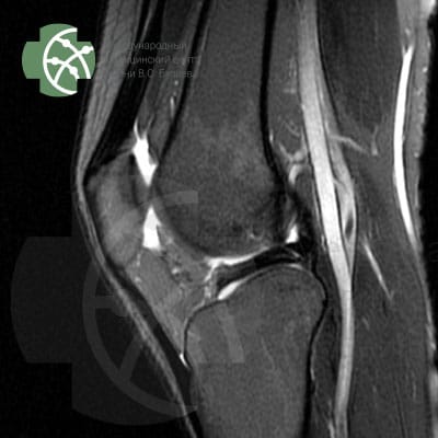MRI of the Knee Joint. Program T2 FS Sagittal 3 mm.
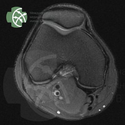MRI of the knee joint. Program PD FS Axial 3 mm.
MRI of the knee joint. Program PD FS Coronal 3 mm
Magnetic resonance imaging of the knee joint without contrast agent
#### Rating:
#### Reviews [2GIS](https://2gis.ru/ufa/search/%D0%BA%D0%BB%D0%B8%D0%BD%D0%B8%D0%BA%D0%B0%20%D0%91%D1%83%D0%B7%D0%B0%D0%B5%D0%B2%D0%B0/firm/70000001044251429/55.983612%2C54.753242/tab/reviews) [Yandex](https://yandex.ru/search/?lr=172&text=%D0%BA%D0%BB%D0%B8%D0%BD%D0%B8%D0%BA%D0%B0+%D0%B1%D1%83%D0%B7%D0%B0%D0%B5%D0%B2%D0%B0+%D1%83%D1%84%D0%B0&src=suggest_B) [Google](https://www.google.com/search?gs_ssp=eJzj4tVP1zc0TCuoKiipSEsyYLRSNagwMU6xNLY0NTU3TDExNUw1tjKoMDZKMTGzNE0yTzRLS0lMSfOSvbDrwu4LOy7sBeJdFzYoXNh4sfnC9gsbLmy9sOnCBgDV6CUR&q=%D0%BA%D0%BB%D0%B8%D0%BD%D0%B8%D0%BA%D0%B0+%D0%B1%D1%83%D0%B7%D0%B0%D0%B5%D0%B2%D0%B0&oq=%D0%9A%D0%BB%D0%B8%D0%BD%D0%B8%D0%BA%D0%B0+%D0%B1%D1%83%D0%B7%D0%B0%D0%B5%D0%B2%D0%BE&aqs=chrome.1.69i57j46i13i175i199j0i13j0i8i10i13i30.2750j0j15&sourceid=chrome&ie=UTF-8#)
#### What you will receive
Medical report with recommendations
We will record the MRI data on DVD-disk. On the disk envelope, we will indicate QR-CODE, which allows you to download the study description 24 hours after the examination.
#### Who needs this service?
Indications for MRI of the knee joint (determined by the trauma doctor during an in-person consultation):
MRI examination is performed after consultation with an orthopedic doctor. The most common indications for MRI of the knee joint:
- traumatic injuries
- preparation for surgery
- signs of joint inflammation;
- suspicion of dystrophic changes
- congenital developmental anomalies
- the need to exclude tumors, metastases;
- questionable data obtained during ultrasound, CT, or X-ray.
#### Who cannot receive such a service? Contraindications for MRI of the knee joint (applicable to the MRI machine in our clinic):
Absolute contraindications for MRI of the knee joint:
- The presence of “embedded” metallic elements, implants, shrapnel, and electronic stimulators in the body that cannot be removed
- Early pregnancy in women
- Chronic diseases of the urinary system, kidney ailments accompanied by reduced excretory functionality. The restriction applies to the procedure **in contrast mode**
- The presence of allergic reactions in a patient to a specific contrast agent
Conditional contraindications:
- Weight over 170 kg
- shock, extremely severe condition of a person
- CNS disorders accompanied by uncontrolled movements
#### How we do it
MRI of the knee can be performed in a lying position, **feet towards the aperture** of the machine, which allows successful examination even for people with claustrophobia (fear of enclosed and tight spaces)
MRI **does not require anesthesia or special preparation**.
Before the procedure, the MRI-technician will check you with a special device to ensure there are no metals in your body or clothing that react to the magnetic field. Then, you will be placed on the MRI table and instructed. You will be given a button in your hand, which can instantly stop the examination if pressed. Then, the knee joint examination protocol, consisting of seven programs, will be initiated.
If necessary to clarify the obtained data, additional research programs may be connected after agreement with you.
#### What is included in the service?
The research protocol in different clinics can vary significantly. MRI is always a compromise between the time spent on the examination and its quality.
We use all recommended programs, ensuring the highest quality of research, allowing for a detailed examination of the knee area without missing pathological changes.
An important aspect is the research step – thickness “sections “, we obtain as a result of the study. The smaller the step and the more slices, the more detailed we can assess the condition of the tissues.
Our MRI knee joint protocol includes the following sequences:
*Program / slice thickness*
PD FS saggital 3 mm
T2 saggiati 3 mm
T2 FS saggital 3 mm
PD FS coronal 3 mm
T1 coronal 3 mm
PD FS axial 3 mm
MERGE saggital 3 mm
---
#### How long will it take?
The MRI protocol of the knee joint in our clinic, including 7 different research programs, will take about 40 minutes.
#### How else can this be done?
Assessing the condition of the knee and knee joint can be done based on symptoms during an examination by a trauma orthopedist. This specialist should see the patient, make a preliminary diagnosis, and after reviewing the examination results, provide a final diagnosis and prescribe appropriate treatment.
In addition to MRI, CT (computed tomography) and ultrasound can be performed.
But it should be understood that CT uses ionizing radiation, the dose of which needs to be strictly controlled; while ultrasound provides much less informative results.
MRI of the knee area – joint and surrounding tissues. Program PD FS sagittal 3 mm.
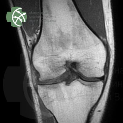MRI of the knee area – joint and surrounding tissues. Program T1 coronal 3 mm.
MRI of the knee area – joint and surrounding tissues Program T2 FS sagittal 3 mm
#### You might need
- Consultation with a Trauma and Orthopedic Specialist
- Second Expert Opinion (description of MRI studies conducted in other clinics)
---
### [MRI of the brain ](https://buzaevclinic.com/services/expert-mri-imaging/mri-brain/)
**Published:** September 20, 2021
**Author:** Aleksej Savelev
**Content:**
# MRI of the brain
MRI of the brain: T1 3D Sagittal
MRI of the brain: T2 Axial
MRI of the brain: T1 3D Sagittal
Magnetic resonance imaging of the brain without contrast enhancement
#### Rating:
#### Reviews [2GIS](https://2gis.ru/ufa/search/%D0%BA%D0%BB%D0%B8%D0%BD%D0%B8%D0%BA%D0%B0%20%D0%91%D1%83%D0%B7%D0%B0%D0%B5%D0%B2%D0%B0/firm/70000001044251429/55.983612%2C54.753242/tab/reviews) [Yandex](https://yandex.ru/search/?lr=172&text=%D0%BA%D0%BB%D0%B8%D0%BD%D0%B8%D0%BA%D0%B0+%D0%B1%D1%83%D0%B7%D0%B0%D0%B5%D0%B2%D0%B0+%D1%83%D1%84%D0%B0&src=suggest_B) [Google](https://www.google.com/search?gs_ssp=eJzj4tVP1zc0TCuoKiipSEsyYLRSNagwMU6xNLY0NTU3TDExNUw1tjKoMDZKMTGzNE0yTzRLS0lMSfOSvbDrwu4LOy7sBeJdFzYoXNh4sfnC9gsbLmy9sOnCBgDV6CUR&q=%D0%BA%D0%BB%D0%B8%D0%BD%D0%B8%D0%BA%D0%B0+%D0%B1%D1%83%D0%B7%D0%B0%D0%B5%D0%B2%D0%B0&oq=%D0%9A%D0%BB%D0%B8%D0%BD%D0%B8%D0%BA%D0%B0+%D0%B1%D1%83%D0%B7%D0%B0%D0%B5%D0%B2%D0%BE&aqs=chrome.1.69i57j46i13i175i199j0i13j0i8i10i13i30.2750j0j15&sourceid=chrome&ie=UTF-8#)
#### What you will receive
Medical report with recommendations
We will record the MRI data on DVD-disk. On the disk envelope, we will indicate QR-CODE, from which you can download the study description 24 hours after the examination.
For an additional fee, we can print the research results on film.
#### Who needs this service?
Indications to MRI of the brain:
- frequent headaches
- diagnosis of brain neoplasms (tumors – benign and malignant)
- identification of the causes of inflammation of the paranasal sinuses (sinusitis, ethmoiditis, frontal sinusitis)
- epilepsy (acute and chronic);
- stroke and assessment of post-stroke condition
- periodic fainting
- meningitis
- Parkinson’s disease, Alzheimer’s
- multiple sclerosis
- vascular disorders
- preoperative and postoperative assessment
- cyst formation
- aneurysms, thrombosis, and other vascular pathologies;
- hematomas and their consequences
- ischemic zones
- increased intracranial pressure
Most often, brain pathology is accompanied by noticeable deviations in a person’s condition, behavior, and reactions. These may include confusion, facial tingling, sensory disturbances, hearing or vision loss, and speech problems.
#### Who cannot receive such a service? Contraindications for MRI:
- Presence of electronic medical devices in the body (pacemaker, etc.)
- Presence of metal elements in the area of study (prostheses, clips, fragments)
- Inadequate patient behavior (panic attack, psychomotor agitation)
- State of drug or alcohol intoxication
- Inability to remain still during the examination (due to severe pain or other reasons)
- The necessity of continuous resuscitation measures (artificial respiration, etc.)
#### How do we do it?
MRI of the brain **does not require anesthesia or special preparation**.
Before the procedure, the MRI-technician will check you with a special device to ensure there are no metals in your body or clothing that react to the magnetic field. Then, you will be placed on the MRI table and given instructions. You will be given a button in your hand, which can instantly stop the examination if pressed. Next, the brain examination protocol, consisting of several programs, will be initiated.
If necessary to clarify the obtained data, additional research programs may be involved after agreement with you.
#### What is included in the service?
The research protocol in different clinics can vary significantly. MRI is always a compromise between the time spent on the examination and its quality.
We use all recommended programs, ensuring the highest quality of research, allowing for a detailed examination of the brain without missing pathological changes.
An important aspect is the research step – thickness “sections “, which we will obtain as a result of the research. The smaller the step and the more slices, the more detailed we can assess the condition of the tissues.
Our brain MRI protocol includes the following programs:
*Program / slice thickness*
3D T1 sagittal / 1 мм
T2 axial / 4 мм
T2 FLAIR axial / 4 мм
DWI\* axial / 4 мм
SWAN\*\* axial / 3 мм
---
\**MRI sequence — DWI и ADC*
**Clinical Application:**
- Early Detection of Ischemic Stroke
- Differentiation Between Early and Late Stroke
- Differentiation between Epidermoid and Arachnoid Cyst
- Differentiation between abscess and tumors with necrosis
- Detection of Cortical Lesions in the Brain
- Differentiation between herpes and diffuse glioma
- Staging of Gliomas and Meningiomas
- Demoralization assessments
\*\* SWAN – diagnosis of brain hemorrhages, helps identify fresh hemorrhages and traces of old hemorrhages – calcifications
#### How long will it take?
The basic MRI protocol of the brain will take about 30 minutes
#### How else can this be done?
The condition of the brain can be assessed by symptoms during a neurologist’s examination.
Instrumental methods of examination will help the doctor make the correct diagnosis.
In addition to MRI, a CT (computed tomography) scan and an ultrasound of the brain can be performed.
But it should be understood that CT uses gamma radiation, the dose of which needs to be strictly controlled; while ultrasound provides much less informative results.
MRI of the Brain: Program SWAN
MRI of the Brain: Program ADC map
MRI of the Brain: Program DWI
#### You may need
- Neurologist Consultation
- Neurosurgeon Consultation
- Second expert opinion (description of MRI studies conducted in other clinics)
- Neuropsychologist Consultation
---
### [MRgFUS for Other Diseases ](https://buzaevclinic.com/kniga-fokusirovannyj-ultrazvuk-pod-kontrolem-mrt-v-nevrologii/mr-fuz-pri-drugih-zabolevaniyah-nervnoj-sistemy/)
**Published:** November 2, 2024
**Author:** Rezida Galimova
**Content:**
# MRgFUS for Other Nervous System Diseases
Thermal impact using MRgFUS opens up broad possibilities for treating many other nervous system diseases. The list of such new indications is rapidly growing. For some of these diseases, traditional stereotactic neurosurgery (radiofrequency thermodestruction) was previously successfully applied DBS), and some diseases, on the contrary, became the object of stereotaxis for the first time thanks to MRgFUS. This chapter will present an analysis of some of the most promising applications of MRgFUS in neurology and psychiatry beyond extrapyramidal pathology, for which the first practical results have already been obtained today
## Neuropathic pain
Neuropathic pain is pain caused by a disease or injury to the somatosensory nervous system at any level. Neuropathic pain is accompanied by unpleasant distressing sensations and emotional experiences that significantly affect quality of life (Davydov et al., 2018). The prevalence of neuropathic pain in the general population, according to various authors, ranges from 0.9 to 17.9% ( Bouhassira et al., 2008).
Medication treatment for patients with neuropathic pain is not always successful. In cases resistant to pharmacotherapy, surgical treatment methods are recommended, which involve affecting the structures of the CNS and peripheral nervous system involved in the development of pain. These include neuromodulation (chronic neurostimulation of peripheral nerves, spinal cord, DBS and others) and destructive interventions
Among the destructive surgeries used for neuropathic pain, the most common are the destruction of the dorsal root entry zone, less commonly chordotomy, commissural myelotomy, cingulotomy. A well-known and effective method of ablative impact on CNS structures in patients with neuropathic pain is thalamotomy, as some thalamic nuclei (central lateral nucleus – central lateral nucleus of the thalamus CL), medial nuclei) are “relay stations” of the somatosensory pathway. Thalamotomy using radiofrequency thermodestruction and radiosurgery, as well as thalamic DBS proved effective in patients with intractable neuropathic pain ( Jeanmonod et al., 1994; Rasche et al., 2006).
A new approach to treating neuropathic pain is the use of the MRgFUS method ( Brown et al., 2015; Jung et al., 2018). An interesting historical fact is that treatment with focused ultrasound was first conducted specifically to relieve pain in a patient with neuropathic pain—long before the use of ultrasound in extrapyramidal disorders. D. Jean monod et al. (2012) were among the first to demonstrate that patients with chronic neuropathic pain experience a 49% pain relief at 3 months and 57% at 1 year after central lateral thalamotomy using MRgFUS. A similar positive effect of MRgFUS is shown in the work D.G. Iacopino et al. (2018) in 11 patients with neuropathic pain. Impact on CL the MRgFUS method was effective in treating trigeminal neuralgia in patients resistant to both medication therapy and other surgical treatments (thermocoagulation, microvascular decompression, gamma knife) Gallay et al., 2020a). It is noteworthy that during ablation CL no deafferentation pain occurs anaesthesia dolorosa), despite the interruption of information flow from pain receptors, which often occurs with other destructive operations on the trigeminal nerve ( Gallay et al., 2020a).
A case is described in the literature of treating and 6-year postoperative observation of a patient with refractory cluster headache – bilateral thalamotomy was performed CL using the MRgFUS method ( Magara et al., 2022). Before the surgery, there were episodes of severe pain with pronounced autonomic symptoms, lasting from 20 to 90 minutes and occurring up to 7 times a day (interictal periods up to 3 months). After the surgery, during the first 3 months, there was a single typical episode lasting 20 minutes, and in the following months, episodes occurred 1–2 times a week without accompanying autonomic symptoms. The patient discontinued basic therapy with ergotamine and caffeine. At the last examination 6 years after the surgery, cluster headache episodes had completely ceased Magara et al., 2022).
In some countries, the use of MRgFUS for chronic pain is officially approved, and currently, in the USA, patients with uncontrolled chronic pain are being recruited to undergo MRgFUS ablation as part of a clinical trial I phases NCT03111277). Other studies are also being conducted in parallel to evaluate the effectiveness and clinical safety of MRgFUS for neuropathic pain.
Presenting our first clinical case of treating neuropathic pain using MRgFUS.
**Patient N., 39 years old** , visited the clinic with complaints of constant aching pain up to 10 points on the VAS, burning sensation, and lack of sensitivity below the level of the nipples, absence of movement in the legs, and impaired pelvic organ function. Has been suffering from type 2 diabetes since 2011, on insulin therapy since 2020. Disabled I groups.
Noted complaints since 2015 after suffering from purulent epiduritis at the C level VII–ThVI, which developed against the background of septicemia. Since 06/16/2015, the patient experienced severe pain under the left shoulder blade, the pain syndrome progressed, and after 1 week, dysfunction of the pelvic organs and lower paraplegia developed. On 06/24/2015, he underwent emergency surgery – laminectomy was performed ThII–ThIV , removal of a paravertebral abscess at the level of ThV and epidural empyema at the C level VII–ThVI. After the surgery, the neurological status showed persistent lower paraplegia and dysfunction of pelvic organs, along with a stable neuropathic pain syndrome with burning, constricting pain and absence of all types of sensitivity below the level of the nipples. To relieve the pain syndrome, the patient took baclofen 25 mg 3 times a day, gabapentin 400 mg 3 times a day with minimal effect. Periodically, up to 4 times a week, tramadol 50–100 mg was used intramuscularly or orally to manage severe pain. When the dose of gabapentin was increased to 2400 mg/ cthe patient noted a deterioration in overall well-being without a reduction in pain syndrome. Also, for the treatment of neuropathic pain, they took: amitriptyline 50 mg/day for 3–4 months, duloxetine 60 mg/ cut – 6 months, pregabalin 150 mg/day – 3 months, noting poor tolerance to the medications in the form of lethargy, drowsiness, sluggishness without a positive effect on the pain. Increasing the dosages of the medications led to an intensification of the aforementioned side effects. The patient regularly, 2–3 times a year, underwent rehabilitation courses in specialized hospitals; in 2017, an epidural electrode was implanted for 2 weeks for chronic epidural stimulation, in 2018 and 2019 – two-level epidural electrode placement with stimulation for 2 weeks, but the neuromodulation provided only a slight reduction in the pain syndrome, which is why the electrodes were not installed on a permanent basis.
Thus, the patient is diagnosed with pharmacoresistant neuropathic pain according to the following criteria:
1\) duration of pain syndrome more than 6 months;
2\) pain intensity of at least 4 points on the VAS
3\) lack of response to 4 proven effective drugs (gabapentin, pregabalin, amitriptyline, duloxetine),
at the same time, the medications were used for at least 3 months, and increasing the dosages of the medications led to side effects that interfered with taking the medication in adequate dosages (despite treatment, the pain decreased by less than 30%)
Due to the minor positive effect of conservative therapy, the patient turned to the V.S. Buzaev International Medical Centre for the treatment of neuropathic pain using MRgFUS. A consultation was held with the participation of the center’s neurosurgeon, neurologist, and psychiatrist, and a decision was made on the possibility of using MRgFUS therapy for neuropathic pain. Informed consent was obtained from the patient for the procedure.
At the time of admission, the patient was using tramadol 50–100 mg intramuscularly daily, gabapentin 400 mg 3 times a day, and baclofen 25 mg 3 times a day for pain relief. The patient rated the intensity of constant pain at 8 points on the VAS, noting periodic increases in pain to 9–10 points, with the most severe pain in the feet. The pain was accompanied by numbness, tingling or prickling sensations, and burning. Upon admission, the general condition was satisfactory, with no notable features in the somatic status, and the patient was moving with the help of a wheelchair.
Neurological status. Cranial nerves – no pathology. Strength and tone of the neck, shoulder girdle, and upper limb muscles are unchanged. Tendon and periosteal reflexes of the upper limbs are brisk, symmetrical, no pathological reflexes. Active movements in the lower limbs are absent, passive movements are possible in full range. Hypotonia and hypotrophy of the lower limb muscles. Knee reflexes are brisk, Achilles reflexes are absent, pathological plantar signs are elicited on both sides. Below the level of the nipples (level ThIV) absence of superficial, deep, and pain sensitivity on the trunk and lower limbs. Does not control pelvic organ function, requires absorbent underwear. According to the questionnaire PAIN Detect the patient scores 13 points
Fig. 9.1. MRI of the patient’s brain N. after surgery (2nd day) a – axial slice. b – coronal slice. Visualized thalamic lesions in the area CL indicated by arrows A neuropsychological study was conducted: according to HADS noted absence of anxiety and depression symptoms (5 and 3 points respectively), according to MADRS – absence of depression symptoms (8 points). No distinct cognitive impairments detected (26 points on the scale MoCA).
Bilateral non-invasive thalamotomy of the central lateral nuclei of the thalamus ( CL) performed without anesthesia, the patient was conscious, which allowed for monitoring the clinical effect and preventing the development of complications in the form of focal neurological symptoms. The destruction sites were projected in the area of the central lateral nuclei of the thalamus ( CL) – on the left measuring 4 × 5 mm, on the right – 4 × 7 mm. No complications or side effects of the surgery were recorded. The duration of the procedure was 3 hours and 15 minutes. After treatment, on the 2nd day, an MRI of the brain was performed: bilateral foci are visualized in the projection CL the specified sizes, no hemorrhages detected, edema around the focus up to 2 mm (Fig. 9.1)
Immediate clinical improvement was achieved after treatment – a reduction in constant pain from 8 to 4 points, and the dosage of gabapentin was reduced to 800 mg/day.
After the treatment, the patient was monitored remotely for 12 months. He kept a pain diary, noting the intensity of the pain syndrome daily. During the first month after surgery, the average pain intensity was 5.9 points on the VAS, and the patient used tramadol 4 times that month. In the second month after treatment, the average pain intensity was 6.3 points on the VAS, with tramadol used 3 times a month. In the third month after treatment, the average pain intensity was 6.7 points on the VAS, with an increased need for tramadol—used 8 times a month. There was a gradual increase in pain intensity: 4th month after surgery—7.2 points on the VAS, 5th month—7.6 points, and 6th month—8.0 points. The need for tramadol gradually increased: in the 4th month after surgery, the patient used it 13 times, in the 5th month—16 times, and in the 6th month—18 times. According to the patient, during the 5th and 6th months after surgery, he used tramadol due to “anxiety, panic, nervousness.” In this regard, venlafaxine was added to the treatment for pain syndrome correction, which the patient took at a dosage of 75 mg/day for 3 months (he refrained from increasing the dosage due to concerns about weight gain, risk of drowsiness, lethargy, sluggishness). Since the pain was mixed (neuropathic and dysfunctional), cognitive-behavioral therapy was recommended by the center’s psychiatrist to reduce pain. After 3 months of cognitive-behavioral therapy, the patient noted a significant reduction in pain syndrome (to 6 points), with tramadol needed only once a month, and this continues to the present. The patient continues to receive cognitive-behavioral therapy with positive effects.
In this case, the target was chosen as the thalamic nucleus CL, since this structure is the main one in transmitting afferent information from the spinothalamic pathways to the subcortical and cortical somatosensory domains, and the posterior part CL (CLp) regulates the cognitive, sensory, and affective aspects of chronic neuropathic pain ( Nüssel et al., 2022). In addition CLp is a convenient surgical target due to its anatomical location and is characterized by low individual variability. According to the literature, ablation specifically CL provided a good lasting effect in the form of reducing neuropathic pain syndrome by an average of 50% in early studies ( Allam et al., 2022).
The effect of treatment was significantly influenced by the patient’s psychological state. By the 6th month, the patient’s sensation of pain increased, yet there was no need for narcotic drugs. During consultations, he noted that he took medications in advance, out of anxiety and fear that “acute pain would start again.” However, the patient categorically refused antidepressants. Due to these characteristics, cognitive-behavioral therapy was proposed, which proved to be quite effective.
Thus, bilateral non-invasive thalamotomy using MRgFUS can be considered a potentially effective method for treating refractory neuropathic pain. To achieve optimal results, it is necessary to consider the psycho-emotional state of patients selected for such treatment. The final decision to include MRgFUS in the list of recommended methods for neuropathic pain requires randomized controlled trials.
## Hypothalamic Hamartoma
Hypothalamic hamartoma (HH) is a rare benign tumor, with a prevalence estimated at 1 in 200,000 children ( Bernasconi et al., 2019). From a morphological perspective, it represents a gangliocytoma—a benign tumor of the sympathetic nerve ganglia with low proliferative potential and minimal nuclear atypia ( Susheela et al., 2013; Louis et al., 2021). The first symptoms of GG can be observed as early as the 1st year of life, although many cases with onset in adulthood have been described ( Beggs et al., 2008).
Hypothalamic hamartoma is characterized by various manifestations, the most common of which are gelastic (laughter-associated) seizures, cognitive impairments, and precocious puberty ( Beggs et al., 2008). In 0.5% of patients with HH, dacrystic seizures are described, characterized by pathological stereotypical crying, tearing, grimacing, sobbing, a sad facial expression, or a subjective feeling of sadness ( Cohen et al., 2021). According to data L.H. Castro et al. (2007), the presence or absence of epilepsy in patients with HH is more determined by the location of the tumor and its connection with the temporal lobe (epilepsy is more common when located in the posterior part of the hypothalamus and the mammillary bodies area) than by the tumor volume
In practice, the classification most commonly used for GG is O. Delalande, M. Fohlen (2003), which classifies GG into 4 types based on MRI data: I type – small formations on a stalk, characterized by a horizontal plane of attachment to tuber cinereum; II type – predominantly intraventricular formations with a vertical attachment plane; III type – spreading in III ventricle and interpeduncular cistern (combination I и II types with both horizontal and vertical attachment planes) IV type is defined as GG, completely located within III ventricle (these are usually very large tumors with a wide attachment to both the mammillary bodies and the hypothalamus, and they invade the interpeduncular cistern). Dynamic observation of patients showed that hamartomas do not progress, but increase in size as the brain develops.
Approaches to the treatment of HH in recent years have undergone significant changes, with a shift in focus from medication therapy towards innovative minimally invasive surgical methods. Anticonvulsants are generally unable to completely control seizures and are considered an important but only symptomatic treatment (Mikhailov et al., 2017). Open neurosurgical resection or disconnection of the hamartoma through pterional, transcallosal, or transventricular access leads to good seizure control but is accompanied by a high rate of complications (neurological, endocrine, cognitive, behavioral) – up to 50%, which limits the use of this method ( Alvarez-Garijo et al., 1983; Machado et al., 1991). A number of studies have been published on stereotactic thermocoagulation of the GG with positive effects: in 84% of cases, seizure control was achieved, and complications were observed in 3–5% of patients without the development of neuroendocrine disorders ( Kameyama et al., 2016).
As a new approach to ensure safe and complete disconnection of the attachment zone of the GG from surrounding structures, the use of laser and radiofrequency thermocoagulation with the help of robotic stereoendoscopy was proposed Kameyama et al., 2016). The effectiveness of this approach was evaluated as high (especially in patients with isolated gelastic seizures and intraventricular hamartomas), with a favorable outcome rate of 78% and a complication rate of 8%. The use of stereotactic laser interstitial thermotherapy demonstrated effectiveness similar to open microsurgery (absence of seizures in 76–81% of operated patients), but with the development of a number of irreversible complications in up to 20% of cases ( Xu et al., 2018). Stereotactic radiosurgery has proven to be effective and safe, with seizure control rates ranging from 60 to 71% and a very low complication rate (2%) (Savateev et al., 2022)
Analysis of available literature data indicates that the success of treating HH is determined not so much by technical capabilities as by understanding the mechanism of epileptic seizure formation, including within the hamartoma, and proper planning of the disconnection of the formation from surrounding structures while preserving adjacent brain formations. In this regard, an innovative treatment method such as MRgFUS may become a successful alternative minimally invasive method for affecting HH to dissect the formation from adjacent tissues. Currently, only 2 articles have been published in the available literature describing 5 clinical observations of patients with HH who underwent treatment using MRgFUS with good results and without the development of side effects ( Yamaguchi et al., 2020; Tierney et al., 2022).
Presenting our clinical case of treating ET using MRgFUS.
**Patient,** 32 years, disease onset at the age of 4, when rare episodes of unnatural laughter first appeared. Parents sought help from a pediatrician at their place of residence, and the condition was considered behavioral traits related to the girl’s character, with no diagnostic studies or treatment recommended. At 16, there was an increase in gelastic seizures with the addition of dacrystic ones, and the development of bilateral tonic-clonic seizures. Anticonvulsant therapy was selected, but seizures persisted for many years without a positive response to medications. At the beginning of treatment, 3 types of seizures were observed: 1) isolated gelastic and dacrystic seizures with preserved consciousness; 2) focal hypomotor with loss of consciousness, accompanied by tearing, oroalimentary and bimanual automatisms, speech perseverations, ambulatory automatisms; 3) focal, evolving into bilateral tonic-clonic. Every 2 years, the patient underwent brain MRI: retrospective evaluation of all images showed a formation in the hypothalamus projection, but it was not timely recognized and described.
Fig. 9.2. MRI of a patient with GG (arrows) before surgery. Fig. 9.3. Electroencephalogram of the same patient before treatment. Complexes of sharp-slow waves are marked with ovals. Since the age of 16, treatment was conducted with oxcarbazepine, levetiracetam, lamotrigine. At the time of admission to the V.S. Buzaev International Medical Centre, the patient was receiving oxcarbazepine 1200 mg/day, levetiracetam 500 mg/day, carbamazepine 400 mg/day. Despite the ongoing therapy, the frequency of seizures remained high, with gelastic seizures occurring 5–6 times a day, focal hypomotor seizures 2–4 times a month, and bilateral tonic-clonic seizures once every 2 months. Fig. 9.2. MRI of the patient with HH (arrows) before surgery. Fig. 9.3. Electroencephalogram of the same patient before treatment. Ovals indicate sharp-slow wave complexes.
Family history: A paternal cousin is under the care of an epileptologist, receiving anticonvulsant therapy. MRI (Fig. 9.2) reveals a T2 hyperintense lesion that does not accumulate contrast agent, along the anterior and right wall III ventricle, measuring 8 × 8 × 8 mm, corresponding to GG II Fig. 9.4. Intraoperative monitoring of the temperature regime in the projection of the optic tract and the right mammillary body (pink line). Fig. 9.5. MRI of the same patient during the MRgFUS procedure (GG indicated by arrows)
Video-EEG monitoring was conducted without sleep deprivation for 94 hours during active and passive wakefulness, sleep, and post-awakening with functional tests (13.10.2022). During passive wakefulness, the main rhythm is represented by regular, stable, modulated a-a rhythm with a frequency of about 9.5–10 Hz and an amplitude of up to 75 µV is recorded in the occipital leads with spreading to the posterior temporal and parietal areas of the hemispheres. Zonal differences are clearly expressed. In the posterior frontal-central regions, a regular stable sensorimotor rhythm of arc-shaped configuration with a frequency of 8.5–9.5 Hz and an amplitude of 60 µV is recorded (Fig. 9.3). During sleep and wakefulness, regional epileptiform activity is recorded in the right posterior frontal and temporal areas in the form of single sharp waves, sharp-slow wave complexes, with a low representation index during wakefulness, and an increase in the index to medium during sleep. Also, during sleep, regional epileptiform activity is recorded in the left temporal area in the form of single sharp-slow wave complexes with a low representation index
Fig. 9.4. Intraoperative monitoring of the temperature regime in the projection of the optic tract and the right mastoid body (pink line) Treatment using MRgFUS was aimed at dissecting the tumor from surrounding tissues. The operation was performed on 12/05/2022. During the treatment, the MRI T2 mode was used to assess the impact FSE in the axial, sagittal, and coronal planes. Additional temperature control was performed in the projection of the optic tract and the mammillary body during each ultrasound exposure (Fig. 9.4). Nine therapeutic ultrasound exposures were conducted with a gradual increase from 10,000 to 22,000 J, lasting from 15 to 25 seconds, reaching temperatures from 52 to 58°C.
Fig. 9.5. MRI of the same patient during the MRgFUS procedure (GG indicated by arrows) In Fig. 9.5, an MRI study during treatment with MRgFUS is presented. Intraoperative imaging did not reveal signs of hemorrhage or off-target heating. Fig. 9.7. MRI of the brain of the same patient 3 months after treatment.
Fig. 9.6. MRI of the same patient 3 hours after treatment. Fig. 9.8. Electroencephalogram of the same patient 3 months after treatment with MRgFUS. A single sharp-slow wave complex is marked with an oval. There were no serious complications during the procedure. One gelastic seizure occurred during the first therapeutic ultrasound treatment. A combination of modes was used to monitor changes after MRgFUS T2 cube и T2 FSE using a 32-channel head coil. Three hours after treatment, two necrosis zones with cytotoxic edema measuring 3 mm each were identified in the projection of the aforementioned hamartoma, surrounded by vasogenic edema up to 4 mm thick (Fig. 9.6)
The patient returned to professional activities 10 days later. She noted increased confidence at work due to the absence of seizures. Currently, she has been under observation at the Center for more than 12 months, with no gelastic or dacrystic seizures detected after treatment, which corresponds to IA class according to the scale of surgical treatment outcomes for epilepsy by J. Engel (1993).
On the follow-up MRI of the brain 3 months after the treatment (Fig. 9.7), there is a complete regression of vasogenic edema. In the projection of the hamartoma, two hyperintense foci in T2 mode measuring 3 × 2 × 3 and 2 × 3 × 4 mm are determined, corresponding to the sonication points performed at the visible edge of the formation.
In Fig. 9.8, an electroencephalogram is presented 3 months after treatment during sleep and wakefulness, showing regional epileptiform activity in the right temporal region as single sharp-slow wave complexes. Compared to Fig. 9.8. Electroencephalogram of the same patient 3 months after treatment with MRI-guided focused ultrasound. A single sharp-slow wave complex is marked with an oval. There is a positive trend in the form of a significant decrease in the index of epileptiform activity in the right hemisphere of the brain compared to the preoperative electroencephalogram.
The described clinical case shows that dissection of the hamartoma using the MRgFUS method was effective in controlling epileptic seizures without any side effects. For a surgically inaccessible HH, this technology allowed the patient to be completely free from epileptic seizures, forced laughter, and crying (during more than a year of observation). Effectiveness may depend on the accuracy of targeting, the extent of thermal damage to the selected target area, and the size of the HH.
## Epilepsy
More than 50 million people worldwide suffer from epilepsy, with drug resistance observed in 25-30% of cases. The method of choice in such situations is surgical correction, which, however, does not always lead to the complete elimination of the epileptogenic focus.
Currently, clinical trials of MRgFUS are ongoing in patients with therapy-resistant focal epilepsy NCT02804230, NCT03417297). The results of pilot studies demonstrate the clinical effectiveness of thermal impact on the anterior nuclei of the thalamus using the MRgFUS method to prevent the generalization of seizure activity in focal epilepsy ( Parker et al., 2019). In the research V. Krishna et al. (2023) the application of the method in 2 patients with therapy-resistant epilepsy followed by an evaluation of clinical outcomes at 3, 6, and 12 months contributed to a reduction in seizure syndrome manifestations: one patient experienced no seizures throughout the observation period, while the second achieved a reduction in the number of seizures from 90–100 to 3–6 per month. One of the patients experienced changes in attention and memory in the postoperative period.
Treatment of patients with resistant forms of epilepsy using MRgFUS may become a routine approach in the coming years, just as this technology is currently used for the correction of movement disorders.
## Hydrocephalus
Hydrocephalus remains a condition that in many cases is associated with an unfavorable clinical outcome. In occlusive hydrocephalus, the separation of spaces for cerebrospinal fluid may require surgical interventions (often repeated) to restore CSF circulation, which increases the risk of intra- and postoperative complications. In the study S. Monteith et al. (2013) on cadaveric material, the potential possibility of creating openings using septum pellucidum ablation and ventriculostomy at the floor projection has been demonstrated III ventricle under MRI-guided focused ultrasound for optimal cerebrospinal fluid dynamics. The principle of the method is based on creating cavitation-induced tissue damage to ensure free circulation of cerebrospinal fluid under dynamic MRI control
To conclude the possibility of using the MRgFUS method for treating patients with hydrocephalus, further experimental and clinical research is necessary.
## Depression and Obsessive-Compulsive Disorder
Therapeutic efficacy and safety of high-intensity MRgFUS in the treatment of depression were evaluated in studies NCT02348411 и NCT03421574. Bilateral ablation of the anterior limb of the internal capsule using MRgFUS ( NCT02348411), conducted on a 56-year-old patient diagnosed with “major depressive disorder with a course refractory to psychotherapy and electroconvulsive therapy” (more than 6 sessions conducted), demonstrates significant and sustained (over 1 year of observation) improvement in Hamilton scale scores (decrease from 26 to 8 points after 1 week and to 7 points after 12 months), Beck scale (decrease from 26 to 12 points after 1 week and after 12 months), and the Global Assessment of Functioning scale (increase from 40 to 85 points after 12 months). The patient’s quality of life was assessed using the questionnaire SF-36: an increase in mental health scale scores from 20.5 to 45.6 was identified ( Kim M. et al., 2018).
In 2015, a team of specialists from South Korea performed anterior capsulotomy using MRgFUS in a pilot study on 4 patients with medication-resistant obsessive-compulsive disorder (OCD) Jung et al., 2015). The procedure proved to be not only safe (without hemorrhages or infarctions) and tolerable (without deterioration of neurocognitive functions) but also effective: all patients showed significant improvement on the Yale-Brown Obsessive-Compulsive Scale. Subsequently, this same group increased their cohort to 11 patients with OCD, reporting improvement in 6 of them ( Kim S.J. et al., 2018).
From July 2017 to November 2019, in 2 clinical studies NCT03156335, NCT03421574) 16 patients with treatment-resistant OCD or major depressive disorder underwent capsulotomy using MRgFUS (University of Toronto, Ontario, Canada). In 4 patients, it was not possible to reach the target temperature for lesioning the intended nuclei, and they were excluded from the study. A detailed clinical report on the first 12 patients (6 patients with major depressive disorder and 6 patients with OCD) who were observed for at least 6 months shows that 4 patients with OCD achieved the planned response criteria—a reduction of ≥35% in the Yale-Brown Obsessive-Compulsive Scale score, and 2 patients with a major depressive episode achieved response criteria with a reduction of ≥50% in the Hamilton Depression Rating Scale score Davidson et al., 2020a, b) Mild side effects (headache, scalp sensory disturbance, etc.) occurred in 7 patients.
Currently, there is a significant historical experience in treating patients with depression and OCD using the method of radiofrequency ablation. Extrapolating the obtained data to the MRgFUS technology may provide a new impetus in improving the treatment of patients with this profile.
## Thrombolysis in Ischemic Stroke
To date, there remains a problem with the inefficiency or impossibility of timely thrombolytic therapy in patients with ischemic stroke. The anterior and posterior cerebral arteries, distal branches of the middle cerebral artery are difficult to access for endovascular intervention and are at high risk of perforation during mechanical manipulations. It has been hypothesized that MRgFUS may improve clinical outcomes of thrombolysis. Since 2011, this method has demonstrated in animals and cadaveric material the fundamental possibility of affecting thrombotic masses in the vessel lumen, as well as suppressing the growth of atherosclerotic plaques by reducing the level of oxidized low-density lipoprotein cholesterol and increasing macrophage apoptosis ( Brahmandam et al., 2022).
In ischemia models, MRgFUS facilitated thrombolysis and angiogenesis by enhancing the expression of angiogenic/anti-apoptotic factors ( Brahmandam et al., 2022). Pilot clinical studies have demonstrated positive results of using high-intensity MRgFUS in the form of artery recanalization after thrombotic stroke, reduction in the number of atherosclerotic plaques in carotid arteries, increased tissue perfusion, and reduced stenosis diameter in patients with atherosclerotic lesions of cerebral arteries ( Zhang et al., 2015; Sun et al., 2019). Thrombus fragmentation was achieved using MRgFUS within 2 to 24 hours after its formation ( Brahmandam et al., 2022). The use of MRgFUS in the occlusion of small brain vessels has advantages over endovascular techniques due to lower invasiveness and higher accuracy in thrombus visualization. These studies are currently ongoing in several stroke clinics worldwide.
## Opening of the Blood-Brain Barrier
All the ablation techniques discussed above fall into the category of high-intensity MRgFUS (power impact around 1000 W/cm²). A standalone non-invasive method for affecting the brain has become low-intensity MRgFUS (most often in the range from 2 to 20 V/ cThis method already has several applications in clinical neurology today.
Low-intensity MRgFUS combined with intravenously administered microbubbles of contrast agent reversibly opens the BBB with high temporal and spatial precision. The combined action of ultrasound and microbubbles promotes the formation of microstreams of tissue fluid, causing the separation of tight intercellular contacts of endothelial cells. It should be added that the direct mechanical impact of the ultrasound wave impulse on biological tissue leads to areas of mechanical tension and compression, which also play a role in the temporary opening of the BBB. The restoration of the BBB after low-intensity MRgFUS exposure occurs approximately within 1 day without any damaging tissue effect (Kholyavin, 2019; Kobus et al., 2016).
The idea of using low-intensity MRgFUS to open the BBB is particularly appealing for the treatment of neurodegenerative diseases. The essence is, firstly, to achieve the penetration of endogenous antibodies (immunoglobulin G (IgG) и IgM) к b-amyloid and other pathological proteins accumulating in the brain as the disease progresses, and secondly, in the activation (in response to ultrasound exposure and opening of the BBB) of astrocytes and microglial cells to break down protein aggregates and enhance neurogenesis ( Mainprize et al., 2016; Baek et al., 2022; Ma et al., 2023). The same approach can be used for other diseases to deliver high-molecular therapeutic complexes or cells to the brain (e.g., viral vector genetic constructs, stem cells, etc.), as well as in neuro-oncology for targeted delivery of chemotherapeutic agents to tumor tissue ( Mainprize et al., 2016).
## Alzheimer’s Disease
Alzheimer’s Disease (AD) is the most common neurodegenerative disease and the leading cause of dementia in modern society. Alzheimer’s Disease is characterized by a progressive decline in memory and other cognitive functions in the elderly. In 2019, there were over 55 million people with dementia worldwide, and according to some forecasts, this number could increase to 150 million by 2050. Nichols et al., 2022). Such alarming epidemiological data determine the exceptional medical and social significance of developing effective treatments for BA
The classic histomorphological features of Alzheimer’s disease are amyloid plaques in the brain parenchyma, which are aggregates of pathologically altered neuronal protein b-amyloid, and intracellular neurofibrillary tangles formed by hyperphosphorylated tau protein of neurofilaments (Illarioshkin, 2003). These changes and the accompanying neuroinflammation trigger a large cascade of molecular events, ultimately leading to the death of hippocampal neurons (the main target of neurodegeneration in AD) and other parts of the CNS. Currently, the exact role b-amyloid burden in the development and progression of clinical symptoms of AD remains controversial and is under active study, however, overall the leading therapeutic approach in most studies is the development of methods for eliminating aggregates b-amyloid from brain tissue. This is the focus of innovative drugs developed in recent years based on monoclonal antibodies to various “maturity” forms b-amyloid and different parts of the molecule ( Mintun et al., 2021; van Dyck et al., 2023). However, the clinical results of treating Alzheimer’s disease with these drugs lead only to a moderate slowing of the rate of neurodegeneration (by approximately 30%) and are associated with serious complications. Therefore, it is important to continue the search for alternative anti-amyloid strategies.
Attempts to use low-intensity MRgFUS with the introduction of microbubbles into the bloodstream to study the possibility of modulating the BBB, effects on cognitive functions, level b-amyloid and tau protein in the brain began 15 years ago in experimental studies on transgenic mice with AD models. It was shown that such exposure can promote “washing out” b-amyloid from the brain parenchyma into the cerebrospinal fluid and lymphatic system, enhance uptake b-amyloid by microglial cells, reduce the level of phosphorylated tau protein, improve memory and behavior in animals, and slow the progression of the disease ( Ma et al., 2023). In experiments with intravenous administration IgG in AD models, the use of low-intensity MRgFUS led to a 39-fold increase in uptake IgG hippocampal cells and a 4-fold increase in neurogenesis in the hippocampus ( Dubey, 2020).
The success of these works led to the initiation of a similar approach in clinical trials for patients with AD. In 2022, the results of a multicenter study were presented NCT03671889 (Rezai et al., 2022). The authors assessed the safety, clinical, and neuroimaging effects of low-intensity MRgFUS targeting the hippocampus, frontal, and parietal lobes in patients with mild AD. Ten participants aged 55–76 underwent 30 separate MRgFUS procedures with follow-up for 6–12 months. All patients experienced immediate BBB opening after MRgFUS exposure and subsequent closure within 24–48 hours. The treatment was well tolerated, with no serious procedure-related side effects. Changes in cognitive functions assessed by various scales after MRgFUS exposure were comparable to those in the control group, but PET scanning demonstrated a noticeable reduction in the average level b-amyloid in the regions of the brain exposed to ultrasound. Thus, it was confirmed that the use of low-intensity MRgFUS on several areas of the brain with temporary opening of the BBB is accompanied by a reduction b-amyloid load and does not negatively affect the progression of AD ( Rezai et al., 2022).
In 2023, the results of an open study were published NCT04118764: 9 patients (average age 70.2 ± 7.2 years, average score on the Mini-Mental State Examination 21.9 points) underwent 3 low-intensity MRgFUS procedures once every 2 weeks with subsequent follow-up for 6 months. The permeability of the BBB in the hippocampus, anterior cingulate gyrus, and precuneus temporarily increased without adverse events. PET image analysis demonstrated moderate reduction b-amyloid load in the right parahippocampal and inferior temporal lobes. However, there was no recorded change in cognitive functions and the level of AD biomarkers in cerebrospinal fluid and blood (p- tau181, b-amyloid 42/40) Meng et al., 2023).
Expanding research in this area will help determine the real clinical significance of MRgFUS in AD and the long-term effects of the treatment conducted.
## Other Neurodegenerative Diseases
Amyotrophic lateral sclerosis (ALS) is a fatal neurodegenerative disease characterized by progressive degeneration of upper and lower motor neurons, neurogenic paresis of skeletal and bulbar muscles, amyotrophies, fasciculations, muscle spasticity, and early fatal outcome (usually due to respiratory failure) (Illarioshkin, 2003). The mechanisms of neurodegenerative process development in patients with ALS remain not fully understood. In experiments on transgenic animals with an ALS model Eisen et al., 2017) and in clinical studies using transcranial magnetic stimulation, neuroimaging, and electrophysiological methods ( Menon et al., 2015) it has been demonstrated that signs of cortical neuronal and glial dysfunction are observed in the early stages of ALS and may precede the degeneration of spinal motor neurons. This indicates the key role of upper motor neurons in the pathophysiology of the disease, so interventions targeting the motor cortex area may be important for slowing or stopping disease progression
In the research A. Abrahao et al. (2019) Four patients with ALS and clinical signs of upper motor neuron dysfunction were included to assess the feasibility of non-invasive access to the motor cortex by opening the BBB using low-intensity MRgFUS. Ultrasound treatment targets were individualized according to fMRI activation of the hand or foot in the primary motor cortex. The average duration of the MRgFUS procedure was 66 minutes (ranging from 43 to 173 minutes), which included obtaining planning sequences, targeting, and the ultrasound exposure itself. Patients tolerated the procedure and intravenous microbubble injections without any side effects; no changes in neurological status were recorded during ultrasound therapy. Follow-up MRI studies in all participants did not reveal parenchymal or subarachnoid hemorrhages, ischemia, gliosis, or worsening of cortical atrophy of the precentral gyrus within 30 days post-intervention. Thus, this study represents an important first step towards creating a platform for the non-invasive delivery of potential neuroprotective agents to the motor cortex area in patients with ALS.
Huntington’s disease (HD) is an autosomal dominant neurodegenerative disorder caused by the expansion of trinucleotide CAG-repeats in the gene HTT, what leads to the synthesis of a pathologically elongated neurotoxic polyglutamine-containing protein huntingtin (Illarioshkin, 2003). Currently, there is only symptomatic therapy for this disease. In the study C.Y. Lin et al. (2019) on a transgenic model of BG low-intensity MRgFUS c the introduction of microbubbles into the bloodstream was used to open the BBB to deliver liposomes containing plasmid DNA of the glial cell line-derived neurotrophic factor gene into the brain GDNF). Such MRgFUS-based therapy significantly improved motor characteristics in mice with a BG model, with the achieved hyperexpression in the brain GDNF led to a significant reduction in the formation of polyglutamine protein aggregates, a decrease in oxidative stress and apoptosis, and improved neuronal survival
It is evident that further research related to the opening of the BBB in neurodegenerative diseases is of great importance for experimental and clinical neurology and will actively develop in the coming years.
## Neuromodulation
Low-intensity MRgFUS induces reversible changes in neuronal excitability and activity of neural networks exposed to ultrasound. Despite significant interest in this promising new area of non-invasive, highly targeted neuromodulation, the mechanisms of the modulatory effect of MRgFUS remain poorly understood. It is suggested that the ultrasonic flow, when interacting with cell membranes, influences the functioning of mechanosensitive ion channels (Kholyavin, 2019). The neuromodulatory potential of low-intensity MRgFUS is also explained by various non-destructive thermal effects (the influence of even small changes in local temperature on transmembrane currents, activity of cellular enzymes, and other parameters determining the pattern of neuronal discharges), as well as cavitation effects caused by ultrasound within the lipid bilayer of neuronal membranes Darrow, 2019).
Experimental studies on various animal species and clinical studies in humans have shown that low-intensity MRgFUS can be used safely and effectively for cortical and subcortical neuromodulation ( Legon et al., 2020). In humans, such ultrasound exposure has been applied to the temporal cortex, primary and secondary somatosensory cortex, primary motor cortex, primary visual cortex, and thalamus. It has been shown that low-intensity MRgFUS affects the amplitude of evoked potentials, power, phase, and frequency of the electroencephalogram, resting-state functional MRI parameters, reaction time ( Lee et al., 2016; Legon et al., 2018, 2020). There are isolated reports on the use of low-intensity MRgFUS in the treatment of serious CNS diseases, such as thalamic ultrasound stimulation in patients with consciousness disorders after severe traumatic brain injury ( Monti et al., 2016).
W. Legon et al. (2020) compared the results of neuromodulation using MRgFUS in humans with well-known stimulation methods such as transcranial magnetic stimulation and transcranial electrical stimulation. In a series of experiments using various protocols and levels of ultrasound energy, it was found that in terms of overall effectiveness, as well as the frequency and severity of side effects, low-intensity MRgFUS is comparable to the other aforementioned non-invasive neuromodulation methods, which have a long history and are recognized as safe for humans
The emergence of the low-intensity MRgFUS method not only opens new therapeutic opportunities in neurology but also allows for the integration of non-invasive innovative neuromodulation technologies into broader practice for use in healthy individuals of various ages and professions (memory and attention modulation, sleep normalization, various training, relaxation techniques, etc.). The experience gained in recent years with MRgFUS in extrapyramidal and other nervous system diseases shows that this technology represents more than just a new method of functional therapeutic impact on the brain based on a stereotactic approach. Essentially, thanks to MRgFUS, we are talking about erasing the boundary between conservative and surgical neurology. This significantly expands the possibilities of providing effective assistance to various categories of patients with severe, often disabling movement, sensory, and behavioral disorders. As discussed in the monograph, MRgFUS represents, on one hand, a certain alternative DBS (taking into account the existing limitations for macroelectrode brain stimulation due to its invasive nature, surgical risks, the need for constant monitoring of the stimulator’s operation mode, etc.), and on the other hand, a qualitatively new level of ablative stereotactic neurosurgery. For example, trial reversible ultrasound interventions allow modeling the effect on a specific area of the brain and finding the most effective target for a particular patient VIM, PTT, VO, ZI and others – in isolation or in combinations). Positive experiences have been gained worldwide and in our clinic with effective two-stage treatment, i.e., safely performing repeat surgeries when symptoms return in patients with ET, PD, and dystonias. The use of several developed methodological techniques (asymmetry of created lesions, timely cessation of impact on the second side with sufficient anti-tremor effect, symptom control during trial sonications) has breathed new life into the idea of bilateral stereotactic ablations in ET and potentially in other movement disorders. Our results indicate that such operations using MRgFUS can be safe both in staged and simultaneous execution, but this requires strict adherence to all requirements and adequate consideration of existing risks, indications, and contraindications. It can be expected that the use of MRgFUS for treating various movement disorders will gradually expand. Simultaneously, methodological foundations and indications for using ablative high-intensity MRgFUS modes will be clarified, even in non-motor neurological disorders – pain syndromes, epilepsy, affective disorders resistant to conservative therapy, hydrocephalus, thrombotic ischemic stroke for arterial recanalization, etc. The initial results of such interventions look promising, although significant additional experimental and multicenter randomized clinical studies are still needed here. A promising variety of MRgFUS is the use of low-intensity ultrasound exposure. It allows for temporary opening of the BBB for the treatment of neurodegenerative and other CNS diseases, and also lays the foundation for implementing a fundamentally new neuromodulation mode in practice. This demonstrates the significance of MRgFUS not only for clinical practice but also for fundamental neurology and neurophysiology. Thus, despite its youth, the MRgFUS method has already firmly established itself and confidently looks to the future.
---
### [About Functional Neurosurgery ](https://buzaevclinic.com/kniga-fokusirovannyj-ultrazvuk-pod-kontrolem-mrt-v-nevrologii/sovremennye-predstavleniya-o-funkcionalnoj-stereotaksicheskoj-nejrohirurgii/)
**Published:** November 2, 2024
**Author:** Rezida Galimova
**Content:**
# Modern Concepts in Functional Stereotactic Neurosurgery
Neurosurgical interventions have been used for over a century to treat various diseases of the nervous system, primarily movement disorders. During this time, more than 8000 publications related to stereotactic surgery of extrapyramidal diseases have been released, the overwhelming majority of which concern PD. More than half of the publications are dedicated to DBS, which gradually took a dominant position among all types of functional stereotactic interventions. At the same time, traditional destructive operations on the basal ganglia have not lost their significance, with a renaissance of interest linked to the emergence of new non-invasive treatment methods such as MRgFUS.
Familiarity with this rapidly developing field of neurology and neurosurgery involves understanding the basic principles of the neural network organization of subcortical brain structures and related structures in both normal and pathological conditions.
## **Fundamental Principles of Functional Stereotactic Neurosurgery for Extrapyramidal Movement Disorders**
The concept of stereotactic neurosurgery is based on the functional topography of the brain, which began to develop as far back as the time of Hippocrates, who described in IV century BC, motor deficit with damage to the opposite hemisphere of the brain. Later, the Roman physician Claudius Galen in the II expanded the understanding of the brain and the foundations of some movement disorders (tremor) in our era. After Galen, there was a 1000-year hiatus in the study of anatomy due to religious prohibitions on dissection of the deceased. The first information about the structure of the brain’s subcortical formations is found in the works of K.F. Burdach (1819) and I.P. Lebedeva (1873), and in the upcoming XX the century saw a rapid development of neuromorphology, complemented by the intensive advancement of brain imaging methods (computed tomography (CT), magnetic resonance imaging (MRI), positron emission tomography (PET)) and achievements in experimental neurobiology and neuropharmacology in recent years, allowing for the creation of a coherent picture of modern concepts of the structural-functional organization of the basal nuclei
The primary function of the basal ganglia is the organization of the involuntary component of motor acts, the formation and implementation of automated motor programs, including ensuring the synchronous activity of various muscle groups and regulating mechanisms for maintaining adequate muscle tone. This function is carried out in close interaction with the cerebellum. In addition to motor control, the basal ganglia also play a role in organizing cognitive activity, learning, and emotional behavior (Illarioshkin, Ivanova-Smolenskaya, 2011)
Fig. 1.1. “Dynamic Activity” Model
basal nuclei in the process of movement control GPe – external segment of the globus pallidus
шара, GPi – internal segment of the globus pallidus SNs – compact part of the black sub-
stations, SNr – Reticular part of the substantia nigra. Here and in Fig. 1.3, 1.4 STN –
subthalamic nucleus. Here: blue arrow – direct pathway, green arrows – indirect
path, red arrows – hyperdirect path, black arrows – other paths in the central
nervous system. Explanations in the text To perform such complex functions, the basal ganglia receive information from various cortical areas and, in turn, project it to the thalamus, as well as to the prefrontal, premotor, and motor cortex (Fig. 1.1). Afferentation to the striatum mainly comes from the hemispheric cortex (motor, sensory, associative, limbic) and the thalamus. Corticostriatal fibers are excitatory and use glutamatergic transmission. Their projections have a clear topographic organization: the putamen is mainly involved in general motor control, the caudate nucleus in the control of oculomotor functions and some cognitive operations, and the ventral part of the striatum is related to the organization of limbic system functions (emotional-volitional sphere). Further, the motor control pathways reach the globus pallidus and the reticular part of the substantia nigra—two main efferent structures of the basal ganglia. Fibers from the globus pallidus and the reticular part of the substantia nigra go to the thalamic nuclei—ventrolateral, ventral anterior, mediodorsal, and centromedian, after which the considered loop closes again on the prefrontal, premotor, primary, and supplementary motor cortex.
Thus, the basal ganglia have bilateral connections with both the cortical areas of the cerebral hemispheres and (through the thalamus) with sensory input structures. The projections of the basal ganglia influence descending motor pathways and, through connections with the superior colliculi, affect oculomotor functions. These numerous, well-branched connections of the basal ganglia provide them with direct involvement in the planning and execution of complex motor strategies.
In the functioning of the entire extrapyramidal system and, in particular, in the pathophysiological mechanisms of movement disorders, the subtle interaction of the direct and indirect pathways is of great importance ( Albin et al., 1989). Direct pathway (see Fig. 1.1) includes direct fiber projections from the striatum to the internal segment of the globus pallidus ( GPi) (and further to the reticular part of the substantia nigra and thalamus). The indirect pathway from the striatum leads to the external segment of the globus pallidus ( GPe) both the subthalamic nucleus, and only after that do the fibers of the indirect pathway reach GPi.
It is believed that the direct pathway ensures the execution of the current motor program, while the indirect pathway simultaneously suppresses competing motor programs (Bril et al., 2022). Based on these concepts, it is not surprising that the direct and indirect striopallidal pathways have different neurotransmitter organizations. The direct pathway uses inhibitory mediators – g-gamma-aminobutyric acid (GABA) and substance P, therefore cortical stimulation of the striatum leads to the suppression of activity in the globus pallidus and the reticular part of the substantia nigra. Since these last two structures have an inhibitory effect on the thalamus, ultimately this action is accompanied by the release of the thalamus from tonic inhibition and the execution of a motor act. In turn, thalamocortical activation induces the premotor and supplementary motor areas, projecting impulses to the motor cortex, brainstem, and spinal cord. The indirect pathway has the opposite effect and reduces the activation of cortical motor neurons. The hyperdirect cortico-subthalamic pathway plays a significant role in the dynamic organization of the functions of the basal ganglia, providing the possibility of direct modulating influences of the cortex on any voluntary movements Nambu et al., 2023). Additional effects within these functional “circuits” are exerted by dopaminergic projections of the compact part of the substantia nigra: they facilitate movements by exciting the direct and inhibiting the indirect pathway ( Albin et al., 1989). From the above, it is evident that the thalamus plays a key role as a synaptic relay for afferent and efferent flows in the subcortical structures of the brain. Thalamic nuclei relay motor signals generated in the cerebellar pathways and basal ganglia directly to the motor areas of the frontal cortex. Moreover, the thalamus is also involved in maintaining wakefulness and central regulation of autonomic functions. Thalamic nuclei can be divided into relay and projection nuclei.
There are 6 groups of thalamic nuclei (Fig. 1.2)
1. Lateral nuclei (their ventral and dorsal parts) are relay nuclei, receiving a limited number of afferents and projecting to specific sensory, motor, and associative cortical areas. They include: the ventral anterior and ventrolateral nuclei involved in motor control; the ventral posterior nucleus, important for somatosensory functions; the medial and lateral geniculate nuclei, mediating auditory and visual afferentation, respectively; the lateral dorsal and lateral posterior nuclei (function unclear); the pulvinar nucleus—the largest in the thalamus, responsible for integrating diverse sensory information.
2. Medial nuclei (the most studied being the mediodorsal) are relay nuclei, also related to motor control.
3. Anterior nuclei – relay, involved in emotion control
4. Intralaminar nuclei are projection nuclei. The largest of them is the centromedian nucleus, projecting to the frontal cortex and the striatum.
5. Midline nuclei – projection, located in the dorsal part of the wall III ventricle
6. Reticular nucleus – located in the lateral part of the thalamus. It is intensely interconnected with specific relay nuclei and is the only thalamic nucleus with an inhibitory function that does not project to the cerebral cortex.
Fig. 1.2. Diagram of thalamic nuclei CM – centromedian nucleus IC – internal capsule, LPO – lateral polar nucleus pallidum – pale sphere, pulvinar – thalamic cushion VA – ventral anterior nucleus Vce – ventrocaudal external nucleus Vci – ventrocaudal internal nucleus VIM – ventral intermediate nucleus of the thalamus VL – ventrolateral nucleus, VOA – ventro-oral anterior nucleus VOP – ventrooral posterior nucleus VP – ventral posterior nucleus, III – III ventricle (according to Goldman, Kelly, 1995, (with changes) All the mentioned structures have close bilateral connections with the most important part of the CNS involved in the organization of movements—the cerebellum. Special attention should be given to the dentate-thalamo-cortical and dentate-rubro-olivary-cerebellar projections, which form the characteristic closed internal “loops” of cerebellar connections. The pathways that are part of the superior cerebellar peduncle, central tegmental tract, and inferior cerebellar peduncle form the so-called Guillain-Mollaret triangle, which is considered key in the genesis of certain types of tremor (ET, Holmes tremor, etc.) Berendse, van Laar, 2007).
Thus, the implementation of motor functions is carried out within the activity of somatotopically organized thalamocortical circuits, including
a) cortical efferents to the striatum, globus pallidus (through direct, indirect, and hyperdirect pathways, affecting differently on GPi), to the subthalamic nucleus and thalamus;
b) afferent thalamocortical projections;
c) cerebellar efferent flows relayed by the thalamus to the motor cortex
In turn, activity GPi and the subthalamic nucleus is controlled by the nigrostriatal dopaminergic pathway. As a result, a clear coordination of various components of a single motor “ensemble” is established, carrying out reciprocal balanced interactions involving the main CNS mediators (GABA, dopamine, acetylcholine, glutamate). The main converging structures within the overall neurocybernetic organization of the cortico-subcortical motor loop are the thalamus (primarily its lateral nuclei) and GPi, as well as the subthalamic nucleus located ventrally between them and zona incerta (ZI) (fig. 1.3)
The essence of stereotactic surgery for movement disorders lies in the targeted impact on key relay structures of the brain that modulate the activity of the extrapyramidal system and facilitate the convergence of its connections with various levels of the CNS (Kandel, 1981)
Figure 1.3. The main converging structures within the general neurocybernetic organization of the cortico-subcortical motor loop are the thalamus (primarily its lateral nuclei) and GPi, as well as the subthalamic nucleus located ventrally between them and zona incerta (ZI) From a pathophysiological perspective, in parkinsonism and other movement disorders, there is an imbalanced activity in the motor pathways, a kind of “brain arrhythmia,” which can be mitigated by the destruction of these relay structures or by inducing the work of their neurons in a specified rhythm through direct electrical stimulation (Illarioshkin, Ivanova-Smolenskaya, 2011 Tarsy et al., 2008). The most significant targets for such interventions aimed at treating tremor and other extrapyramidal movement disorders are the ventral intermediate nucleus of the thalamus ( VIM) (it modulates any rhythmic hypersynchronous discharges in the motor pathways of the CNS passing through the thalamus, regardless of their primary genesis), other motor nuclei of the thalamus, posterior ventral sections GPi, subthalamic nucleus ZI, dentate nucleus and other structures, as well as their connections (Illarioshkin, Ivanova-Smolenskaya, 2011; Lyons, Pahwa, 2005). It is also assumed that the destruction or electrical stimulation of certain structures disrupts the oscillators formed in the brain (groups of “pacemaker” neurons) that generate tremor (Illarioshkin, Ivanova-Smolenskaya, 2011). Recently, it was discovered that targeting the subthalamic nucleus during stereotactic interventions blocks signal transmission through it and restores cortically-induced inhibition GPi via the direct path (see Fig. 1.1), which ensures you crelease of planned movement by disinhibition of thalamocortical activity ( Nambu et al., 2023).
## **History of Stereotaxis**
The name of the stereotactic method (abbreviated as stereotaxis) comes from the Greek words stereos – «volumetric, spatial» and taxis – «movement” (collectively – moving in space). The method is a combination of techniques and manipulations that allow, with the help of special devices, to precisely target a predetermined brain structure for therapeutic purposes. Currently, such an effect can be achieved using an electrode (radiofrequency thermodestruction DBS), Cannulas for cryoablation or chemical ablation, gamma knife (radiation ablation, radiosurgery) or MRgFUS (ultrasound ablation)
In Russia, interest in stereotactic surgery was documented as early as in XIX century. Thus, in 1889, Professor of Anatomy at Moscow University, D.N. Zernov, demonstrated the encephalometer he created at a meeting of the university’s Physical and Mathematical Society. It was designed for anatomical measurements and stereotactic operations on the brain’s surface structures (Zernov, 1892). In the same year, a patient with Jacksonian epilepsy underwent local trepanation of the skull in the area of the left Rolandic sulcus at a Moscow surgical clinic, and a brain abscess was drained. D.N. Zernov determined the localization of the Rolandic sulcus using his encephalometer. Several similar operations were conducted, all on the surface, in the cerebral cortex area, not in the subcortical brain structures. Subsequently, the encephalometer was successfully used in other Moscow clinics. Professor D.N. Zernov, together with his stu-
with N.V. Altukhov using an encephalometer, detailed encephalometric maps were created for men, women, and children, as well as tables of the average location of the basal ganglia (Altukhov, 1891). At the beginning of the 20th century, the outstanding domestic neurologist Professor G.I. Rossolimo used the D.N. Zernov encephalometer
during brain surgeries, he subsequently improved the encephalometer and in 1907 called it the “brain topograph”
Only 20 years after D.N. Zernov, a neurosurgeon V. Horsley and engineer R.H. С larke the first stereotactic frame was created, described by them in the journal ” Brain» (Horsley, Clarke, 1908). These researchers were the first to introduce the term “stereotaxis.” The stereotactic apparatus they created was used only on animals, and the first stereotactic brain atlases of experimental animals were released. Unfortunately, after the initial success, the authors ceased collaboration due to arising disagreements, and work with the stereotactic frame was halted. In the 1920s E. Saks used the device V. Horsley и R.H. S larke for animal experiments. Student R.H. S larke O. Mussen proposed using this device in people for galvanic heating of brain tumors through a 5 mm burr hole, but no operations were performed by him. Many years later, this device was found by relatives O. Mussen and transferred to the Montreal Neurological Hospital Museum. One of the first cases of using the stereotactic method dates back to 1918: Captain N. Ferguson extracted a bullet fragment from the depth of the brain using a guiding device, with the bullet localized using X-rays, and the extraction was performed with forceps mounted on a supporting arc
Despite certain successes, the pioneering works of all the aforementioned authors did not receive further development and remained unnoticed for a long time. Only in 1950, a neurologist E.A. Spiegel and neurosurgeon H.T. Wycis conducted the first stereotactic surgeries on the subcortical structures of the brain using their stereotactic apparatus and stereotactic atlas of the human brain ( Spiegel, Wysis, 1952). Stereotactic apparatus E.A. Spiegel и H.T. Wycis had high accuracy. According to the creators, in control experiments, in 12 out of 20 cases, the error in hitting the target point did not exceed 1 mm. By 1962, these scientists performed about 100 operations (dorsomedial thalamotomies and pallidoanzotomies) on more than 70 patients with Parkinson’s disease and achieved tremor reduction in 77% of cases with 2% mortality.
During the same period, other neurosurgeons were also working on similar projects, particularly a group led by J. Talairach in Paris. Their first stereotactic surgery J. Talairach conducted on December 7, 1948, on a 72-year-old patient – it was a thalamotomy for the treatment of severe trigeminal neuralgia. In 1949 J. Talairach et al. published the first work on stereotaxis ( Talairach et al., 1949), later they developed a coordinate system of the human brain. In 1950 J. Talairach performed my first stereotactic intervention (thermocoagulation of several brain areas, including the globus pallidus, etc.) for movement disorders in a patient suffering from hemiballismus
In Japan H. Narabayashi in 1949, he designed an original stereotactic apparatus, and in 1951, he performed the first stereotactic surgery on a patient with Parkinson’s disease ( Narabayashi, 1953). In 1957, he founded a private clinic where hundreds of stereotactic surgeries were successfully performed on patients from various countries around the world.
It may seem that stereotaxis was developed for the treatment of movement disorders (primarily Parkinson’s disease) and only later began to be used for psychiatric indications. However, already in the first lectures E.A. Spiegel и H.T. Wycis about the method they called “stereoencephalotomy,” it was stated that the basis for developing this procedure was the need to improve leucotomy, which was then used by them and other doctors in the USA in cases of schizophrenia, depression, obsessive states, etc. Studies of the dissected lobotomized brain ( Freeman, Watts, 1947) showed that retrograde Wallerian degeneration, developing after leukotomy, mainly affected the thalamus, particularly its dorsomedial nucleus. Based on this, E.A. Spiegel и H.T. Wycis concluded that the favorable effect of lobotomy on emotional reactivity was due to the induction of degeneration of the dorsomedial nucleus of the thalamus; this served as the basis for targeting this area during their first stereotactic procedure. According to the assumption E.A. Spiegel и H.T. Wycis, damage directly at the thalamic level could achieve the same result as a lobotomy but without the side effects caused by the destruction of large areas of the frontal lobes. The first targets of destructive operations were the dorsomedial parts of the thalamus in patients with obsessive-compulsive disorder, schizophrenia, and other mental illnesses. Later E.A. Spiegel и H.T. Wycis extended the stereotactic method to the treatment of pain, epilepsy, and only after that to movement disorders (Huntington’s disease, Parkinson’s disease, dystonia)
From the very beginning, the leading approach to stereotactic treatment of Parkinson’s disease was pallidotomy. The history of this operation dates back to the 1950s, when I.S. Cooper accidentally ligated the anterior choroidal artery to stop bleeding during an attempt at pedunculotomy in a patient with PD, leading to an unintended infarction of the globus pallidus and related structures. The pedunculotomy operation was canceled, but, surprisingly I.S. Cooper, tremor and rigidity in the patient’s limbs contralateral to the infarct significantly decreased ( Cooper, 1953). In the future I.S. Cooper began intentionally ligating the choroidal artery and operated on 50 patients in this way, which allowed for the elimination of tremor in 65% of patients with Parkinson’s disease. Although the technique of ligating the choroidal artery to create a pallidal infarction did not gain widespread acceptance due to its contradictory benefits and high complication rate (including hemiparesis), in the 1950s, stereotactic pallidotomy began to be performed for Parkinson’s disease. The Swedish neurosurgeon working in those years L. Leksell moved the destruction zone to the posteroventral parts of the globus pallidus and achieved a reduction in the severity of all major parkinsonian symptoms ( Leksell, 1966), however, his observations went unnoticed for a long time
When analyzing the results of pallidotomy, it gradually became clear that this operation for Parkinson’s disease has a greater impact on rigidity and less effectively suppresses tremor (and it was the tremor that primarily attracted the attention of neurologists and neurosurgeons at that time) R. Hassler suggested that the ventro-oral group of thalamic nuclei converges pallidothalamic, cerebellothalamic, and vestibulothalamic pathways directed to the premotor cortex, after which in 1951 R. Hassler и T. Reichert performed stereotactic destruction of the ventro-oral posterior nucleus of the thalamus ( VOP) c to suppress tremor ( Hassler, 1953). After the publication of the first works R. Hassler many neurosurgeons with Parkinson’s disease began to try targeting the thalamus (mainly the ventrolateral sections) instead of the globus pallidus. This was also facilitated by another coincidence: in 1957 I.S. Cooper began using chemopallidectomy with novocaine injection into the medial parts of the globus pallidus, and in a patient with significant tremor suppression after the presumed pallidotomy, years later, an autopsy revealed that the lesion was located not in the globus pallidus but in the thalamus. After 1961 I.S. Cooper performed about 3000 stereotactic cryothalamotomies, resulting in tremor suppression in 89% of patients and reduction of rigidity in 66% of patients, with the development of various complications in 10% of operated patients and a mortality rate of 1.3% ( Cooper, 1969). Surgeons performing thalamotomies gradually reduced the size of the destruction focus and shifted it posteriorly – to the area VOP и VIM. As a result, the number of thalamotomies for PD began to increase exponentially.
Thus, the development of stereotaxis occurred mainly through empirical methods. The well-known American neurosurgeon R. Kelly wrote: “Throughout the history of functional neurosurgery, theories have been developed to justify empirical observations” ( Kelly, 1995).
Since the 1950s and until the second half of the 1960s, stereotactic surgeries (thalamotomy and pallidotomy) were the main method of treating Parkinson’s disease. By that time, the global literature had reported on the experience of more than 38,000 such interventions. In reality, the number of surgeries conducted was significantly higher, as not all neurosurgeons reported their work and summarized their experiences. Neurosurgeons attempted to improve results by performing destructions in other parts of the thalamus (reticular nucleus, posterior ventral and dorsomedial nuclei, centromedian nucleus). In 1963 E.A. Spiegel и H.T. Wycis proposed a campotomy (subthalamotomy) surgery c focus of destruction in ZI, the Forel fields and prerubral area. During this period of rapid stereotaxis development, a large number of stereotactic devices were proposed L. Leksell, J. Gillingham, T. Riechert, F. Mundinger, J. Talairach, T. Wells, L.V. Laitinen. All of them gradually improved; for example, E.A. Spiegel и H.T. Wycis in a short time, created 5 modifications of their own design of the stereotactic apparatus
The introduction of levodopa medications into clinical practice led to a sharp decrease in the number of neurosurgical operations performed on patients with Parkinson’s disease—the pendulum swung towards pharmacological treatment. However, the predictable development of levodopa-induced complications (fluctuations, dyskinesias) and the weaker effect of levodopa on tremor compared to its effect on rigidity and hypokinesia showed that there is no pharmacological “panacea” for Parkinson’s disease. Renewed interest in surgical treatment for Parkinson’s disease began in the mid-1980s after reports L.V. Laitinen on the beneficial effect of destructive interventions on the medial segment of the globus pallidus ( Laitinen et al., 1992). Around the same time, more advanced neuronavigation systems and ablation techniques emerged, solidifying the place of stereotaxis in the arsenal of doctors treating patients with extrapyramidal movement disorders.
Compared to patients with Parkinson’s disease, the number of patients with other extrapyramidal movement disorders, for which ablative stereotactic interventions have been successfully performed, is relatively small. Among them, dystonia, essential tremor, and some symptomatic forms of tremor, double athetosis, and tics should be mentioned first.
The role of stereotactic ablations of subcortical nuclei in the history of dystonia treatment is extremely significant: for 40 years (until the advent of botulinum therapy and DBS) these surgeries were practically the only effective method to reduce the severity of severe, often extremely painful muscle spasms and pathological postures (Kandel, Voitina, 1971; Kandel, 1981; Shabalov, 2002). According to E.I. Kandel, the diagnosis of dystonia itself was already a direct indication for surgery.
The use of stereotactic destructions of subcortical structures in dystonias was initiated in the mid-20th century by the pioneers of this neurosurgical method E.A. Spiegel и H.T. Wycis, as well as I.S. Cooper. The largest generalized experience of ablative surgeries in patients with various forms of dystonia was presented in the early 1980s by E.I. Kandel (1981) and I.S. Cooper et al. (1982). As the main target structure, depending cdepending on the form of the disease, various nuclei of the ventrooral thalamic group, structures of the subthalamic area (Fields of Forel) were most often selected H1 и H2) and their combinations (Shabalov, 2002) R.R. Tasker et al. (1982) upon careful analysis of the results of nearly 200 stereotactic ablations in patients with dystonias, it was found that the most effective was the destruction in the basal parts of the ventrooral group of thalamic nuclei
According to various authors, the effectiveness of stereotactic interventions with significant regression of symptoms occurred in 66–85% of dystonia cases (up to 30 years of observation). A good result was considered either almost complete disappearance of hyperkinesias and dystonia of the axial muscles and limbs, or a reduction in symptom severity by 60–70%. Higher effectiveness of stereotactic interventions was noted in primary dystonia compared to secondary forms (Kandel, 1981). According to E.B. Sungurov (1997), who conducted a follow-up study (up to 23 years) on patients operated on at the Research Institute of Neurology, long-term results were somewhat better in patients with focal forms of dystonia (symptom regression 74%) than in patients with generalized form (67%). It should be noted that the complication rate in the described series of observations was quite high—ranging from 10.0 to 21.4%
In focal forms of dystonia, especially in cervical dystonia (CD) (one of the most frequently operated), E.I. Kandel and other authors successfully applied the destruction of the Cajal nucleus (Kandel, Voitina, 1971; Kandel, 1981). This nucleus is part of the medial longitudinal fasciculus system and has extensive connections with the ventrooral group of thalamic nuclei and the system responsible for turning the head and eyes toward the source of stimulation. Destructions are performed both on the Cajal nucleus itself and on the interstitial-thalamic pathways in the subthalamic area or in the medial parts of the ventrooral group of thalamic nuclei (ventrooral internal nucleus, ventrooral medial nucleus). During the operation, to avoid the risk of damaging the medial longitudinal fasciculus and oculomotor nuclei, the use of microelectrode recording (MER) techniques and more precise targeting of structures involved in dystonia is mandatory (Shabalov, 2002). A positive effect after ablation of the Cajal nucleus was observed in more than 65% of CD cases, often developing only several months after the operation (Vasin et al., 1985)
The experience of stereotactic ablations in patients with ET, symptomatic tremor, tics, athetosis, chorea, and ballism is significantly less (Kandel, 1981; Shabalov, 2002; K andel, 1989). In ET, the intervention strategy is fundamentally similar to that for tremor variants of PD, and the long-term results of the intervention can be quite favorable (Shabalov, 2002). However, due to the relative benignity of this condition, patients less frequently “reached” destructive surgeries. Today, the leading role in the treatment of these pathologies is held by DBS, but recently the situation has gradually started to change with the emergence of the MRgFUS method (see chapter 6)
## Human Brain Atlases
During a stereotactic surgery, a neurosurgeon cannot do without a human brain atlas. The first stereotactic atlas of the human brain, as mentioned above, was created E.A. Spiegel и H.T. Wycis in 1952. It consisted of photographs of brain slices every 5 mm in three planes.
Atlas of the Brain of Experimental Animals, Created by V. Horsley и R.H. С larke in 1908, as well as the stereotactic technique used in animal experiments, are not applicable to humans. In experimental animals, this technique is based on relatively constant relationships of brain structures with external bony landmarks, whereas in humans, such constancy does not exist. Therefore, in humans, during stereotactic brain operations, only intracerebral points can be used as landmarks, relative to which the localization of other desired intracerebral structures can be established.
In 1959, an atlas was published G. Schaltenbrand и P. Bailey. The coordinate system in this atlas is tied to the intercommissural line. The atlas contains many diagrams based on a large number of studied specimens; these diagrams standardize the coordinates of the main subcortical nuclei used as target points in stereotactic operations. In 1977, the 2nd edition of this atlas was published in 3 volumes under the editorship of G. Schaltenbrand, W. Wahren (1977). This atlas turned out to be very successful and is widely used by neurosurgeons to this day.
There are 4 generations of atlases: early brain cortex maps, printed stereotactic atlases, early digital atlases, and the most advanced digital brain atlas platforms, as well as 5 directions in electronic atlases covering the last 2 generations. In terms of content, new electronic atlases are divided into 8 groups considering their application, parcellation, modality, multiplicity, scale, ethnicity, anomalies, and their combinations. The development of atlas content in these groups is conducted in 23 different directions.
## Ablation Methods
The final stage of each stereotactic surgery, except DBS, is ablation (destruction) in the рас cread by the neurosurgeon at the target point. The primary requirement for a destructive method of impact is controllability—the ability to regulate the volume of destruction during the operation and immediately stop the destruction if undesirable side effects occur. Another significant condition is the ability to destroy a predetermined volume of brain tissue, ensuring the stability of the lesion size. The perifocal reaction of brain tissue to the destruction site is important. Among all possible methods of contact (invasive) ablation of brain substance that have been tested over decades, currently, only radiofrequency thermodestruction is mainly used in neurosurgery. Lesions caused by thermal destruction methods induce relatively minimal perifocal reactions, allowing them to be considered biologically inert.
Destructive methods also include radiosurgery (gamma knife) and MRgFUS, which do not require trepanation of the skull and direct invasive contact with brain tissue. A feature of the radiosurgical destruction method is that the effect of the operation does not appear immediately but after a certain latent period following the focused gamma radiation, lasting several months ( Duma et al., 1998; Young et al., 2010; Niranjan et al., 2017). The maximum effect in 80% of patients develops approximately 1 year after the intervention, which is a major drawback of this technology. Despite the non-invasive nature of the procedure, the radiosurgical method is not safe, and complications (such as radiation necrosis of brain tissue, etc.) can also be delayed ( Young et al., 2010).
Advantages and disadvantages of the new MRgFUS technology, providing non-invasive ablation of specific targets in brain matter through the impact of a focused beam of ultrasound waves, are presented in the following chapters of the monograph.
## Origins of the Domestic School of Stereotactic Neurosurgery
The pioneer of domestic stereotaxis is Professor Eduard Izrailevich Kandel, an outstanding neurosurgeon, to whose 100th birthday this monograph is dedicated. A brief biographical sketch of Professor E.I. Kandel is presented in Appendix 1.
Since the early 1950s, E.I. Kandel began developing stereotactic surgery for movement disorders (primarily for Parkinson’s disease, essential tremor, and dystonia) initially at the N.N. Burdenko Neurosurgery Research Institute, and later at the Institute of Neurology. He created a stereotactic apparatus of original design, developed a system for calculating the target point based on ventriculograms, and also implemented and advanced the method of cryodestruction using liquid nitrogen Kandel, 1965). A cryocannula of their own design was created by Professor E.I. Kandel in collaboration with Academician A.I. Shalnikov, which underwent 7 modifications and was widely used in operations for patients with extrapyramidal pathology.
In subsequent years, E.I. Kandel began using the stereotactic method for the removal of intracerebral hematomas, stereotactic clipping of cerebral aneurysms, cryodestruction of brain tumors, pain relief surgeries, and more. The list of diseases for which E.I. Kandel and his students successfully applied stereotactic cryodestruction is quite impressive: Parkinson’s disease, essential tremor, dystonia, cerebral palsy, Tourette syndrome, post-traumatic hyperkinesias, and others.
The unique experience of E.I. Kandel, the research school he established, and the classic monographs and guides he wrote have significantly contributed to the development of this field in Russia and all the countries of the former Soviet Union, as well as attracting many talented young doctors to the world of stereotactic neurosurgery. Such operations were performed in clinics in Moscow, St. Petersburg, Ryazan, Yekaterinburg, Samara, and others. These works laid the foundation for the further development of stereotaxis and the emergence of new effective technologies for the functional treatment of extrapyramidal disorders in our country.
## From Destructive Stereotaxis to Electrical Brain Stimulation
Method of stimulating subcortical structures with electrical impulses of different frequencies ( DBS), in its modern form, which appeared just over 30 years ago, it represents a logical continuation of the long history of the development of functional and stereotactic neurosurgery (Illarioshkin, 2013a)
As early as 1954, the American neurosurgeon R. Heath performed pioneering operations for electrode implantation in the brain using stereotactic techniques. The targets R. Heath there were limbic and other nonspecific brain systems, but his experiments on inducing emotional reactions and other “social” responses through deep electrical stimulation were ethically controversial and did not develop further Moan, Heath, 1972). Over the next two decades, the main indication for brain electrostimulation was chronic central pain syndromes (stimulation of the hypothalamus, thalamus, dorsal columns of the spinal cord). One of the first works in which brain electrical stimulation was proposed for the treatment of tremor was a publication by the team of the Institute of Experimental Medicine of the USSR Academy of Medical Sciences led by Academician N.P. Bekhterev Bechtereva et al., 1975). Another milestone in the surgical treatment of tremor was an article by American neurosurgeons, who in 1980 described the moderate effect of electrical stimulation of the midbrain and basal ganglia in 5 patients with intention tremor against the background of multiple sclerosis Brice, McLellan, 1980).
In the late 1970s to early 1980s, studies appear I.S. Cooper on the use of electrical stimulation of various parts of the brain (with a focus on the cerebellar cortex and thalamus) in dystonia, epilepsy, and spasticity ( Cooper, Upton, 1985). And although most of the positive results he presented were not confirmed by other authors, an important outcome of these works was the creation of the first direct current generators for cerebral stimulation. It is precisely I.S. Cooper first indicated that the reversible nature of chronic electrical stimulation can help avoid complications typical of bilateral thalamotomy
Modern Era of Application DBS began in 1987–1991 after the pioneering publications of the French neurosurgeon A.-L. Be nabid, demonstrating extremely favorable results of high-frequency electrical stimulation VIM and the subthalamic nucleus in patients with PD ( Benabid et al., 1987, 2000). Shortly after that, many researchers noted the high effectiveness DBS in the treatment of PD, ET, dystonia, and other diseases. To date, the method DBS has firmly gained widespread recognition due to its significant “flexibility” in target selection, reversibility of the achieved effect, and the possibility of non-invasive modification of the effects of electrical stimulation, as well as safety and, in particular, a substantially lower risk of pseudobulbar syndrome in bilateral interventions compared to traditional destructive surgeries (Illarioshkin, 2013a; Wårdell et al., 2022).
In our country, the pioneer in the field of brain electrical stimulation was Professor V.A. Shabalov, who worked at the N.N. Burdenko Research Institute of Neurosurgery. Since 1993, he actively developed surgical neuromodulation for extrapyramidal movement disorders and pain syndromes (Shabalov, 2002; Shabalov, Tomsky, 2003; Shabalov, Isagulyan, 2010). A few years later DBS for movement disorders was implemented at the Research Center of Neurology and later in other institutions. As of early 2024, the method DBS are actively used by 11 specialized neurosurgical clinics in various cities of the Russian Federation
## Microelectrode Recording
Microelectrode recording in stereotactic surgeries has been the “gold standard” in developed countries for 25 years. Signal recording is conducted by inserting ultra-thin electrodes into the area of interest, allowing for the registration of brain cell activity and differentiation based on the obtained signals. Mapping zones during microelectrode recording determines the optimal area for electrode implantation, and the ability to perform trial stimulation helps avoid undesirable side effects. Registration is carried out using an electric current (from 5 to 100 µA) at a very high frequency (Nizametdinova et al., 2016)
Stereotactic surgery involves precise localization of targets
areas; however, deep functional structures in different people have their individual characteristics. The MER technique eliminates possible errors during destructive operations, as well as at the stage of final electrode implantation for DBS. Microelectrode recording not only allows for the visualization of unique neuronal activity based on real-time diagrams, but also
«listen to sound signals corresponding to specific physiological structures (Fig. 1.4)
Fig. 1.4. Example of typical patterns of neuronal activity obtained using
MRI during electrode implantation surgery for DBS into the subthalamic nucleus
in a patient with PD. Recording started 9 mm before reaching the target coordinates of the point
goals. The length of the subthalamic nucleus along the specified trajectory is
approximately 4 mm. Th – thalamus, SN – black substance The MER procedure is performed intraoperatively during stereotactic interventions under local anesthesia, allowing contact with the patient during the procedure. By performing active or passive movements, it is possible to modulate the activity of neurons in the corresponding sensorimotor zone involved in the execution of the movement, and recording neuronal activity using MER allows the surgeon to select the most significant functional area for further destruction or stimulation.
## Modern Stage of Functional and Stereotactic Neurosurgery
Nowadays, methods of stereotactic interventions on the brain continue to improve, and the range of indications is constantly expanding. The stereotactic method is widely used for electrode implantation, delivering a beam of radiation to the desired area of the brain, performing targeted ablations for movement disorders, brain tumors, epilepsy, vascular malformations, pain syndromes, and other conditions.
### Destructive procedures
The modern approach to destructive stereotactic operations on the brain has been formed based mainly on the use of radiofrequency thermodestruction, which for improved navigation can be performed using MER techniques. This section of the chapter is dedicated to a brief overview of invasive (contact) stereotactic destruction of the basal ganglia. In XXI century, non-invasive technologies for ablative stereotaxis also emerged – gamma knife (radiosurgery) and MRgFUS. The gamma knife is rarely used today in the treatment of PD, ET, and other movement disorders, whereas the following chapters of the monograph are dedicated to a detailed examination of the increasingly popular MRgFUS method worldwide.
The main targets for destruction in extrapyramidal pathology are the ventrolateral nuclei of the thalamus (thalamotomy) and the globus pallidus (pallidotomy).
#### Pallidotomy
Played a significant role in improving this intervention and expanding its application in PD L.V. Laitinen, who “rediscovered” the outcomes of surgeries L. Leksell and moved the target point to the posteroventral sections GPi. The operation was named posteroventral pallidotomy (PVP). In its foundational work L.V. Laitinen et al. (1992) analyzed the results of surgical treatment of patients with Parkinson’s disease operated on from 1985 to 1990: it was found that the posteroventral pallidotomy effectively suppresses tremor, rigidity, and bradykinesia. The mechanism of action of the posteroventral pallidotomy is that the destruction of the posteroventral sensorimotor area GPi contributes to the elimination of the pathological model of neuronal activity GPi and releases or “normalizes” thalamocortical activity, causing a reduction in PD symptoms
Indications for MRI-guideded focused ultrasound include preserved cognitive functions without significant brain atrophy in patients with Parkinson’s disease who have motor fluctuations, medication-induced dyskinesias, dystonias, and muscle cramps during the “off” period, as well as tremor. Ideally, the patient should have asymmetrical symptoms. Unilateral MRI-guideded focused ultrasound in elderly patients is preferable to bilateral DBS to the area of the subthalamic nucleus Hariz, Bergenheim, 2001).
A.E. Lang et al. (1997) conducted an analysis of DBS outcomes presented in 11 studies. The immediate results were assessed using the Unified Parkinson’s Disease Rating Scale ( Unified Parkinson’s Disease Rating Scale, UPDRS) 3–6 months after surgery: on average, motor functions improved by 28% during the “off” period, while the “on” period showed almost no change; daily activity in part II UPDRS improved both during the “on” period (by 28%) and the “off” period (by 25%). The most dramatic effect was observed in relation to drug-induced dyskinesias: they decreased by 77% on the contralateral side of the operation and by 43% on the side of the operation. In addition to hypokinesia and rigidity, PVP was also effective for tremor. It was noted that unilateral PVP does not allow for a reduction in the daily dose of levodopa and does not affect speech, gait, or freezing.
There is a limited number of studies analyzing the long-term results of MRI-guideded focused ultrasound in patients with Parkinson’s disease. It has been noted that the regression of contralateral dyskinesias from the operation persists for a long time, whereas ipsilateral dyskinesias may recur as early as 1 year after the operation ( Goodman et al., 1998). A number of authors confirm a sustained improvement on the side contralateral to the intervention (especially regarding dyskinesia and tremor, to a lesser extent regarding bradykinesia) after unilateral MRgFUS ( Lang et al., 1997; Kimber et al., 1999). When observing 10 patients over 4 years, a decrease in the effect of focused ultrasound was noted: for instance, improvement in motor functions decreased from 27% during the first year of observation to 7% after 4 years ( Kelly, 1995).
Until now, certain discussions continue in the literature regarding the optimal location of the lesion during pallidotomy ( Laitinen, 1998).
The risks of side effects after MRI-guideded focused ultrasound in patients with Parkinson’s disease are lower than after thalamotomy; it is no coincidence that elderly patients tolerate MRI-guideded focused ultrasound better Kimber et al., 1999). Posteroventral pallidotomy in any hemisphere can lead to frontal-type mental changes (in 25-30% of patients), memory impairments, damage to the optic tract, paresis, depression, and PVP in the dominant hemisphere can cause dysarthria. Some side effects may become apparent only days or weeks after PVP (especially concerning dysarthria and memory impairments). Observations show that adverse effects correlate with more anterior and posterior damage GPi. As a rule, cognitive impairments after MRgFUS are minimal in patients without signs of dementia, so a thorough neuropsychological examination is recommended before surgery.
Serious complications include hemorrhage (most often associated with the destruction site) and brain infarctions. The risk of hemorrhage is somewhat higher in patients operated on using MER to confirm the target point (2.7% after MER vs 0,5% без МЭР) ( Palur et al., 2002).
In the conducted meta-analysis of MRgFUS results performed in 7 neurosurgical centers (170 patients with Parkinson’s disease), the mortality rate averaged 1.8% ( Palur et al., 2002).
A small number of clinics have experience performing bilateral MRI-guideded focused ultrasound in patients with Parkinson’s disease ( Scott et al., 1998). More significant regression of PD symptoms after such surgeries, unfortunately, is often outweighed by an increased frequency of dysarthria, hypophonia, dysphagia, hypersalivation, and cognitive impairments.
Thus, the effectiveness of MRgFUS for Parkinson’s disease is beyond doubt: it is one of the few procedures with a class A evidence level. Unilateral pallidotomy is comparable in effectiveness to unilateral DBS GPi и DBS subthalamic nucleus, but is inferior to bilateral DBS subthalamic nucleus ( Intemann et al., 2001).
In the Neurology Research Center, the results of unilateral MRgFUS procedures were studied in 14 patients with Parkinson’s disease complicated by drug-induced dyskinesias. Among them, 11 patients had the akinetic-rigid form of the disease, and 3 had the mixed form. The patients included 6 men and 8 women, aged 56 to 69 years (average age 65.4 years), with a disease duration of 5–11 years (average 7.2 years), and stage 3–4 according to the Hoehn and Yahr functional scale. The severity of drug-induced dyskinesias, assessed by part IV UPDRS, averaged 3.7 points. No significant cognitive and somatic disorders were identified in the patients. In 9 patients, the procedure was performed on the left side and in 5 patients on the right; ablations were conducted using the method of radiofrequency thermodestruction with the use of a planning station Radionics. Patients were examined during the “on” and “off” periods 1 week and 6 months after surgery using UPDRS (parts II, III, IV), Schwab and England scales, the 39-item Parkinson’s Disease Questionnaire ( The 39-Item Parkinson’s Disease Questionnaire, PDQ-39).
One week after pallidotomy, regression of drug-induced dyskinesias on the contralateral side was observed in 100% of patients, ranging (according to the severity of dyskinesias in points) from 44 to 82% (average 68%), and on the ipsilateral side in 50% of patients (average reduction in severity by 43%). In pallidotomy, a positive effect was achieved in 100% of patients in the form of reduced rigidity and bradykinesia, and in 50% of cases, a reduction in the dose of levodopa was achieved (average of 15%). Six months after the operation, a reduction in drug-induced dyskinesias was observed contralaterally by 32–64% (average 52%), but ipsilaterally, the severity of dyskinesias returned to preoperative levels. Improvement in UPDRS (parts II, III) amounted to 49% during the “off” period and 45% during the “on” period. The use of pallidotomy significantly improved daily activity and quality of life indicators: by 30% on the Schwab and England scale, by PDQ-39 – at 31%. Overall, the most positive impact of MRgFUS on motor functions (including tremor) was observed on the contralateral side of the operation, while improvements on the ipsilateral side were less significant and diminished within the next 6 months. The dose of levodopa returned to previous levels in all patients 6 months after the operation. No significant postoperative complications were observed: 3 patients experienced mild speech disturbances of the dysarthria type, which regressed 2–3 weeks after the operation.
As stated above, historically, pallidotomy played a significant role in treating patients with dystonias. However, nowadays, the number of pallidotomies for dystonic disorders has practically diminished due to the advancement of technology DBS, since it is chronic electrical stimulation that provides the possibility of safe bilateral impact (which is usually required in most forms of dystonia). Classical unilateral pallidotomy is occasionally used in patients with lateralization of dystonic hyperkinesis in the clinical picture, especially in the absence of signs of generalized motor disorders. However, the introduction of the MRgFUS method into clinical practice has revived interest in pallidotomy for patients with dystonia at a new technological level (see chapter 8)
#### Thalamotomy
Leading neurosurgical centers worldwide for the treatment of tremor most commonly use ablation of the ventrolateral group of thalamic nuclei. At the same time, there is data on the use of other target points (pallidum ZI, Forel’s fields, subthalamic nucleus, dentate nucleus of the cerebellum)
According to E.I. Kandel (1981), the ideal candidate for stereotactic thalamotomy is a patient under 65 years of age, with ineffective or intolerable conservative treatment, preserved intelligence, no significant organic brain changes, and predominant tremor on one side. The list of diseases with various types of tremor for which thalamotomy is performed is diverse: these include Parkinson’s disease, essential tremor, dystonic, kinesia-specific, post-traumatic and post-stroke tremor (including rubral), intention tremor with cerebellar lesions, and others.
The most effective targets are considered to be the ventro-oral group of thalamic nuclei ( VOA (ventro-oral anterior nucleus), VOP) и VIM. Destruction of these nuclei is accompanied by persistent suppression of tremor and reduction of rigidity in the contralateral limbs. Currently VIM is considered the main target point in such interventions. Before conducting VIM-thalamotomies for the purpose of neurophysiological mapping of boundaries and assessment of individual extent VIM, as well as the identification of “tremor” neurons, the MER procedure can be performed using special intraoperative neuromonitoring systems
In the Scientific Center of Neurology, from 1965 to 2023, 2800 thalamotomies were performed for tremor and mixed (tremor-rigid) forms of Parkinson’s disease and essential tremor. Significant reduction of tremor in contralateral limbs was observed in 92% of patients, and reduction of rigidity in 90%. It is important to note that in the long-term postoperative period, 65% of patients maintained the effect of the operation for more than 5 years, and half of the patients for more than 10 years. Functional improvement was assessed at 1–2 points on the Hen-Yahr scale in 70% of patients. Complications of the operations included contralateral hemiparesis (0.8%), pseudobulbar speech and swallowing disorders (4%), psychopathological changes (5.8%), hemiballism (0.5%), hemorrhage in the destruction area (2.5%). Postoperative mortality was 0.8%
Early results from the Research Center of Neurology and numerous literature data indicate that after the second surgery (thalamotomy on the other side), the complication rate in patients with Parkinson’s disease sharply increases: for instance, pseudobulbar disorders develop in 25–36% of cases. Therefore, the need for bilateral destructions is currently a contraindication for surgery.
It has been noted that patients with generalized or segmental dystonia who have previously undergone thalamotomy show a lesser degree of clinical improvement after stimulation GPi (Levin et al., 2022). Therefore, performing thalamotomy for primary dystonia is currently not recommended.
Due to the widespread adoption of the technology DBS over the past 30 years, the frequency of thalamotomies in patients with tremor has steadily declined. The renaissance of ablative stereotaxis for tremor (parkinsonian, essential, dystonic) is associated with the emergence of the non-invasive MRgFUS method (see chapters 6–8)
### Deep Brain Stimulation
Chronic electrical stimulation of deep brain structures using implanted electrodes is part of the group of surgical neuromodulation methods. The essence of the technology is that an electrode is stereotactically implanted into a specified target using a special CT/MRI-guided program, fixed in the skull bones, and connected via a connector to a neurostimulator implanted subcutaneously (Shabalov, 2002; Tarsy et al., 2008). Modern systems for DBS allow programming, modifying, saving specified stimulation parameters (amplitude, frequency, pulse duration, etc.) or pausing it using a magnetic switch. The standard stimulation frequency is 110–150 Hz, pulse duration is approximately 60 μs. A fundamental feature DВ S is minimal damage to brain tissue and the possibility of non-invasive correction of the effects of electrical exposure, as well as fewer complications in bilateral operations on the subcortical structures of the brain (Shabalov, 2002; Levin et al., 2022)
Traditionally, it is believed that DBS neutralizes the pathological hyperactivity of neurons in the stimulation zone, forming a depolarization block (Illarioshkin, 2013a). Available data from fundamental and clinical research indicate additional mechanisms of impact DBS, related to synaptic modulation in the basal ganglia area and activation of a specific set of both afferent and efferent axons, which alters the overall synaptic response in target neurons (Bril et al., 2022). In DBS intervention is carried out in spontaneous pathological patterns by “imposing” new activity at the nodal points of the neural network (Illarioshkin, Ivanova-Smolenskaya, 2011; Tarsy et al., 2008). Influence shown DBS on synaptic plasticity through activation of glial cells ( Fenoy et al., 2014). Certain neuroprotective (including long-term) effects of this intervention are also suggested (Brill et al., 2022)
#### Parkinson’s Disease
Bilateral stimulation of the subthalamic nucleus or GPi is used in the therapy of advanced stages of Parkinson’s disease in patients responding to levodopa-containing therapy, in cases where conservative treatment does not achieve adequate results ( Vingerhoets et al., 2002). Stimulation VIM used to suppress tremor (Parkinsonian, essential, dystonic, etc.) when conservative therapy is ineffective and there are significant functional and social limitations
More commonly used in PD DBS subthalamic nucleus, as targeting this area allows for influencing both tremor and other motor symptoms of the disease—hypokinesia, muscle rigidity, and drug-induced dyskinesias. The basis for targeting the subthalamic nucleus was the understanding of its hyperactivation’s role in the pathogenesis of movement disorders in PD. Suppression of excessive activity of the subthalamic nucleus against the background of DBS leads to enhancement (through GPi and nonspecific thalamic nuclei) activating influences on the premotor/supplementary motor cortex, as evidenced by PET and functional MRI data, accompanied by a reduction in hypokinesia (Levin et al., 2022)
A detailed analysis of the follow-up of bilateral subthalamic stimulation surgeries in patients with Parkinson’s disease revealed that the severity of tremor decreases by an average of 74%, rigidity by 66%, bradykinesia by 59%, postural instability by 17%, and gait disturbances by 37% ( Gervais-Bernard et al., 2009). Anti-tremor effect DBS the subthalamic nucleus in PD is quite stable for 5 years or more ( Tarsy et al., 2008). Stimulation of the subthalamic nucleus does not affect the pharmacodynamics of levodopa; however, its average daily dose can be reduced by about 50% after surgery (and in some patients, completely discontinued), which is explained by the decreased need for the drug due to the regression of the main clinical manifestations ( Fox et al., 2018). Severity of dyskinesias in DBS subthalamic nucleus decreases by 60–80% (mainly due to the reduction of the daily dose of levodopa), and the duration of the “off” period – by 33–90% ( Gervais-Bernard et al., 2009; Limousin, Foltynie, 2019). The economic efficiency has been proven in a number of studies DBS subthalamic nucleus: the overall treatment costs increased by 32% by the end of the first year of stimulation, but decreased by 54% by the end of the second year compared to pre-surgery expenses (Levin et al., 2022)
According to the experience of the Research Center of Neurology, in a large group of operated patients with a mixed form of Parkinson’s Disease, 92.8% of patients against the background of DBS complete suppression of tremor was achieved in the subthalamic nucleus, and partial suppression in the remaining patients. In a group of 50 patients with the akinetic-rigid form of PD, after 6 months of conducting DBS subthalamic nucleus noted a decrease in the average total score for UPDRS (parts II и III) during the “off” period from 88.2 to 44.3 and during the “on” period from 41.2 to 22.4, improvement in daily activity scores on the Schwab and England scale by 20% and quality of life by PDQ-39 – на 31%.
As experience accumulates in conducting DBS in patients with PD, it was suggested that earlier intervention, younger patient age at the time of surgery, and shorter disease duration are predictors of better postoperative outcomes (Brill, 2020; Suarez-Cedeno et al., 2017). In initiated research ( EARLYSTIM and others), which included working-age patients with PD under 60 years old with a disease duration of about 7 years and a recent onset of fluctuations, demonstrated the advantages of surgery in almost all parameters compared to medication therapy, with early DBS subthalamic nucleus alleviated neuropsychiatric non-motor fluctuations and allowed better control of hyperdopaminergic behavior without significant manifestations of apathy, depression, or anxiety ( Schuepbach et al., 2013; Lhommée et al., 2018). In 2020, the 5-year results were published DBS at the earliest stage of PD (stage 1–2 on the Hoehn and Yahr scale), showing that early stimulation of the subthalamic nucleus combined with medication provides better control of motor symptoms compared to medication alone, mainly by slowing the progression of tremor Hacker et al., 2020). Simultaneously early DBS subthalamic nucleus allows for simplifying the therapeutic regimen for patients whose disease progresses from early to mid-stage, significantly reducing the proportion of patients requiring polypharmacy over 5 years; possibly due to reduced pharmacological load early DBS the subthalamic nucleus may reduce the risk of developing or worsening dyskinesia in PD ( Hacker et al., 2020). Long-term safety of early intervention demonstrated DBS subthalamic nucleus (Levin et al., 2022; Hacker et al., 2020). It should be emphasized that the issue of earlier patient selection c BP for conducting DBS the subthalamic nucleus remains controversial due to the high likelihood of misdiagnosis (which occurs in at least 25-30% of early-stage PD cases), the risk of various surgical complications, etc., therefore further research is needed to determine the role of stimulation surgery in this category of patients
In patients with PD who predominantly have tremor, chronic stimulation is successfully used VIM. Immediate and Long-term Results DBS VIM were comparable to the effect of thalamotomy with fewer persistent neurological complications, due to being less invasive DBS and the reversibility of the effect of electrical stimulation ( Benabid et al., 1991, 1993).
Another target for DBS in patients with PD is GPi. In PD, pallidal stimulation is particularly effective for controlling contralateral levodopa-induced dyskinesias, although it also improves tremor, muscle rigidity, and hypokinesia. Chronic electrical stimulation of this structure is characterized by a significantly lower frequency of postoperative complications compared to pallidotomy. It has been noted that with pallidal stimulation, the effect is more variable than with subthalamic stimulation (possibly due to the larger size of this structure), and some patients with initially good tremor control may lose it within 1–3 years after surgery ( Durif et al., 2002). Bilateral DBS GPi affects symptoms on both sides, which increases its effectiveness compared to unilateral pallidotomy (Shabalov, 2002; Shabalov, Tomsky, 2003)
#### Essential tremor
No less effective VIM-stimulation in the treatment of patients with ET, as reflected in numerous publications ( Benabid et al., 1993; Pahwa et al., 2006; Altinel et al., 2019). In a number of prospective studies with an average follow-up duration of 1 to 7 years, it was found that against the background of DBS VIM the severity of hand tremor can decrease by 50–91%, and head and voice tremor by 15–100% (Levin et al., 2022). For head and voice tremor, the results of bilateral surgeries were significantly better, which are quite safe for this category of patients ( Tarsy et al., 2008). Although the improvement achieved with ET is long-lasting, in some cases the effect of the surgery may decrease over time, requiring periodic adjustment of the neurostimulation regimen
In some patients c Is good control of tremor in the dominant hand sufficient in terms of improving overall functioning, and can they be offered unilateral DBS. In such cases, there is no need for the additional surgical risk associated with implanting a second electrode in the contralateral hemisphere of the brain
Positive effect DBS VIM in patients with ET is confirmed by the experience of the Neurology Research Center: favorable treatment outcomes in our cohort of patients were objectified using tremorography and other physiological tests (Fig. 1.5, 1.6)
Fig. 1.5. Patient with ET. Two segments of a tremorogram with a duration of 1 second are presented during the recording of postural tremor with a load of 1000 g before (a) and after (b) DBS VIM. Before the surgery, oscillations with a frequency of approximately 5 Hz are visualized according to accelerometry data (top), as well as rhythmic and synchronous bursts of electromyographic activity at the same frequency (middle and bottom). After the surgery, disordered low-amplitude oscillations are observed according to accelerometry data (top), with no visible tremor-related electromyographic activity in the flexors and extensors of the hand (middle and bottom, respectively) Fig. 1.6. Patient with ET: Archimedean spiral drawing test with the right hand before (a) and after (b) DBS VIM.#### Dystonia
Unilateral or bilateral stimulation GPi is currently considered the treatment of choice for severe pharmacoresistant primary dystonia (various forms) in adults and children over 7 years old (Shabalov, 2002; Levin et al., 2022). Bilateral pallidal DBS leads to a reduction in the severity of dystonic hyperkinesia by 33–90% with minimal side effects ( Krause et al., 2020; Kamel et al., 2021). A criterion for a favorable prognosis is the reduction in the severity of movement disorders in the first days of neurostimulation; subsequently, the reduction of hyperkinesis continues and reaches a maximum 9–12 months after the surgery. The effectiveness of bilateral pallidal electrical stimulation in primary generalized dystonia is evidenced by the results of all conducted prospective randomized studies Vidailhet et al., 2005; Alterman, Snyder, 2007), indication for DBS the appearance of the first signs of disability is considered. It should be noted that during pallidal DBS in patients with dystonia, a rather intensive stimulation mode is used, which contributes to the faster depletion (within 2–3 years) of the impulse generator battery and the need for replacement or recharging of the device
In patients with primary diabetes mellitus, conducting DBS GPi accompanied by a reduction in the severity of hyperkinesis by 50–70% and a decrease in pain syndrome by 50–60% ( Krauss, 2007). If pallidal stimulation is insufficiently effective, it may be recommended DBS thalamic nuclei. The question of neurosurgical treatment in patients with CD should be considered when at least 3–5 consecutive injections of botulinum toxin preparations are ineffective
A relatively new target for dystonia is the subthalamic nucleus, the stimulation of which began to be used later than the stimulation GPi. One of the reasons researchers were searching for an alternative GPi targets, became the fact that some patients with dystonia against the background DBS GPi such an undesirable phenomenon as “Parkinson-like” hypokinesia may occur ( Tisch, 2018). In the meta-analysis of published results of chronic electrical stimulation GPi (16 studies, 30 patients) and the subthalamic nucleus (3 studies, 12 patients) in isolated dystonia showed a slight advantage DBS subthalamic nucleus in reducing the severity of hyperkinesis ( Wu et al., 2019). The results were better with a shorter duration of dystonia. Y. Liu et al. (2019) noted the same short-term effectiveness of stimulation of both nuclei ( GPi and subthalamic nucleus) in primary dystonia, with a slightly higher number of side effects during subthalamic stimulation. The effectiveness of subthalamic stimulation over at least 3 years of observation was confirmed in a large cohort of patients with severe medication-refractory primary dystonia ( Ostrem et al., 2017). According to B. Sun et al. (2007), the subthalamic nucleus may be a more attractive target for stimulation compared to GPi, since: 1) symptomatic improvement in DBS the subthalamic nucleus occurs immediately after surgery and allows for quicker selection of the necessary stimulation parameters; 2) the intensity of subthalamic stimulation is lower, which conserves battery life; 3) the results of subthalamic stimulation obtained by the authors in patients with primary and tardive dystonia were better compared to the results published in the literature DBS GPi. Positive effect DBS the subthalamic nucleus in various forms of dystonia can have a long-term effect – from 7 to 14 years ( Li H. et al., 2021; Wu et al., 2021).
Common side effect DBS subthalamic nucleus in dystonia is the development of dyskinesias, which can be controlled by careful reprogramming of the stimulation mode ( Tisch, 2018).
In cases where dystonic tremor predominates in the clinical picture, preference is given to stimulation VIM (Shabalov, 2002; Lyons, Pahwa, 2005). Effectiveness DBS VIM in dystonic tremor is generally comparable to that of tremor of other etiologies, however, in some cases, the tremor remains resistant and requires pallidal or combined VIM + GPi) stimulation.
One of the prognostic criteria for the effectiveness of subcortical nuclei electrostimulation in patients with primary dystonia is the presence of specific mutations. The best results DBS noted with DYT1-dystonia; a good effect of pallidal stimulation is also observed in myoclonic dystonia DYT-SGCE (DYT11) и при X-linked dystonia-parkinsonism ( DYT3) (Artusi et al., 2020; Tisch, Kumar, 2021). Less favorable response to DBS GPi described for the form of dystonia DYT-THAP1 (DYT6) (Levin et al., 2022)
In secondary dystonia, the results of pallidal stimulation are not as convincing: the degree of reduction in the severity of hyperkinesias ranges from 5 to 30%, which is attributed to differences in the pathogenesis of movement disorders in primary and secondary forms. In some cases of secondary dystonia, positive results can be obtained by stimulating other brain structures (thalamic nuclei, subthalamic nucleus GR e), however, such reports are rare. Exceptions include cases of late dystonia, where promising results have been noted with DBS pale globe ( Tisch, 2018).
#### Tics and Tourette Syndrome
Deep brain stimulation is an effective procedure for treating severe pharmacoresistant tics, including in patients with Tourette syndrome. During chronic brain electrostimulation in this category of patients, various neurosurgical centers worldwide targeted 9 different subcortical targets in GР e и GPi, dorsomedial thalamus, subthalamic nucleus, nucleus accumbens, anteromedial part of the internal capsule, etc. ( Hariz, Robertson, 2010). As a result of surgical treatment, tics were reduced by 25–85%, according to various authors. In our country, priority results for this pathology have been obtained at the Scientific Center of Neurology. From 2012 to 2023, we operated on 10 patients with Tourette syndrome who had disabling motor and vocal manifestations of the disease that were not controlled with standard pharmacotherapy (Tyurnikov, 2022). The condition of the patients before and after surgery was assessed using the Yale Global Tic Severity Scale ( Yale Global Tic Severity Scale, YGTSS). As a target DBS used GPi. In all cases, the results of stimulation were positive: the severity of tics according to YGTSS on average decreased by 41% (from 15 to 85%), and these results remained relatively stable when observing operated patients for up to 12 years. No significant complications from the surgery were recorded
It should be noted that many questions regarding the surgical treatment of Tourette syndrome remain unanswered, particularly the effectiveness DBS regarding various “non-tic” manifestations of the disease, predictors of a good response to stimulation, its parameters, the choice of the optimal target, and more. Even for the same target, such as the globus pallidus, there is no evidence base on which part of this structure needs to be stimulated for maximum suppression of tics – anteromedial or posteromedial. Therefore, until now DBS is considered an experimental treatment method for Tourette syndrome
### Future Prospects DBS and a combination of stereotactic methods
Continuous development of the method DBS continues at present and is related both to the improvement of navigational and neurosurgical techniques and the stimulators themselves, as well as to the differentiated selection of new targets for electrode implantation depending on specific diseases ( Wårdell et al., 2022). Over the past few years in the field of DBS a number of important innovations were implemented Merola et al., 2021). Sensory electrodes have been developed to optimize settings using feedback from local brain potentials (algorithm “adaptive DBS» for personalized therapy), asynchronous high-frequency pulse sequences have been proposed for more effective retuning of dysfunctional brain neural networks, simpler programming algorithms have been developed, platforms for remote management of neurostimulation via telemedicine have been implemented, and tools for assessing the volume of tissue activated inside and outside the target have been introduced. Surgical precision has increased due to intraoperative MRI/CT and robotic surgery for submillimeter electrode placement. These and other new technologies significantly improve outcomes DBS (Levin et al., 2022)
At the same time DBS – this is just one of the tools in the broad arsenal of modern functional stereotactic neurosurgery. Our experience confirms the emerging trend in recent years in the surgical treatment of Parkinson’s disease and other extrapyramidal movement disorders—a more balanced approach to the possibility of using not only stimulation but also (when necessary and considering potential risks) destructive functional neurosurgery. For example, unilateral pallidotomy affecting the posteroventral sections GPi in patients with Parkinson’s disease can be considered an effective and safe surgical method for treating drug-induced dyskinesias, and unilateral VIM-thalamotomy – tremor. Unilateral destructive operations compared to the method DBS do not require the use of expensive stimulation systems and constant patient monitoring for stimulation parameter adjustment, no risk of infectious complications and hardware issues, this procedure is safe and suitable for elderly patients, and it is accessible for patients living far from specialized movement disorder centers
Currently, many authors agree that destructive surgeries for extrapyramidal pathology are quite viable, especially when it comes to unilateral interventions. It is evident that the further development of non-invasive MRgFUS technology (see below) significantly expands the potential of ablative surgery. Ultimately, the rational choice of surgical treatment method for a specific disease in a particular patient (considering the available conditions), the experience of the surgeon and the relevant center, as well as the possibility of combining different technologies, become crucial.
---
### [MRgFUS for Parkinson's Disease ](https://buzaevclinic.com/kniga-fokusirovannyj-ultrazvuk-pod-kontrolem-mrt-v-nevrologii/mr-fuz-pri-bolezni-parkinsona/)
**Published:** April 2, 2024
**Author:** Rezida Galimova
**Content:**
# MRgFUS for Parkinson’s Disease
## Parkinson’s Disease: General Information
Parkinson’s disease is the second most common neurodegenerative disorder (after Alzheimer’s disease), primarily affecting older adults (Levin et al., 2022). The lifetime risk of the disease is 8.5% for men and 7.7% for women, and the overall prevalence of PD in the population is 120–150 per 100,000 people ( Driver et al., 2009). In most cases, Parkinson’s disease is sporadic; however, in about 10% of patients, the disease may be hereditary (Illarioshkin, 2015)
Current understanding of the mechanisms of Parkinson’s Disease includes protein misfolding and accumulation a-synuclein spreading trans-synaptically along neuronal pathways via a prion-like mechanism, as well as the interaction of a number of exogenous (toxic, infectious-inflammatory) and genetic factors acting against the background of age-related depletion of the cell’s protective mechanisms (Illarioshkin, 2015; Heras-Garvin, Ste fanova, 2020; Coleman, Martin, 2022; Yacoubian et al., 2023). Pathognomonic morphological markers of Parkinson’s disease (PD) are the degeneration of dopaminergic neurons in the substantia nigra pars compacta of the midbrain and the appearance of eosinophilic cytoplasmic inclusions—Lewy bodies—in dying neurons (Illarioshkin et al., 2019). It is believed that the manifestation of the first symptoms of PD requires the death of more than half of the dopaminergic neurons of the substantia nigra Chahine et al., 2023). The hidden (latent) stage of neurodegeneration in the development of Parkinson’s disease lasts, according to approximate estimates, on average from 10 to 25 years (Illarioshkin, 2013b)
Loss of neurons in the substantia nigra of the midbrain is accompanied by chronic dysfunction of the nigrostriatal, mesolimbic, and mesocortical dopaminergic pathways, which, combined with the involvement of other neurotransmitter systems (noradrenergic, serotonergic, cholinergic, GABAergic), leads to the disintegration of multilevel neural networks not only in the basal nuclei but also throughout the brain ( Jenner et al., 2013). Moreover, during the latent stage of PD, the earliest changes with the appearance of pathological aggregates a-synuclein is noted not in the substantia nigra, but in the olfactory bulbs, neurons of the dorsal nucleus of the vagus nerve, raphe nuclei and reticular formation, as well as in the cells of the intestinal plexuses and other parts of the peripheral autonomic nervous system (Illarioshkin et al., 2019 Braak et al., 2003). All this underscores the complexity of the molecular pathogenesis of Parkinson’s disease and explains the numerous challenges associated with treating this condition.
The clinical picture of Parkinson’s disease (PD) is well studied. The most characteristic motor symptoms of PD include hypokinesia, muscle rigidity, resting tremor, and postural disturbances (falls, freezing, posture changes, etc.) (Levin et al., 2022). The primary target for MRgFUS and functional stereotactic interventions is pronounced, disabling tremor. In PD, tremor is asymmetric from the onset of the disease, distal, with oscillations of 4–6 Hz (sometimes more frequent), usually starting in the hand, most pronounced at complete rest, and decreases with movement of the affected limb (Illarioshkin, Ivanova-Smolenskaya, 2011). Particularly characteristic of PD is the resting tremor of the fingers, described as “pill-rolling” or “coin counting,” which may be the first manifestation of the disease in 60–70% of patients. Once it begins in the hand, the tremor gradually spreads to the ipsilateral leg, then to the contralateral limbs (hand, leg), and in the later stages of PD, tremor of the lower jaw, muscles of the lower face (lips, chin), and some other body parts is typical. Parkinsonian tremor intensifies with active movements of other body parts, mental strain, stress, and anxiety.
**Table 1.** Differential Diagnosis of Essential and Parkinsonian Tremor
Table 1. Differential Diagnosis of Essential and Parkinsonian Tremor In some patients with Parkinson’s disease, resting tremor is combined with kinetic tremor of the same frequency. Moreover, sometimes postural tremor may be the first manifestation of the disease or even the leading type of tremor in Parkinson’s disease (Illarioshkin, Ivanova-Smolenskaya, 2011; Levin et al., 2022). In such cases, it is particularly difficult to differentiate between Parkinson’s disease and essential tremor (Table 7.1)
In addition to typical motor manifestations, Parkinson’s disease is characterized by a wide range of non-motor symptoms, many of which may occur even in the latent stage of the neurodegenerative process (Illarioshkin, Levin, 2017; Levin et al., 2022). These include autonomic insufficiency (constipation, urinary disorders, erectile dysfunction, regulation disorders of salivation and sweating, orthostatic hypotension, etc.), affective symptoms (depression, anxiety, apathy), sleep disturbances (difficulty falling asleep, fragmented night sleep, daytime sleepiness, REM sleep behavior disorders, etc.), cognitive decline (up to dementia in the late stage of Parkinson’s disease), sensory symptoms (hyposmia, pain, impaired contrast color perception). Non-motor manifestations of Parkinson’s disease, which play an important role in the diagnosis of the disease, often have an even greater impact on the quality of life of patients with Parkinson’s disease than motor function disorders.
Traditional concepts of the progression of clinically manifest Parkinson’s Disease suggest the identification of several stages that consistently follow one another and are most clearly described by the Hoehn and Yahr functional scale (Appendix 4). Within this scale, stage 3 (bilateral symptoms with postural instability) lasts the longest—≥10 years; it primarily determines the various “scenarios” of clinical syndrome development, patient quality of life, and long-term prognosis (Illarioshkin, 2013b)
Parkinson’s disease is characterized by pronounced phenotypic heterogeneity. Several forms of PD are recognized: mixed (most common), akinetic-rigid (10–15% of cases), and tremor-dominant (about 5% of cases). In patients with predominantly tremor phenotypes, non-motor symptoms and complications of dopaminergic therapy develop more slowly, and the process usually takes a milder course (Illarioshkin, Ivanova-Smolenskaya, 2011). Considering the diagnostic challenges of such a polymorphic neurodegenerative disease MDS clear and detailed criteria for diagnosing PD are proposed Postuma et al., 2015). They are presented in Appendix 3.2.
Although the diagnosis of Parkinson’s disease remains clinical, various modern neuroimaging methods that allow the identification of certain characteristic changes in the brain are increasingly being used in the diagnostic process (Illarioshkin, Levin, 2017; Illarioshkin et al., 2019; Levin et al., 2022)
The most significant markers are:
• the disappearance of nigrosome-1 – the largest cluster of dopaminergic neurons in the dorsolateral part of the substantia nigra ( Chau et al., 2020). The research is conducted on ultra-high-field MRI scanners (at least 3 T) in T2\* modes SWI;
• reduced binding of dopamine radiopharmaceuticals in the striatum, indicating damage to nigrostriatal neurons. These changes can be detected using PET with fluorodopa and SPECT with presynaptic dopamine transporter markers ( de Natale et al., 2018);
• hyperechogenicity of the substantia nigra, detected using transcranial sonography on expert-class ultrasound machines (provided the patient has a sufficient temporal ultrasound “window”) (Illarioshkin et al., 2019)
A number of these methods have already been included in the relevant recommendations of the international expert community (Illarioshkin, Levin, 2017; Berardelli et al., 2013; Postuma et al., 2015; Tao et al., 2019), what contributes to the improvement of early diagnosis of Parkinson’s disease
As of today, Parkinson’s disease therapy is symptomatic. Developed therapeutic strategies are aimed at correcting the neurotransmitter imbalance in the CNS that develops with this disease, primarily focusing on compensating for dopamine deficiency in the nigrostriatal system. The universally recognized “gold standard” for treating Parkinson’s disease is levodopa, a biological precursor of dopamine (Illarioshkin, 2009; Illarioshkin, Levin, 2017; Levin et al., 2022). Levodopa therapy has significantly changed the lives of patients with Parkinson’s disease, providing a pronounced clinical effect and maintaining work capacity over many years of continuous therapy. Moreover, the introduction of levodopa-based drugs at the turn of the 1960s-1970s and the initial results of its use in clinical practice led to a noticeable decrease in the number of destructive surgeries on the subcortical nuclei for Parkinson’s disease worldwide.
As practical experience accumulated, it became clear that the stable effect of levodopa medications gradually decreases over the years, and patients develop predictable drug complications from the therapy—daily symptom fluctuations and drug-induced dyskinesias (Illarioshkin, 2009 Jenner et al., 2013). Fluctuations represent changes in the severity of parkinsonism symptoms, which can be either predictable in relation to levodopa intake or unpredictable. The main types of motor fluctuations include the wearing-off effect of levodopa, the “on-off” syndrome, delayed “on,” freezing, missed doses, uneven effect of levodopa throughout the day, worsening after a dose of levodopa, and yo-yoing. In many cases, motor fluctuations are accompanied by fluctuations in non-motor symptoms—autonomic, sensory, mental, etc. Chaudhuri et al., 2006). Motor dyskinesias (peak-dose dyskinesias, biphasic dyskinesias, etc.) can occur at different phases of the functional state of patients and reflect profound disturbances in receptor pharmacodynamics. The development of fluctuations and dyskinesias is primarily explained by the short half-life of levodopa (approximately 60 minutes) and the progressive death of nigrostriatal endings, which disrupts the buffering properties of the remaining nigral neurons and their inherent ability to tonically release dopamine stored in terminals (Levin et al., 2022; Stocchi et al., 2008). In these conditions, pulsating stimulation of postsynaptic dopamine receptors in the striatum is formed. The distortion of sensitivity of the postsynaptic receptors is also significant D2-receptors under conditions of desensitization and periodically repeated suprathreshold stimulation ( Jenner et al., 2013). The development of complications in chronic levodopa therapy for Parkinson’s disease is also facilitated by factors of peripheral pharmacokinetics (reduced absorption and bioavailability of levodopa due to impaired gastrointestinal tract motility, competition of levodopa with dietary amino acids in overcoming the gastrointestinal barrier and the blood-brain barrier, etc.) (Levin et al., 2022)
Risk factors for complications of long-term levodopa therapy include young age (which should always be considered when determining treatment tactics, including timely referral for surgical treatment), as well as low body weight, female gender, genetic predisposition, and the presence of the akinetic-rigid form of the disease (Illarioshkin, 2013b). Symptom fluctuations and dyskinesias significantly hinder the daily activities of patients with PD and reduce quality of life (Illarioshkin, 2009)
Other antiparkinsonian drugs also play a significant role in the treatment of Parkinson’s disease—dopamine receptor agonists, amantadines, enzyme inhibitors of levodopa metabolism—monoamine oxidase B (MAO-B) and catechol- O-methyltransferases (COMT) (including as part of combination drugs), central anticholinergics. Various correctors are also used to combat various manifestations (clonazepam and other benzodiazepines, anticonvulsants, local injections of botulinum toxin, etc.). In advanced stages of PD, complex multicomponent therapy is usually used, which includes (in various combinations and dosages) many of the aforementioned medications (Illarioshkin, Levin, 2017)
If motor disorders are not effectively controlled, more complex methods of delivering dopaminergic medications may be used—for example, continuous administration of levodopa suspension into the duodenum/jejunum via a micro-gastrostomy using a special intestinal pump, or continuous subcutaneous administration of the dopamine receptor agonist apomorphine via an apomorphine pump ( Reese et al., 2012). However, these technologies are quite complex, expensive, and not always accessible, and they are associated with a large number of serious complications, which limits their use. In recent years, a number of new antiparkinsonian agents and/or new forms of their delivery into the body for the treatment of Parkinson’s disease have been proposed (a drug with a complex mechanism of action safinamide, inhalation and sublingual apomorphine, forms of levodopa for inhalation and subcutaneous administration, etc.), but all of them, while filling certain niches in drug therapy for Parkinson’s disease, also cannot radically affect the course of the disease.
Thus, at a certain stage in the progression of Parkinson’s disease, the resources of drug therapy become exhausted and no longer provide a sufficient level of functional activity for the patient. As a result, the discussion of the possibilities of neurosurgical assistance comes to the forefront. Methods of functional and stereotactic surgery have been applied in patients with Parkinson’s disease since the early 1950s (Kandel, 1981) and today include both ablative (destructive) surgeries and surgical neuromodulation – a technology DBS. Most often, for ablative functional interventions in Parkinson’s disease, radiofrequency thermodestruction is currently used, targeting the thalamic nuclei, globus pallidus, subthalamic nucleus, lenticular loop, and other targets; combined effects are also used (pallidothalamotomy, destruction of the dorsolateral part of the subthalamic nucleus + pallidofugal fibers + zona inserta and so on) (Kandel, 1981; Levin et al., 2022; Laitinen et al., 1992; Okun, Vitek, 2004). Such operations for PD in 82–90% of cases are accompanied by the disappearance or noticeable reduction of tremor and muscle rigidity on the side contralateral to the performed lesion (Kandel, 1981 Kandel, 1989; Alti nel et al., 2019). Randomized prospective studies have shown the possibility of maintaining the achieved clinical effect for 10 years or more ( Fine et al., 2000; Hariz, Bergenheim, 2001; Vitek et al., 2003). Recurrence of symptoms after destructive surgeries is usually observed in the first few months, and repeat surgeries in such cases are not very effective. The surgery does not prevent further progression of motor and non-motor manifestations of PD – hypokinesia, axial symptoms, cognitive, emotional-volitional, and autonomic disorders
Bilateral destructive treatment of Parkinson’s disease using the standard method has not become widespread due to the high risk of dysarthria, dysphonia, and cognitive impairments ( De Bie et al., 2002). With the advent of non-invasive ablation techniques (Gamma Knife), the situation has somewhat changed. In one study, 4 patients with Parkinson’s disease after bilateral thalamotomy using the Gamma Knife did not experience any neurological disorders ( Duma et al., 1998); a similar experience is also available from other authors ( Ochiai, 2021). Nevertheless, due to a number of serious limitations of this technology (lack of intraoperative control, delayed effect, risk of radionecrosis of brain tissue), the use of gamma knife for widespread application in patients with PD is currently not recommended.
Leading positions in the surgical treatment of PD are held by the method DBS. Its main advantages are functionality (adaptation of the stimulation mode depending on the patient’s condition), the possibility of bilateral impact, and the absence of irreversible brain tissue damage. The main indications for use DBS in patients with PD serve:
a) advanced stages of the disease with disabling, poorly controlled triad of motor disorders (tremor, hypokinesia, rigidity) – in this case, the subthalamic nucleus is usually stimulated
b) pharmacoresistant tremor (stimulation preferred VIM);
c) pronounced side effects of levodopa therapy (in this case, stimulation is most often performed GPi) (Levin et al., 2022; Limousin et al., 1995, 1998; Hacker et al., 2018; Merola et al., 2021).
In chronic stimulation of the subthalamic nucleus (the main target for DBS) a reduction in tremor by 53–81%, hypokinesia in limbs by 19–60%, and overall severity of movement disorders by 25–66% is achieved (Levin et al., 2022). Clinical effectiveness of bilateral DBS c from the perspective of impact on Parkinson’s disease motor symptoms, it is comparable to bilateral ablation surgeries, but DBS has a significantly lower risk of side effects Blomstedt et al., 2006). Positive effect DBS is also manifested by the possibility of reducing the dose of dopaminergic agents (Gusha et al., 2018). The advisability of earlier invasive deep brain stimulation in patients with PD is being discussed Suarez-Cedeno et al., 2017). One of the advantages DBS is the possibility of its application after previously performed destructive surgeries
As mentioned above, despite its appeal, the method DBS is characterized by significant drawbacks (invasiveness, presence of foreign bodies in the body, influence of magnetic fields on the stimulator’s operation, infectious and hemorrhagic complications, limited battery life, etc.), which in some cases may hinder its use in clinical practice, especially in elderly patients
Neurosurgical aspects of PD are detailed in Chapter 1.
The new term in the functional treatment of Parkinson’s disease, significantly expanding the possibilities of providing radical assistance to patients with advanced stages of the disease, is the use of the innovative MRgFUS technology.
## Treatment of Parkinson’s Disease with MRgFUS
Treatment with MRgFUS was initially approved for Parkinson’s disease with a clinical picture dominated by tremor ( Bond et al., 2017), in 2021, the application of the method for correcting muscle rigidity and drug-induced dyskinesias was described Eisenberg et al., 2020).
Starting from the first studies, the primary target for ultrasound exposure in tremor phenotypes of Parkinson’s disease was VIM (Bond et al., 2017). This structure was recognized as safer compared to GPi or cwith the subthalamic nucleus. Since destructive surgeries in PD patients are traditionally limited to one side to avoid complications, thalamotomy using MRgFUS has become a significant alternative for patients with asymmetrical symptoms who have contraindications for DBS. Unilateral VIM-thalamotomies using the MRgFUS method noted a reduction in the severity of parkinsonian tremor according to CRST by 62% during a 3-month observation period, but the degree of impact on tremor hyperkinesis with such ablation in patients with PD was less than in patients with ET ( Elias et al., 2016; Bond et al., 2017).
Long-term results of unilateral VIM-thalamotomies using the MRgFUS method for PD were evaluated in the study A. Sinai et al. (2022). The study included 26 patients with predominantly tremor phenotypes of the disease, observed over 1–5 years (median follow-up 36 months). Treatment led to the immediate disappearance of tremor on the contralateral side in 23 patients and a 90% reduction in 3 patients. Significant improvement was also noted in 15 patients with leg tremor, 2 patients with chin tremor, and 1 patient with head tremor. Over 5 years, the median scores for CRST и UPDRS (both overall and in relation to the “treated” half of the body) significantly decreased compared to the baseline level ( p < 0,0001). In 2 patients during the observation period, the tremor returned completely, and in 8 patients, a partial recurrence was noted. According to the authors, MRgFUS thalamotomy performed for tremor in patients with Parkinson’s disease also allows delaying the prescription of levodopa Sinai et al., 2022).
The obvious disadvantage of using VIM as a target for MRgFUS is the inability to affect hypokinesia and muscle rigidity – the main symptoms of PD
In 2014, the experience of using another target for Parkinson’s disease was published – the pallidothalamic tract ( pallidothalamic tract, PTT) (Magara et al., 2014). Targeting PTT (Fields of Forel H1 and H2) are performed in the area where the pallidothalamic fibers of the lenticular fasciculus and lenticular loop converge on the way to the ventroanterior and ventrolateral nuclei of the thalamus, therefore such destruction combines the advantages of thalamotomy and pallidotomy ( Aufenberg et al., 2005). Pallidothalamotractotomy (another name for this operation is campotomy) at the level of Forel’s fields H1 and H2 allows for maximum release of thalamocortical pathways from the inhibition of the globus pallidus with a small ablation area, leaving the thalamus intact. This approach appears more physiological. Use of PTT as a target, in addition to suppressing tremor, it positively affects rigidity and hypokinesia – the severity of these symptoms decreases by an average of 70–73% after surgery ( Gallay et al., 2021).
The effectiveness and safety of MRgFUS using pallidothalamotractotomy were demonstrated in the treatment of 47 patients with advanced-stage Parkinson’s disease and the presence of drug-induced dyskinesias and fluctuations against the background of long-term levodopa use Gallay et al., 2020b). Notably, 15 patients in this study underwent bilateral intervention. As a result, there was an 84% reduction in tremor, 70% reduction in muscle rigidity, and 73% reduction in distal hypobradykinesia. Moreover, the procedure led to the complete disappearance of levodopa-induced dyskinesias and a significant reduction in pain syndrome severity ( p < 0,001), dystonia ( p < 0,001) and behavioral disorders in the REM sleep phase ( p < 0,01), a 55% reduction in the average dose of levodopa. Patients also reported overall symptom relief: 82% in the contralateral side of the operation, 69% throughout the body. There was no statistically significant improvement in speech disorders, axial rigidity, and gait. It is important to note that in the subgroup of patients with bilateral pallidothalamotomy, similar results were observed one year after examination, without serious complications. The authors discuss the need for preoperative assessment of the thalamocortical network to improve treatment outcomes ( Gallay et al., 2020б).
In messages from other authors, a very favorable effect of unilateral pallidothalamic tractotomy using MRgFUS at the level of Forel’s fields H1 and H2 in patients with mixed/tremor forms of Parkinson’s disease is also noted. Thus, F. Godinho et al. (2019) at the 2-year postoperative follow-up, a significant reduction in contralateral resting tremor was observed (by 65.7%; p < 0,001), rigidity (by 87.8%; p < 0,001), bradykinesia (by 68%; p < 0,001) and axial symptoms (by 24.2%; p < 0,05) in a state of “shutdown”. Indicators regarding III UPDRS decreased by 43.9% (p < 0,001), gait improved by 31.9% (p < 0,05), walking time for a distance of 7 m decreased by 43.2% (p < 0,05), and the pain decreased by 33.4% (p < 0,01), at the same time, the neuropsychological status did not change. A statistically significant improvement in the quality of life of operated patients was also noted
After the above-mentioned first experience of bilateral pallidothalamotractotomy in predominantly tremor phenotypes of PD, published M.N. Gallay et al. (2020b) the number of such reports is gradually increasing. In one study, after staged (with a 1-year interval between ablations) bilateral pallidothalamotractotomy over a 36-month observation period, a 91% reduction in tremor severity was noted p = 0,006), distal muscle rigidity – by 67% ( p = 0,006) and distal hypokinesia – by 54% ( p = 0,01) compared to the condition before MRgFUS against the background of conservative therapy ( Gallay et al., 2021). After 1 year following the impact on the second side, the overall score on UPDRS decreased in the “off” state by 52% compared to the initial preoperative indicator in the “on” state ( p < 0,007). No effect of bilateral surgery on postural symptoms was noted. Dyskinesias were relieved in all 4 patients, dystonic manifestations in 4 out of 5 patients, and sleep disturbances in 3 out of 4 patients. There was also an 89% reduction in pain and a decrease in the levodopa dose from 690 ± 250 to 110 ± 190 mg. However, an adverse consequence of the bilateral surgeries was a 58% worsening of speech disorders (hypophonia, tachyphemia) (p = 0.06) Gallay et al., 2021).
Currently, patients with the akinetic-rigid form of PD who have undergone treatment using MRgFUS are being monitored Horisawa et al., 2021a). The results of unilateral pallidothalamic tractotomy in 14 such patients were quite convincing: 1 year after the surgery, there was a decrease in the total score in part III MDS-UPDRS from 45 ± 4.6 to 32.9 ± 4.8 ( p = 0,005), muscle rigidity score – from 4.4 ± 0.5 to 1.7 ± 0.4 points and hypobradykinesia – from 10.4 ± 1.5 to 5.2 ± 1.4 points ( p = 0,011). Posture improved by 20% p = 0,038), but no statistically significant changes in gait were noted. A reduction in the severity of dyskinesias was also demonstrated – by 79.2% ( p = 0,0012) and muscle dystonia – by 91.7% ( p = 0,041) (Horisawa et al., 2021a).
MRgFUS destruction of the subthalamic nucleus in patients with PD, with effectiveness similar to pallidothalamotractotomy, was accompanied by a greater number of complications ( Martínez-Fernández et al., 2020). In a cohort of 27 operated patients, a decrease in the average score was noted after 4 months in part III MDS-UPDRS from 19.9 to 9.9 p < 0,001). At the same time, a number of adverse side effects were registered, such as non-drug-induced dyskinesia in the form of ballism and chorea ( n = 12), paresis n = 5), speech disorders n = 15) and walking ( n = 13). In 6 patients, some of these disorders were noted even after 1 year ( Martínez-Fernández et al., 2020).
Currently, registered clinical trials of the MRgFUS method are ongoing worldwide, aimed at assessing movement disorders in PD, using targets PTT and the subthalamic nucleus ( NCT03454425, NCT04728295, NCT04744493, NCT04692116).
## Personal Experience in Treating Patients with Parkinson’s Disease Using MRgFUS
In Russia, the treatment of Parkinson’s disease using MRgFUS was initiated by us in May 2020 at the V.S. Buzaev International Medical Centre (Ufa) (Galimova et al., 2020, 2022; Akhmadeeva et al., 2022)
### Forms of Parkinson’s Disease with Predominance of Tremor
Treatment using the MRgFUS method was conducted in 82 patients with Parkinson’s Disease (62 men, 20 women), where tremor (T-PD) predominated in the clinical picture. In all cases, there was significant disability among patients, as long-term antiparkinsonian drug therapy no longer adequately controlled the severity of movement disorders.
The average age of patients was 64.5 \[55,0; 70,5\] years (men – 65.0 \[52,5; 70,0\] years, women – 63.0 \[61,0; 72,0\] years) (Fig. 7.1). Men and women did not differ significantly by age ( p = 0,95 by the Wilcoxon criterion). Stage 2 on the Hoehn-Yahr functional scale was present in 37 patients, stage 2.5 in 16, stage 3 in 28, stage 4 in 1
Inclusion criteria:
• diagnosis of PD, established according to recommendations MDS (Appendix 3.2);
• age over 30 years
• lack of sufficient control of movement disorders when using basic medications (levodopa, etc.) in adequate doses for more than 3 months
Fig. 7.1. Density distribution diagram of patients by age and gender. Exclusion criteria were standard for this type of treatment: significant cognitive decline, contraindications for MRI, increased skull bone density, etc. (see Chapter 4)
In all cases, we performed the intervention on only one side due to the increased risk of neurological complications described in bilateral ablation surgeries in patients with PD
Initially, as a target for MRgFUS in patients with T-PD, we used VIM. This was due to the fact that the choice of this target was the standard for the manufacturer’s controlling experts. As mentioned in the previous section, over time, publications appeared about a second promising target – PTT at the level of the Forel fields H1 and H2, which allowed us to use this core for treating patients with PD. Out of 82 treatment cases, the impact on PTT using the MRgFUS method was performed in 12 cases, impact on VIM – in 34 cases, and the combination VIM + PTT used in 36 cases. In further analysis, due to the small group size PPT as an isolated target and with the aim of a detailed assessment of the impact results on this new target, data from the RRT groups and VIM + RRT were combined and compared with data from the group with isolated exposure to VIM.
Coordinates of the impact points are shown in Fig. 7.2. The median KUPKT value was 0.48 \[0,41; 0,58\].
The intervention in the area of the left thalamus was performed on 51 patients, and on the right thalamus side on 31 patients. The first operation on the chosen side was performed on 73 patients, and reoperation on the chosen side on 9.
**Upcoming results** All operated patients were examined according to the approved protocol with clinical and neurological assessments conducted at least at 1, 3, 6, and 12 months. The results of unilateral treatment using MRgFUS were evaluated based on the dynamics of motor symptoms in part III UPDRS. According to the normality test p = 0,007 according to the Shapiro-Wilk criterion), the data did not follow a normal distribution, so we used non-parametric methods for analysis, such as the Wilcoxon test (including the paired test for “before” and “after” observations in the same patients)
Fig. 7.2. Sonication map conducted using MRgFUS in patients with ET-PD. Distances are measured relative to the point PC (posterior commissure) According to the subjective assessment scale of surgical outcomes proposed by us (see Chapter 5), an “excellent” result was achieved in 56 patients with T-PD, a “good” result in 24, and a “satisfactory” result in 1. In 1 more case, no result was obtained due to the presence of cranial hyperostosis in the patient (the CT density index was borderline at 0.35, but it was not possible to achieve a temperature above 50°C at the focal point using focused ultrasound waves, and after 10 trial sonications, the patient was removed from the table without side effects)
Median of the total score for the section III UPDRS in our cohort of patients with T-PD was 54 before treatment \[43; 65\], after treatment – 31 \[24; 39\], differences are statistically significant ( p < 0,00001). The median difference in scores before and after the surgery was 21.5 \[13,0; 31,0\], what amounted to 40.1 \[30,2; 51,7\]% improvements compared to the initial state. The treatment results of T-PD using a unilateral operation with the MRgFUS method are presented graphically in Fig. 7.3.
In men and women, treatment outcomes expressed as the ratio of the difference between initial and obtained values in terms of III UPDRS to the initial value (in percentage), were not statistically significantly different – 40.0 \[31,8; 52,0\] и 43,9 \[29,6; 51,5\]% accordingly ( p = 0,68).
Fig. 7.3. Results of ET treatment using MRgFUS as assessed by part III UPDRS (r = 1.1 × 10⁻¹² by the Wilcoxon criterion) We conducted an analysis of the effectiveness of MRgFUS in patients with T-PD depending on the chosen target – VIM or PTT. In Fig. 7.4, it can be seen that improvement in the assessment of part III UPDRS was clearer when using PTT as a target. In the group VIM improvement was 32.0 \[24,5; 40,2\]%, when combining targets VIM + PTT – 50,0 \[40,3; 57,5\]%, in the group PTT as an isolated target – 40.1 \[37,2; 58,7\]%, differences are statistically significant ( p < 0,001 by the Kruskal-Wallis criterion)
Median improvement in the assessment of part III UPDRS in the group using PTT how the targets amounted to 47.9 \[38,8; 57,6\]%, without using PTT – 32,0 \[24,2; 40,2\]%. In absolute terms, the median improvement in terms of III UPDRS in the group PTT составила 29 \[21; 34\] points, and in the group without PTT – 13,5 \[10,2; 21,0\] points, differences are statistically significant ( p < 0,0001 by the Wilcoxon criterion)
**Long-term results.** The median follow-up duration for patients with tremor operated on using MRgFUS was 376.0 \[107,5; 612,0\] days. Partial return of tremor with less severity compared to the preoperative state occurred in 18 patients. In the group PTT such a recurrence was observed in 5 patients, in the group without RCT – in 13.
Fig. 7.4. Improvement achieved in patients with ET-PD after unilateral application of MRgFUS, as assessed by part III UPDRS depending on the target. a – density diagram. b – median and range of values ( p = 0,00013) by the Kruskal-Wallis criterion) In 9 out of 18 patients, treatment was repeated (7 men, 2 women; median age 63 \[41; 69\] years). In all 9 patients, the procedure was successful. Of these, 5 patients underwent sonications PTT, in 3 patients – sonications VIM and in 1 patient – ablation of both targets ( PTT + VIM). As a result, an “excellent” outcome after the repeat surgery was noted in 7 cases, and a “good” outcome in 2.
In 8 patients with recurrent tremor, the symptoms were not significant enough to indicate repeat treatment, and they were kept under observation. One patient declined further intervention.
Below is a typical clinical example with repeat intervention due to tremor recurrence.
**Patient R** ., 1950 Year of birth. Diagnosis: PD, mixed form, with predominant tremor in the left limbs, stage 3 according to Hoehn and Yahr; motor fluctuations (“on-off,” morning akinesia); freezing during “off” periods, camptocormia; autonomic disorders (constipation); hyposmia. Complaints of severe stiffness and tremor in the hands (more on the left), especially at the end of levodopa’s effect, short duration of levodopa’s effect (up to 2 hours), constipation, freezing while walking. The tremor significantly hinders performing household tasks and hobbies: the patient is involved in furniture repair and restoration, and the tremor in the left hand prevents working with small parts.
Has been ill since 2010, starting with tremor in the right leg. Soon, tremor in the right hand appeared, and after 1 year, in the left limbs. The disease gradually progressed, and over the years, increasing stiffness, difficulties in self-care, and posture and gait disturbances were noted. Diagnosed with Parkinson’s disease by a neurologist at the place of residence, levodopa medications were prescribed with a good initial effect, but after a few years, the response to the medication worsened and the aforementioned motor fluctuations appeared.
Family history – the mother had hand tremor, diagnosis was not established.
Comorbidities: hypertension II stages. At the time of visiting the clinic, he was taking the following medications: levodopa/carbidopa 250/25 mg 4–5 tablets per day, amantadine sulfate 100 mg 2 times a day, metoprolol 25 mg once a day, enalapril 5 mg once a day
Neurological status before surgery, “off” period. The patient is left-handed. Marked hypomimia. Pronounced bradylalia, hypophonia. Significant chin tremor (up to 1 cm in amplitude). Full range of active movements, sufficient strength. Generalized marked oligobradykinesia. Noticeable increase in muscle tone in the limbs of extrapyramidal type ( D>S). Tendon reflexes are active, symmetrical, with no pathological foot or hand reflexes. There is a constant pronounced large-amplitude resting tremor in the left hand, and a constant medium-amplitude resting tremor in the right hand; constant resting tremor in the right leg, periodic in the left. Postural-kinetic tremor of medium amplitude in the hands, more pronounced on the left. Coordinative tests in the hands are performed with moderate kinetic tremor on the left, mild tremor on the right, heel-knee test without disturbances. Gait with freezing, at a slow pace, reduced step width and foot lift height. Stable in the Romberg test, with postural instability when pushed (up to 3 steps). Postural disorders (camptocormia). No disturbances in superficial and deep sensitivity. Motor and sensory components of speech, gnosis, praxis are not impaired. Assessment by MDS-UPDRS 149 points (part I – 26 points, part II – 32 points, part III – 91 score, part IV – 0 points)
Period of “activation.” Moderate hypomimia. Moderate bradylalia, hypophonia. Episodic chin tremor. Moderately expressed hypokinesia, more pronounced during dynamic tests in the right hand. Muscle tone in the limbs is moderately increased in an extrapyramidal manner ( D>S). Intermittent moderately pronounced medium-amplitude resting tremor of the left hand, resting tremor of smaller amplitude in the right hand, intermittent resting tremor in the legs. Mild postural-kinetic tremor persists in the hands ( S>D). Walking, trunk stability, and postural changes are almost the same as during the “off” period
Cognitive Functions: Assessment Scale MoCA 24 points (mild cognitive impairment)
Result of MRgFUS. On 21.07.2023, the patient underwent right-sided ablation PTT и VIM. Duration of therapy was 2 hours and 20 minutes. The focus in the projection PTT on the right 2 hours after surgery – 3 × 4 × 6 mm, in projection VIM – 2 × 2 × 3 mm. Perifocal edema up to 4 mm. No complications or side effects of therapy. Significant clinical improvement achieved – reduction in the severity of hypokinesia and muscle rigidity in the left limbs (by 80%), reduction in resting tremor and postural-kinetic tremor in the left arm and leg (by 90%) in the “off” state. Total score in part III MDS-UPDRS decreased from 91 to 59.
One month after the treatment, the patient experienced a partial return of tremor in the left hand, more in the wrist.
Neurological status 6 months after surgery, “off” period. Moderate hypomimia, bradylalia, hypophonia. Chin tremor up to 1 cm in amplitude. Moderate general oligobradykinesia, hypokinesia in the right limbs (minimal in the left). Muscle tone in the limbs is increased in an extrapyramidal pattern, significantly more on the right (minimal on the left). Constant pronounced resting tremor in the right hand and right leg, no resting tremor in the left leg, periodic, large-amplitude tremor in the left hand, which increases during stress tests. Moderate constant postural tremor in the right hand and right leg, no postural tremor in the left leg, intermittent, large-amplitude tremor in the left hand. Moderate kinetic tremor during the finger-nose test on the left, mild on the right. No disturbances in the Romberg test, instability persists when pushed (5 steps). Gait with freezing, at a slow pace. Camptocormia. Evaluation by MDS-UPDRS 129 points (part I – 25 points, part II – 28 points, part III – 76 points, part IV – 0 баллов).
19.12.2023 the patient underwent a repeat right-sided MRgFUS ablation PTT и VIM; lesion in the projection PTT on the right in 2 hours – 5 × 3 × 7 mm, in the projection VIM – 5 × 3 × 6 mm. Perifocal edema up to 5 mm. No complications or side effects of therapy. Clinical improvement achieved – elimination of resting tremor and postural-kinetic tremor in the left hand (100% during the “off” period). Noted reduction in symptom severity in part III MDS-UPDRS from 76 to 58 points
Fig. 7.5. MRI of the brain of patient R., born in 1950, in the axial plane. a – immediately after the 1st stage of thalamotomy using the MRgFUS method, mode Long-term results. The patient was examined 1 and 5 months after the first surgery and 1.5 months after the repeat surgery. During follow-up visits, the patient underwent brain MRI (Fig. 7.5). Neurological examination revealed that on the contralateral side of the body (left arm, left leg) the total score for MDS-UPDRS did not change compared to the assessment conducted immediately after the repeat surgery. The patient himself noted a significant improvement in quality of life: he was able to fully return to his hobby (furniture repair and restoration) and was once again able to confidently hold a spoon while eating. The antiparkinsonian medication therapy remained the same due to the pronounced motor symptoms in the right limbs.
Kaplan–Meier Outcome Analysis R-studio версия 2021.09.2 B382) after the use of MRgFUS in men and women is presented in Fig. 7.6: no significant differences in long-term results for this indicator between the compared groups were found ( p = 0,64). In Fig. 7.7, Kaplan-Meier curves are presented depending on the specific target, and in Fig. 7.8 and Table 7.2, an analysis of long-term treatment results using the MRgFUS method in patient groups is presented using PTT as a target and without using PTT. As seen in the figures and table, the impact on PTT allowed to achieve significantly better long-term results than treatment without targeting this area ( p = 0,039).
Fig. 7.6. Kaplan–Meier curves: symptom recurrence depending on
mosti from the floor in operated patients with T-BP ( p = 0,64).Fig. 7.7. Kaplan–Meier curves: symptom recurrence depending on the target used during MRgFUS exposure p = 0,11)Fig. 7.8. Kaplan-Meier Curves: Symptom Recurrence Based on Use or Non-Use PTT as a target p = 0,039).Complications. In 73 patients, treatment with MRgFUS was successful without side effects. During the MRgFUS procedure, the following complications were noted: headache, which led to the early termination of the manipulation ( n = 1); headache, not causing premature termination of the procedure ( n = 3); arterial hypertension ( n = 5); temporary suppression of consciousness to stupor ( n = 1); arterial hypotension in response to lidocaine administration during stereotactic frame placement ( n = 2). After the procedure, the indicated disorders were not detected.
All observed immediate and long-term postoperative complications of treatment are presented in Table 7.3. In the 1st month
after the surgery, apraxia occurred in 6 out of 45 patients in the group PTT and in 2 out of 37 patients in the group without using as a target PTT (differences are statistically insignificant); in 2 patients in the group PTT there was dysarthria, 1 had speech slowness, 1 had numbness of the tongue tip. At least 1 complication was observed in 9 out of 45 patients in the group PTT and in 3 out of 37 patients in the group without using as a target PTT (the differences are statistically insignificant). Most neurological symptoms decreased by the 6-month follow-up. After 1 year, apraxia in the group PTT persisted in 2 patients, in the group without PTT – also in 2 patients. In 1 patient in the group PTT normotensive hydrocephalus developed, which was most likely not related to the treatment provided, as symptoms appeared 6 months later
Table 7.2. Symptom recurrence depending on impact on PTT in Parkinson’s disease Table 7.3. Immediate and Long-term Complications of MRgFUS Treatment in Patients with Parkinson’s Disease Depending on Impact on PTT### Akinetic-rigid form
Four patients underwent treatment using MRgFUS for the akinetic-rigid phenotype of PD (AR-PD). The target was the RPT. We preferred to preserve the subthalamic nucleus in case of necessity DBS with the stimulation of this nucleus in the future
General analysis of the results of unilateral pallidothalamic tractotomy using MRgFUS in a cohort with AR-PD showed that operated patients achieved a reduction in the severity of motor symptoms during the “off” period according to part III UPDRS (for 21–32 points), reduction in the severity of therapy-related movement complications in terms of IV UPDRS (from 2 to 9 points), as well as a reduction in the dose of medications taken (levodopa/carbidopa 250 + 25 mg) – from a maximum of 11 tablets (2750 mg of levodopa) to (in 1 patient) 2 tablets (500 mg of levodopa) per day without clinically significant dyskinesias
Since such observations are rare in the world, we provide 2 brief descriptions of operated patients with AR-BP from our cohort
**Patient R.** , 64 years, since 2004, has been suffering from AR-BP. The disease began with stiffness in the left arm, which gradually progressed. The diagnosis was established only after 5 years. Treatment began with pramipexole, after which the patient noted a noticeable improvement in condition. Since 2012, levodopa (levodopa/carbidopa 250 + 25 mg) was started. A good effect was achieved, but gradually the duration of levodopa’s action decreased (dose-wearing-off effect), necessitating constant increases in frequency of intake. By the time of consultation at our clinic, the patient was receiving antiparkinsonian medications according to the following regimen: levodopa/carbidopa (250 + 25 mg) 1 tablet 10 times a day, morning levodopa/carbidopa/entacapone (100 mg of levodopa), pramipexole 24-hour action (1.5 mg). The main complaint of the patient and his relatives was the inconsistent and short-term (less than 2 hours) effect of levodopa. The patient also had widespread choreiform hyperkinesis throughout the waking period. He was almost completely immobilized, unable to stand up or move during “off” periods. Additionally, the patient did not adhere to the levodopa regimen (complex therapeutic scheme).
Neurological status was checked 1.5 hours after taking levodopa (“on” state) and 2 hours after taking it (“off” state). In the “off” state, the patient could not move independently. During these times, his wife took care of him, helping him sit, stand, and walk. Numerous freezing episodes were noted during initiation and attempts to move, with marked hypomimia. Speech was moderately slowed, with bradylalia. Muscle tone in the limbs was significantly increased in an extrapyramidal pattern. The patient could not raise his arms independently for hypokinesia tests, performing tests only with arms down. In the right hand, bradykinesia and hypokinesia were maximal. The patient could only perform light pronation-supination at 30°-40°. In the left hand, the range of motion was somewhat greater. In the legs, hypokinesia with a decrement in amplitude and speed of movements was observed; he could not perform distal movements with the right leg, and movements with the left leg were limitedly possible. No tremor was noted. Posture was slightly impaired, with a forward lean. Evaluation by individual subscales UPDRS compiled by: in part I – 10 points, in terms of II – 16 points, in terms of III – 82 points, in terms of IV –15 баллов.
The duration of the “on” period and the patient’s clinical condition during this time were assessed after taking different doses of medication. With a standard single dose (levodopa/carbidopa 250 + 25 mg), the “on” period began after 1 hour and 10 minutes, with the duration of the “on” period lasting 1.5 hours. The patient experienced pronounced widespread choreic hyperkinesia in the trunk and limbs throughout the “on” period, except for the last 10 minutes. At this point, muscle stiffness in the right lower limb emerged and gradually intensified against the background of existing hyperkinesia in the trunk and left limbs. The duration of the “on” period after taking 3/4 of a tablet was 45 minutes. Clinically, moderate choreic hyperkinesia was observed. The duration of this period after taking 1/2 tablet was about 20 minutes. In this case, there was incomplete “on” response, with moderate stiffness in the right leg. Sometimes, the “on” period did not occur after taking 1/2 tablet.
For 3 months, the patient was prepared and consulted using telemedicine technologies. During the preparation period, the medication regimen was gradually adjusted: the single dose of levodopa was reduced, and the daily dose of dopamine receptor agonists was increased. Additionally, MAO-B inhibitors and amantadines were prescribed. However, the adjustment did not improve the patient’s condition. As before, there were pronounced dyskinesias with small doses of levodopa and significant akinesia after a short “on” period. Amantadines caused visual hallucinations. The patient refused to take atypical antipsychotics (prescribed to reduce the severity of dyskinesias). It was proposed to refer the patient for DBS or the installation of a pump with intestinal administration of levodopa gel, but the patient refused these invasive procedures
Fig. 7.9. MRI of patient R., 64 years old, immediately after treatment with the MRgFUS method. The focus of destruction is indicated by an arrow. Due to the lack of effect from medication treatment and the patient’s refusal to undergo DBS 6 In April 2022, the patient underwent pallidothalamotractotomy using the MRgFUS method. The operation was performed on the left side (for the most compromised right limbs), targeting the PTT.
The treatment duration was 176 minutes. The average maximum energy reached 30,750.25 ± 6246.06 J (range 20,096–35,731 J) with an average maximum temperature of 58.8 ± 3.1° C (range 54–62° C). Location PTT: on the mid-commissural line in the anterior direction, 10.0 mm lateral from the intercommissural line, 7.5 mm to the thalamoventricular border, and 2 mm below the intercommunicating line in the dorsoventral direction. Target center PTT on the left was adjusted approximately 3 mm lateral to the mammillothalamic tract, above and medial to the subthalamic nucleus. The second target was 0.5 mm posterior, 1 mm higher, and 2 mm lateral to the first target. The patient’s MRI scans immediately after the surgery are shown in Fig. 7.9. The patient tolerated the treatment satisfactorily, without side effects.
As a result of the treatment, improvement was achieved with a decrease in the total score in part III UPDRS from 82 to 50, in part IV – from 15 to 6. After treatment, the patient was observed for 48 hours, assessing their neurological status. In the “off” state after MRI-guided focused ultrasound treatment, the patient could stand independently and move with slight support from a cane, occasionally freezing at the start of walking and during turns. Moderate hypomimia was noted against the background of facial symmetry. Speech was moderately slowed. Muscle tone was moderately increased according to Fig. 7.9. MRI of patient R., 64 years old, immediately after MRI-guided focused ultrasound treatment. The destruction focus is indicated by an arrow. Extrapyramidal type (in the right hand – 1 point, in the left – 3-4 points). Tone in the right leg showed no pathological changes, in the left – increased to 2-3 points. During tests for hypokinesia, a decrease in amplitude and speed was recorded on the right side to 1 point, on the left – to 3-4 points. Tremor was not observed. During examination in the “off” state, the total score for part III UPDRS made up 50, in part IV – 6.
Within 1 week after treatment with MRgFUS, the neurological status was assessed after taking 1/2 tablet of standard levodopa/carbidopa (250 + 25 mg). After a single dose, the “on” period occurred after 1 hour and lasted 2.5 hours. Dyskinesias of mild severity occurred only in the left arm and leg while walking and sitting.
One month after treatment with MRgFUS, overall motor activity remained at the same level during the “off” period as after the treatment. The patient could move, take care of himself, and go outside. During the “off” period, the patient learned to overcome freezing by using visual and auditory non-pharmacological aids for movement (chanting a march and bright floor markings). The duration of the effect of 1/2 tablet of medication increased to 2.5 hours, and it was possible to reduce the dose of medications taken (levodopa/carbidopa 250 + 25 mg) from 11 tablets (2750 g of levodopa) to 6 tablets (1500 g of levodopa) per day. The severity of dyskinesias associated with medication intake noticeably decreased after treatment with MRgFUS. They are observed only in the left hand and left leg and during psycho-emotional stress. The patient gained 5 kg in weight, his posture changed, he became more stable, and his mood improved throughout the day.
**Patient B** , 54 years, ill for 7 years. The first signs of the disease were stiffness and limited mobility in the right arm, followed by gait disturbances in the form of dragging the right leg, and the gait became slightly limping. The diagnosis was made after six months, and the patient was prescribed pramipexole at a standard dose of 1.5 mg/day, with a moderate positive effect noted as partial regression of stiffness and hypokinesia. Due to the gradual progression of the disease and the addition of postural instability, levodopa medications were added after 1 year with a pronounced positive effect. The patient was monitored in person for 1.5 years. At the time of the last examination, he was taking levodopa/carbidopa (250 + 25 mg) at 1/2 tablet 4 times a day, extended-release pramipexole 1.5 mg once, amantadine sulfate 100 mg at 3 tablets a day, rasagiline 1 mg. The patient was troubled by motor and non-motor fluctuations in the form of dose wearing off, and he categorically refused to increase the dose of levodopa, citing possible negative effects on the gastrointestinal tract. Increasing the dose of other antiparkinsonian drugs (amantadine, pramipexole) did not lead to the expected effect. Adding a COMT inhibitor (Stalevo) was not possible for the patient due to economic reasons.
During the observation period, the patient was consulted by a leading neurologist specializing in Parkinson’s at the Research Center of Neurology (the diagnosis and options for adjusting the antiparkinsonian regimen were confirmed), and examined by a functional neurosurgeon at the N.N. Burdenko National Medical Research Center of Neurosurgery – at the time of examination, indications for conducting DBS not detected
The patient was examined in two states – 1.5 hours after taking 3/4 of a tablet of levodopa/carbidopa (250 + 25 mg) to assess the condition during the “on” period and 6 hours later to determine the neurological status during the “off” period.
During the “off” period, 6 hours after taking the specified dose of levodopa, moderate hypomimia was observed, speech was slightly dysarthrophonic, muscle tone in the limbs was increased in an extrapyramidal manner: 4 points in the right limbs, 2 points in the left limbs. Moderate bradykinesia, hypokinesia in the right limbs during tests – 4 points, left – 2–3 points. No tremor. Gait is slow, pace and step height are reduced, occasional freezing. Postural instability – 2–3 steps. Posture is slightly altered with a forward lean.
«The “on” state occurred 1.5 hours after taking 3/4 of a tablet of levodopa/carbidopa (250 + 25 mg), with slight hypomimia observed, speech unchanged, muscle tone in the limbs increased in an extrapyramidal manner: 2 points in the right limbs, normal in the left. Bradykinesia, hypokinesia in the right limbs during tests – 1–2 points, in the left – 1 point. No tremor. Walking at a good pace, without freezing. Postural instability persisted for 2–3 steps. Posture slightly altered with a forward tilt.
Evaluation by MDS-UPDRS compiled by: in part III during the “off” period – 48 points, during the “on” period – 32 points, regarding IV – 6 points
Due to the lack of an acceptable effect from medication treatment and refusal to conduct DBS the patient underwent left pallidothalamic tractotomy using MRgFUS on June 21, 2022. The duration of therapy was 1 hour and 50 minutes. The patient tolerated the MRgFUS treatment satisfactorily, without complications, with improvement in part III MDS-UPDRS from 48 to 28 points, in part IV – from 6 to 4 points
During the examination 1 day after the treatment during the “off” period, there was an 80% reduction in akinesia (hypo- and bradykinesia) in the right limbs, a 90% reduction in rigidity in the right lower limb, a 50% reduction in proximal rigidity in the right upper limb, a 95% reduction distally, a 70% reduction in axial rigidity, and an improvement in posture compared to the motor state before therapy.
During the subsequent 1.5-year follow-up, functionality and daily household and social activity were maintained even during the “off” periods. The daily dose of antiparkinsonian drugs was reduced – amantadine was discontinued. The patient gained 6 kg, straightened up, felt subjectively more stable, and experienced improved sleep and mood.
Our initial experience in treating such patients is quite favorable and indicates that symptom relief in AR-PD can be achieved without complications and with good immediate results. However, to date, there is little data on the long-term outcomes of treating patients with non-tremor forms of PD using MRgFUS, so further research in this area is needed
---
### [24-hour Ambulatory Blood Pressure Monitoring (ABPM) in Ufa ](https://buzaevclinic.com/services/heart-team-cardiology-vascular-surgery/ambulatory-blood-pressure-monitoring/)
**Published:** December 10, 2023
**Author:** Aleksej Savelev
**Content:**
# 24-hour Blood Pressure Monitoring (ABPM) in Ufa
We are pleased to announce that before the new year, we managed to launch another method of instrumental heart diagnostics — 24-hour blood pressure monitoring (ABPM)
Very often, patients experience conditions where their blood pressure is normal during the day, but when measured in the evening or in the morning after sleep, it turns out to be high.
A cardiologist has many questions:
- could it be nocturnal arterial hypertension?
- maybe prescribe the patient a medication to lower blood pressure,
only at night?
- should this increase in blood pressure be treated?
- Does it rise at this time every day?
Answers to many of these questions can be provided by 24-hour blood pressure monitoring!
In what other situations is 24-hour blood pressure monitoring indicated?
- if arterial hypertension is suspected
- for hypertension therapy control
- to identify the cause of high blood pressure (stress, snoring, etc.)
- when blood pressure increases during pregnancy
- for periodic fainting, to exclude excessive lowering of blood pressure;
- to address the issue of suitability for high-risk professions such as pilot, driver, train engineer, and тд.
Who controls their blood pressure controls their future!
Appointment for Examination
---
### [MRgFUS Procedure ](https://buzaevclinic.com/kniga-fokusirovannyj-ultrazvuk-pod-kontrolem-mrt-v-nevrologii/operaciya-mr-fuz/)
**Published:** November 2, 2024
**Author:** Rezida Galimova
**Content:**
# MRgFUS Procedure
The novelty of MRI-guided focused ultrasound (MRgFUS) technology involves the continuous improvement of all intraoperative processes to reduce the risk of dangerous situations. In addition to the standard protocol provided by the equipment manufacturer, we have developed a series of special measures for this purpose.
## MRI safety, item fixation, and climate control
Alongside standard measures prohibiting magnetic and electronic devices in the MRI room, we have proposed securing all additional equipment with climbing slings of strict length to loops fixed to the floor (Fig. 5.1). This is done to prevent oxygen concentrators, suction devices, or defibrillators from being brought into the Gauss zone in an emergency, as their entry into the magnetic field poses a serious risk of injury to both the patient and staff. At the same time, patient aspiration during the procedure is very dangerous and requires quick actions. If a suction system is not nearby, the patient would need to be removed from the table, which takes a lot of time because the patient is rigidly fixed to the focused ultrasound table. In such a situation, any delay is dangerous for the patient. In 6 cases, we needed to use a vacuum suction device. For this, the suction device secured with slings was brought to a safe distance from the table, and then the necessary sanitation was performed using an extended silicone tube. The slings are made as long as safely possible.
Figure 5.1. Securing additional equipment with climbing slings of strict length to loops nailed to the floor The placement of loops for securing equipment also deserves attention. To prevent slings from hanging in the air, it is better to place the loops on the floor so that when an employee brings equipment into the operating room, they do not create a tripping hazard for others, i.e., the rope should not lie across the path. In our case, the door loops are on the left, and the MRI machine is on the right of the door, so placing the loops on the left allows the equipment to be brought to the patient’s feet while ensuring staff have unobstructed access to the patient. The MRI room always maintains a certain air temperature to prevent equipment overheating. The patient lying almost motionless for several hours can lead to hypothermia. To prevent this, we recommend the patient wear thermal underwear and warm socks. During the procedure, the patient should be covered with a warm blanket or throw; hot water bottles should be kept ready if necessary.
## Patient positioning
Proper patient positioning is a very important aspect that determines the comfortable and safe conduct of treatment.
The process of brain surgery using MRgFUS is conducted with the patient lying inside the MRI machine on a special table, which includes a helmet with ultrasound wave transmitters and a mechanical positioning block that moves and fixes the ultrasound source for precise targeting (Fig. 5.2). The patient table is also equipped with additional accessories that help keep the patient’s head in one position during treatment.
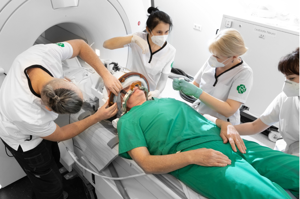Figure 5.2. The team positions the patient before surgery using MRgFUS The patient’s head with a membrane and stereotactic frame is securely fixed to the attachment and to the MRgFUS table helmet with 1024 piezoelectric ultrasound wave generators. The membrane is connected to the transducer, and the space between the helmet and the membrane is filled with water cooled to 15–17°C and degassed. Using 3 guiding screws on the MRgFUS table, the helmet’s position relative to the head is adjusted so that the focus is no more than 0.5 mm from the target in all 3 planes. During positioning, it is necessary to smooth out any folds forming on the membrane as much as possible. If this is technically difficult, at least remove air bubbles by tapping motions. The importance of this manipulation is due to the fact that during treatment planning, these folds will be outlined by the computer, and the ultrasound transmitters in their projection will be excluded from the sonication process. The more folds there are, the more active elements will be turned off, and consequently, the lower the likelihood of achieving the planned temperature.
For the safety of the procedure, the patient is always given an emergency stop button in their hand. Right before the procedure begins, the neurosurgeon or neurologist once again explains when it should be used.
To protect against the noise effect of MRI, it is recommended to insert earplugs. Special soft-elastic cushions are placed under the lumbar spine and knee joints. Many patients cannot relax the cervical spine and continue trying to hold their head “in the air,” despite understanding the presence of firm fixation. To overcome this reaction, we recommend placing a thin rectangular foam pad between the neurosurgical frame and the skin: the sensation of pressure on the back of the neck and occiput allows the patient to relax their head.
Some patients begin to notice the onset of pain syndrome at the sites of attachment of the neurosurgical frame after positioning and water intake. If the intensity of the pain is above 5 points on the visual analog scale (VAS), a pain reliever is re-administered; if the intensity of the pain is less than 5 points, pain relief patches or anesthetic cream can be used.
In the case of relatives being in the operating room (the “open operating room” concept – see chapter 3), a chair, headphones, and a warm blanket are prepared for them in the MRI room.
## Briefing and Checklist
Before the operation begins, readiness is checked by reading aloud our developed [checklist](https://buzaevclinic.com/downloads/buzaevclinic-mrgfus-safety-checklist-rus.pdf). This is necessary to ensure the safety of the patient, the medical team, and relatives, as well as to properly check the readiness of the equipment and the correctness of the planned intervention side. After the briefing, the neurologist conducts a full examination of the patient on the table, recording it on video camera. This examination and video recording are very important for assessing the dynamics of changes at all subsequent stages of treatment using MRgFUS.
[Our developed MRgFUS safety checklist ](https://buzaevclinic.com/downloads/buzaevclinic-mrgfus-safety-checklist-rus.pdf).
## Performing the MRgFUS Procedure
At the very beginning, the MRI-diagnosis doctor synchronizes the MRI equipment and the focused ultrasound table using localization scanning of the patient’s brain. Then, the MRI is controlled by the focused ultrasound workstation, performing a series of images in T1 mode, oriented in the coordinate system of the anterior and posterior commissures of the brain ( AC–PC). The obtained data is automatically loaded into the focused ultrasound console system.
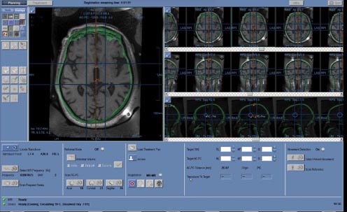Figure 5.4. Obtaining a hybrid CT and MRI image At the next stage, the neurosurgeon plans the treatment by combining previously taken CT images with MRI to determine the position of the target point, to which the transducer is moved using the mechanical positioning block of the focused ultrasound table. In Fig. 5.4, the skull bones are marked in green, overlaid on the black-and-white MRI image. The selection and calculation of the target ablation area (target) in the brain are carried out depending on the patient’s diagnosis. Before starting treatment, fiducial markers are placed on the hybrid image in brain areas with good structural contrast (marked with 5 crosses in Fig. 5.5). The markers are necessary to track possible head movement within the stereotactic frame. If movement is detected in the area of the fiducial markers, the system interrupts operation to avoid affecting the non-target zone. Five markers are placed in each of the 3 projections – axial, sagittal, and coronal.
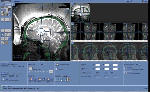Figure 5.5. Placement of Fiducial Markers Frontal sinuses and other structures containing air, calcium, as well as folds of the MRI-guideded focused ultrasound membrane are marked on the planning console to exclude the passage of ultrasound waves. In Fig. 5.6, red circles indicate calcifications; cavities of irregular shape outlined by a red line are membrane folds; the midline is shown in orange; the blue circle represents the boundaries of the area where ultrasound exposure can be performed without mechanically repositioning the helmet.
Figure 5.6. Marking of “prohibited” areas for ultrasound During the MRgFUS procedure, the target is heated by ultrasound beams – this process is called *sonication* . The area of tissue exposed to temperature and the duration of exposure form an equivalent thermal dose, which determines the degree of thermal damage. Depending on the bone tissue density coefficient, the neurosurgeon calculates the energy delivered by the ultrasound exposure (J), power (W), and duration (s) of exposure to achieve the required temperature. During sonication, constant MRI measurement of temperature parameters at the target point is conducted every 3 seconds, with the transmission to the workstation screen of the size and shape of the thermal spot, temperature change graphs (maximum and average values), acoustic spectrum, and control reflecting cavitation during treatment. These graphs help the neurosurgeon monitor the temperature increase; if they see a deviation from expected parameters, they can stop the sonication by pressing a special button. The interval between sonications is determined by the cooling duration of the circulating water
The entire treatment process is divided into three stages:
1. *Calibration Align)* – conducting sonications at temperature parameters from 40 о From up to 45ºC in three main dimensions, the neurosurgeon ensures that the system heats precisely at the selected point and the focus has a rounded shape.
2. *Focus Verification Verify)* – sonication with the target point temperature reaching from 45 о Up to 50ºC to confirm that the impact on the selected point is accompanied by the required reversible clinical effect
3. *Treatment Treat low/high)* – several sonications reaching a temperature of 55 о To up to 60 ºC to achieve the necessary and irreversible effect.
### Calibration Align) – anatomical verification
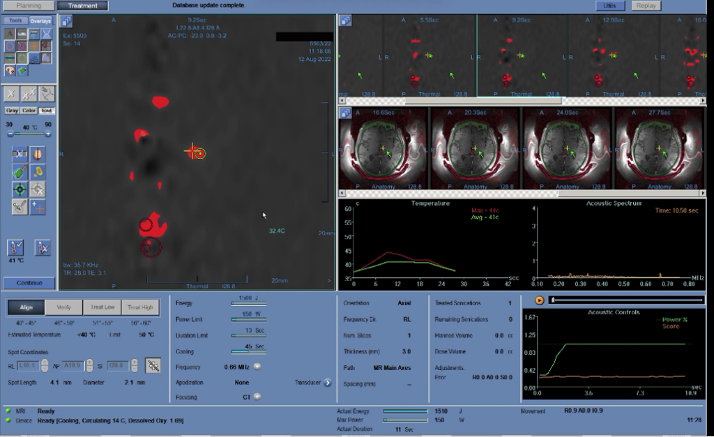Figure 5.7 Stage of Anatomical Verification At the first stage, the neurosurgeon heats the target area to low temperatures (40–45ºC) and assesses the accuracy of the hit (displacement of the heated spot from the target). Thus, the device is calibrated in all three planes. In Fig. 5.7, the temperature graph during sonication is in the center, to the left on the MRI image is a red spot; the area marked with a cross is the heated section, and the nearby circle is the target. If the heated area is not at the target location, the neurosurgeon adjusts the aim and repeats the sonication. Corrections are made until any errors are eliminated.
Observing the temperature increase at the desired point, the neurosurgeon determines the parameters of the applied ultrasound energy
the energy (J), power (W), and duration (s) of exposure to achieve the required temperature he will use in the next two stages of treatment
### Focus Verification Verify) – physiological verification
After the neurosurgeon ensures that the target is formed correctly, they move to the next stage—preliminary heating of the target area to a temperature that deactivates neurons without damaging them (45–50ºC) for a few seconds.
With such thermal exposure, a temporary reversible “shutdown” of brain cells occurs. During this short period, the neurologist assesses the patient’s response—changes in neurological status. At this stage, it is crucial to carefully evaluate the patient’s condition dynamics and the appearance of the slightest side reactions. A positive response is considered a reduction in the amplitude or frequency of tremor, decreased muscle rigidity and bradykinesia (in the case of Parkinson’s disease), and tension in the neck muscles (in the case of cervical dystonia). At the verification stage, it is difficult to achieve complete symptom disappearance due to low exposure temperatures, but an improvement of even 20–30% indicates the correctness of the chosen area.
If the expected improvement is not achieved during physiological verification or side effects occur, the target point or sonication parameters are adjusted. The neurosurgeon has sufficient opportunities to move the target point at this stage; however, each repetition of sonication causes reversible changes in brain tissue, complicating subsequent interventions. Sometimes, with a very high skull density coefficient (0.7–0.8) and thin cranial bones, even if the neurosurgeon has considered this, a significant thermal spot may form at the verification stage. Therefore, the choice of the target point should be made correctly and preferably on the first attempt.
The functional deactivation of neurons with ultrasound allows for modeling the future result of definitive therapeutic heating. Monitoring is conducted at each stage of the procedure to precisely know which area is being affected, what the thermal effects are, whether changes need to be made to the procedure parameters, and what the outcome will be immediately after the operation.
### Treatment treat low/high) – therapeutic effect
After achieving the expected but temporary result during physiological verification, final treatment is performed by applying ultrasound waves to the target area of the brain, heating it to a temperature of 55–60°C. The MRgFUS procedure 97 Irreversible effects are repeated to expand the ablation area and consolidate the result.
After each sonication, a neurologist assesses the patient’s condition with video recording. The neurologist, neurosurgeon, and MRI diagnostician jointly make decisions on further steps based on the obtained data in a continuous consultation mode.
After the irreversible ultrasound treatment is performed, a final assessment of the treatment results is conducted through a neurological examination on the table and an MRI study of the brain. The MRI results and changes in the patient’s neurological status are discussed with the patient and their relatives. If a satisfactory result and sufficient size of the MRI focus are achieved, the treatment is usually concluded.
### Neurological monitoring during the MRgFUS procedure
The team performing the MRgFUS procedure should always conduct a neurological examination of the patient between sonications. Different centers have adopted varying extents of patient examination. In some centers, complete neurological testing is not conducted during the MRgFUS procedure: for example, doctors may only check the effect of sonication on tremor or dystonia symptoms and then proceed to the next sonication, assessing further dynamics. This approach may be practical in terms of cost-effectiveness and the image of a “quick” neurosurgeon. However, the examination is needed not only to assess the effectiveness of treating tremor or other individual symptoms. An adequate amount of neurological testing during the MRgFUS procedure is very important for several reasons
1\. Treatment Effectiveness. The observed elimination of tremor or other movement disorders after sonication indicates a correctly chosen target, allowing the neurosurgeon to continue treatment as planned or to complete it
2\. Safety. A complete neurological examination can help identify unexpected neurological deficits (for example, due to atypical anatomy in a specific patient), which might be missed if limited to simple tests for tremor or muscle rigidity. For instance, if a mild paresis manifests in the leg during test sonication and the surgeon does not properly assess the condition of the lower limbs, the application of ultrasound in therapeutic mode could exacerbate this issue and potentially make it irreversible.
3\. A complete neurological examination allows for a discussion with the patient and relatives about the further course of the operation. It may be advisable to draw the attention of the patient and relatives to neurological “findings” that appear during treatment, so that in the case of a suboptimal effect, the procedure can be concluded with their agreement to avoid more serious complications.
In our medical center, we conduct a complete examination between sonications according to a developed algorithm, which takes no more than 15 minutes and provides very important information to assess the balance of treatment effectiveness and safety and to stop in time. Experience with more than 200 MRgFUS procedures has allowed us to form and propose the following necessary and sufficient protocol for neurological examination between sonications.
1\. Assessment of smell through a survey.
2\. Vision assessment, preliminary visual field testing. Inquiry about visual acuity.
3\. Tracking an object. Assessment of double vision. Examination for ptosis and strabismus.
4\. Facial Sensitivity Assessment
5\. Examination of the face at rest, detection of facial asymmetry, drooping of the corner of the mouth. Facial symmetry. Tests with forehead wrinkling, frowning, closing eyes tightly, showing teeth, puffing cheeks, stretching lips.
6\. Approximate hearing assessment
7\. Voice pitch (excluding dysphonia). Swallowing saliva.
8\. Speech examination for dysarthria, difficulties in pronouncing words and sounds. Tongue protrusion (no deviation)
9\. Muscle strength testing, examination of the range and speed of active movements. Raising arms up while lying down, flexion-extension in the joints, pronation-supination in the wrists, adduction-abduction of fingers. Range and speed of movements are assessed separately. For careful evaluation of even minor paresis, upper and lower Barre tests are used while lying on the back.
10\. Muscle Strength Assessment. Evaluation of resistance to movement produced by the patient. The measurement is subjective, from 0 to 5 points, where 0 points – no movement; 1 point – slight movement; 2 points – movements possible without overcoming the force of gravity on the limbs; 3 points – the patient performs active movements but does not resist the doctor; 4 points – the patient resists but is weaker than the doctor; 5 points – full strength.
11\. Study of passive movements and determination of muscle tone.
12\. Study of Hyperkinesia Severity and Coordination. Examination of the upper and lower limbs at rest and during stress tests (subtracting from 100 by 7, reciting letters in reverse order, squeezing and releasing the contralateral hand). Finger-to-nose test for identifying intentional and kinetic tremor, pointing test. Heel-to-knee test. Examination of hyperkinesias (tremor and others) with arms raised above the head while lying down and with arms in front of the chest. Drawing spirals and lines. Test with a plastic spoon, test with a glass, test with a plastic bottle of water (allows assessment of tremor severity by liquid level fluctuations). Tests with additional objects, such as manipulating earplugs.
13\. Checking the sensitivity of the skin in symmetrical points on the right and left sides of the body, from distal to proximal areas (the examination is conducted using plastic non-magnetic sticks or toothpicks). In our series of observations, 2 out of 200 patients showed a positive Barre test after trial sonication. This allowed us to change the target (shift the point of impact and conduct sonication more medially to avoid damaging the pyramidal pathways) and avoid complications. Additionally, several patients experienced numbness of the tongue tip or lower lip when the thalamus was targeted; in this case, the target was shifted forward to avoid damaging the sensory nucleus and prevent persistent functional impairment.
### Therapeutic monitoring during the MRgFUS procedure
During the operation, the nurse monitors blood pressure. If it increases, antihypertensive drugs are administered. In case of headache, pain relievers are recommended (e.g., ketorolac 30 mg/ml), for the prevention of brain tissue edema – dexamethasone 4 mg/ml intravenously, and for the prevention and treatment of nausea and vomiting, ondansetron 2 mg/ml can be additionally administered. If necessary, humidified oxygen is supplied through nasal cannulas, and an aspirator is used in case of vomiting. As mentioned above, it is advisable to secure the equipment to the floor (contains ferromagnetics).
### Procedure for Removal from the Focused Ultrasound Table
After completing the treatment, the patient remains on the table while the water drains from the system. The membrane is detached from the transducer, and the neurosurgical frame is removed from its attachment to the table. Only after this is the patient allowed to get up from the table and transfer to a transport chair-stretcher. In the dressing room, the silicone membrane and neurosurgical frame are removed, the attachment sites are treated, and bactericidal plasters are applied. The intravenous catheter is left in place for a few more hours for use if necessary.
### Postoperative Stage
The patient remains under observation in the clinic for another 7–8 hours, with a follow-up MRI of the brain performed 2 hours after treatment and a repeat examination by a neurologist and neurosurgeon. The follow-up MRI is conducted in T2 mode FSE with 2 mm slices in the axial and coronal planes, as well as in modes SWAN, DWI, T1 FSPGR with 4 mm slices in the thalamus region in the axial plane. After MRI, depending on the severity of changes in the brain, dexamethasone is prescribed (intramuscularly or intravenously) to reduce the severity of edema. Procedure MRgFUS 101
The day after treatment, the patient, accompanied by a relative, returns to the clinic for a follow-up examination. A complete neurological examination is performed, and a follow-up MRI of the brain is conducted. On this day, further monitoring, regimen, the need for rehabilitation activities, and a schedule for subsequent consultations are discussed. Considering the non-invasive nature of the procedure, patients do not require a long recovery period, but it is recommended to follow a protective regimen without excessive stress for 2 weeks under the supervision of close ones.
In the postoperative period, patients take glucocorticosteroid medications for at least 2 weeks to prevent excessive swelling in the treatment area. All patients undergo follow-up brain MRI every 3 months according to a standard protocol.
## Standardization of the Treatment Process Using MRgFUS
Due to the novelty of the MRgFUS method, staff preparation is required in addition to their basic training necessary for accreditation. Standardizing the process ensures consistent quality of care and provides the most reliable data for research.
Based on our experience, we have developed a clear standardized protocol for performing surgery using MRgFUS, presented in Appendix 2.
## Target determination for MRgFUS
Two methods for determining the target for MRgFUS are proposed:
1\) indirect targeting based on atlas and patient anatomy data on MRI brain scans, using the approximate location of the nucleus
2\) direct tractographic approach, where targeting is personalized based on MR tractography ( Bruno et al., 2021).
Indirect targeting is a permissible approach to thalamotomy using MRgFUS, allowing for the immediate identification of the correct target in most cases. The direct tractographic approach is currently a valuable aid in assessing potential deviation in cases where an immediate clinical response is not achieved. Direct coordinates, for example, are proposed to be measured for the dentatorubrothalamic tract based on diffusion tensor MRI imaging.
Figure 5.8. Coordinate systems of MRI, FUS station, and patient Currently, the precise determination of the impact point is a major challenge. The issue is that the localization of the sonication target is conducted relative to the line connecting the anterior and posterior commissures ( AC–PC), what is the universal coordinate system for patients in functional neurosurgery. At the same time, MRI has its own coordinate system RAS, which is determined by the position relative to the isocenter of the device; additionally, the FUS station has its own coordinate system relative to the focal point. All three coordinate systems relate differently depending on the equipment’s position. To solve this problem, we created a special program in the language R, which allows recalculating coordinates depending on the positions of the zero point of the MRI and FUS station for more accurate target determination and reducing the possibility of error. Figure 5.8 shows an example of the relationship between these coordinate systems, where RAS – these are the coordinates relative to the MRI isocenter, transducer focal – coordinates of the FUS station, and AC–PC target – coordinates relative to the posterior commissure in the plane AC–PC.
Figure 5.9 shows the treatment sonication map for all patients who underwent treatment using the MRgFUS method, on MRI in the axial plane passing through the line AC–PC.
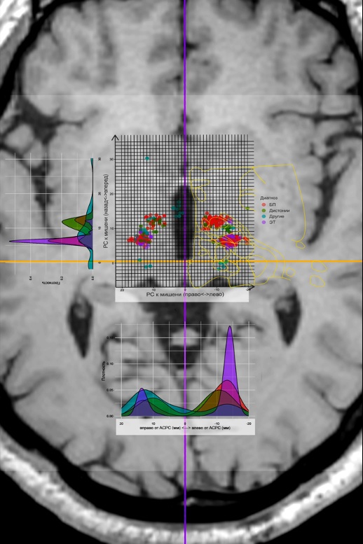Figure 5.9. Map and density diagrams of patient sonications in the axial plane passing through the line AC-PC.## Calculation of the Exact Target Temperature During Sonication
An important parameter for the effectiveness of treatment using MRgFUS is the temperatures reached at the point of impact. During sonication, the temperature increase occurs at different rates and depends on many parameters described by S. Gagliardo et al. (2019). The authors proposed calculating energy delivery curves for each treatment stage based on CT perfusion to determine ultrasound energy and ensure better control over the procedure’s outcome. However, they did not propose a clear formula for calculating heating energy.
As already mentioned above, during MRgFUS, the neurosurgeon uses specific parameters of energy, power, and duration of ultrasound waves to regulate the intensity of heating. During the first sonication at the calibration stage, the power is usually set at 250 W and the duration at 10 s, corresponding to an energy output of 2500 J. For subsequent sonications, the temperature setting is adjusted as follows. The neurosurgeon increases the power to such an extent that heating does not occur too quickly, as rapid heating can easily exceed the safe temperature limit for reversible effects. In other words, the power should be large enough to heat the area to the desired temperature within the set time, but the heating must be done at a rate that allows the neurosurgeon to stop the sonication in case of overheating. Excessive power can instantly overheat the target area of brain tissue. An unfavorable factor is that many patients feel the sonication: with increased power, headaches often intensify, and the patient may interrupt the treatment or even refuse to continue the operation. Too little power will not allow the area to be heated in the set time, but additional sonication may increase unnecessary tissue swelling—albeit temporary, but altering the neurological status at the time of the procedure.
At the same time, the duration determines the heating. More time allows more energy to be delivered to the area, increasing the heat. Too short a time won’t provide enough energy to heat the tissues, while an overly lengthy procedure may be poorly tolerated by the patient and lead to headaches. In this sense, reducing the treatment duration increases its tolerability for the patient. There are cases where the maximum power tolerated by the patient or generated by the device allows the area to be heated to the required temperature only by extending the sonication. The energy required to heat to the necessary temperature is calculated as the product of power and time. However, different patients require varying times to heat an area to a specific temperature.
Energy loss is influenced by KUPKT, or scull score, which is measured in values from 0 to 1. As the bone ultrasound transmission coefficient decreases, greater power and time are required to achieve the necessary temperatures. The bone ultrasound transmission coefficient is calculated using CT of the skull bones with the MRgFUS planning software package
In addition to the skull bones, water with varying concentrations of oxygen bubbles, which absorb ultrasound, stands in the way of the ultrasound. This issue is resolved by standardized cooling of the water, which sharply decreases oxygen solubility with a drop in temperature. The condition of tissues along the ultrasound path also alters its conductivity. There are other factors affecting the delivery, scattering, and reflection of energy. Therefore, using the KUPCT index as the sole guide is impossible (especially considering its inherent error).
The identification of clinically significant factors is useful for constructing a temperature model based on power, time, CT perfusion, and several other factors. Modern computer programs allow the use of complex algorithms and dependencies for model construction in the presence of multiple factors and uncertainty. To solve this problem, we created a special program that includes the following parameters: patient ID, sonication sequence number, achieved temperature (°C), power (W), sonication duration (s), early termination (1 – yes, 0 – no), measured energy (J), CT perfusion, CT machine used to determine CT perfusion Siemens, GE, Toshiba, Philips), data on the response to the first align-sonication, gender, and age of the patient. To determine dependencies, we built 2 models – linear and neural network.
The linear model was built in R package RStudio 2021.09.2 сборка 382, R version 4.2.1 (2022-06-23) regular team lm. As a result of the analysis of the obtained data, it was hypothesized that the linear model does not perfectly predict the temperature due to possible nonlinear dependencies. A neural network-based regression solution could be suitable for building the model.
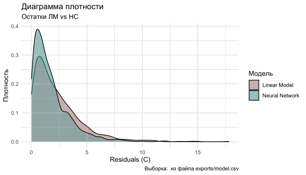Figure 5.10. Density plot of residual distribution (difference between predicted and actual temperature) Neural network model was created in R package RStudio 2021.09.2 сборка 382, R version 4.2.1 (2022-06-23), what were open libraries for production-level machine learning used for TensorFlow. Details of the neural network model construction and the analysis conducted with its help can be provided upon request.
When analyzing the data models, the maximum prediction error for the linear model was 17.8°C, and for the neural network model, it was 12.1°C. Thus, the neural network model for temperature calculation proved to be better. Figure 5.10 shows the density distribution diagram of the residuals, where it is evident that the neural network model is closer to zero error. The corresponding calculations can be practically implemented during the planning and execution of treatment using MRgFUS with the table we proposed (Figure 5.11).
Figure 5.11. Table Excel for automatic temperature calculation Thus, the temperature of the target during ultrasound heating is influenced not only by power, energy, and time as sonication parameters but also by the CT perfusion index, as well as the patient’s gender and age. The parameters of the first sonication during the calibration stage and the response to it are useful for predicting the parameters of subsequent sonications. The linear model allows predicting the temperature with a mean absolute error MAE) = 2,78O S, and the coefficient of determination R2 = 0,71, whereas the neural network – with a mean absolute error of 1.93 O S and R2 = 0,76. A disadvantage of the linear model is its lower accuracy, but it is easily accessible and convenient in the operating room, as it does not require additional software.
## Features of the Cohort of Patients We Operated On
The principles of work organization and action algorithms outlined above were successfully applied by our team at the first center in the Russian Federation for treatment using MRgFUS: from 2020 to 2023, 86 patients with PD (including 82 with predominant tremor and 4 with akinetic-rigid form of the disease), 37 patients with ET, 18 patients with various forms of dystonia, as well as 1 patient each with neuropathic pain and hypothalamic hamartoma, were treated at the V.S. Buzaev International Medical Centre (Ufa). From the first day, the work of the MRgFUS center was conducted under the scientific and methodological guidance and in close cooperation with the Research Center of Neurology and the National Society for the Study of Parkinson’s Disease and Movement Disorders.
The age of operated patients (Fig. 5.12) in the main groups with movement disorders differed statistically significantly ( p < 0,001 by the Kruskal-Wallis criterion): the average age of patients with PD was 63 \[55; 70\] year, patients with ET – 54.50 \[36,25; 64,75\] year, patients with dystonia – 46.0 \[39,0; 53,5\] years. There were no age differences between men and women
The median duration of the operation (Fig. 5.13) was 98.7 \[74,7; 132,6\] min, including in the PD group – 97.2 \[73,6; 126,4\] min, in the ET group – 96.1 \[72,8; 126,4\] min, in the dystonia group – 117.1 \[79,1; 139,2\] min, differences are statistically insignificant (p = 0.53). The median number of sonications (Fig. 5.14) was 11 \[10; 14\], with slight variations between groups. The maximum temperature of the sonication focus during the operation (Fig. 5.15) was 61.6 \[59,8; 63,0\]°C, maximum sonication energy (Fig. 5.16) – 20,035 \[14 030; 32 330\] J, there were no significant differences between the groups.
Figure 5-13. Age of patients with movement disorders treated with MRgFUS. 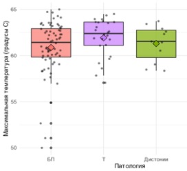Figure 5.14. Maximum temperature of the sonication focus during surgery in different groups. Figure 5-15. Surgery time in different groups. Figure 15. Maximum sonication energy in different groups. 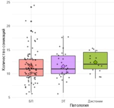Figure 5.16. Number of sonications in patients across different groups ## International Internet Operating Room: Application of MRgFUS in Telemedicine Mode
Usually, for the theoretical training of doctors and the development of their manual skills, special training programs from the manufacturer are used. However, the best practice is to have a dedicated specialist for supervision and consultation during the first operations ( Broering et al., 2020). Many manufacturers do not allow their treatment programs to be initiated without authorized assistance and supervision, especially when it involves new technologies. According to the manufacturer’s standard policy, it is advisable to conduct the first 20–25 operations under the supervision of a specialist, after which the team receives “solo” status, i.e., permission to perform the procedure independently.
Travel restrictions during the pandemic COVID-19 led to a large-scale transition of educational programs at various levels to online digital and distance learning. However, the implementation of such approaches for entirely new, complex technologies may be associated with serious and quite understandable difficulties. The initiation of the practical work of our MRgFUS center coincided with the peak of the pandemic: in April 2020, the equipment installation was completed InSightec ExAblate 4000 (in the surgery waiting list were 32 patients), but at that moment it became clear that the personal arrival of experts from the manufacturer was impossible due to quarantine restrictions. In this regard, we were the first in the world to develop and implement a treatment program using MRgFUS with international remote online monitoring, assessing the effectiveness and safety of this telemedicine approach, as well as the immediate results of the operations performed
The study included the first 94 patients who underwent thalamotomy using MRgFUS in our center, including 27 patients with ET and 67 patients with PD (tremor-dominant phenotypes). Under online control, 38 patients received treatment and without it (group ” cоло”) – 56 patients. The study began on May 5, 2020, and continued for 1 year and 9 months. The center’s team was certified to perform independent operations (” colo”) after performing 38 MRgFUS procedures.
To present the results we obtained both within this study and in further independent work (see chapters 6–8), it is necessary to introduce a number of defining terms. By “attempt,” we mean the situation when a patient is placed on the MRgFUS table and the first ultrasound exposure is performed. We define the “success” of the procedure as a 30% reduction in the severity of tremor according to CRST and/or by UPDRS (part III) on the side contralateral to the performed operation
We also used the following subjective scale: “excellent” effect – complete elimination of movement disorder symptoms and achievement of all major surgical goals; “good” effect – only minor symptoms remain after MRgFUS treatment; “satisfactory” effect (“compromise”) – a certain positive result was achieved during the procedure, but it was stopped because the expected risk of complications from continuing ultrasound exposure was higher than the expected benefit. These 3 categories are considered “successful.” A “failed” result was recorded if the intervention did not significantly impact the severity of the disease’s clinical manifestations. In no case of MRgFUS application did we observe a deterioration in the patient’s condition.
## Telemedicine Remote Expert Monitoring
The world’s first telemedicine implementation of the MRgFUS technology took place on May 5, 2020. We ensured the virtual presence of company experts InSightec Paula Regg (United Kingdom), Tessa Case (Spain), Daniil Molchanov (Russia), as well as project curators – President of the National Society for the Study of Parkinson’s Disease and Movement Disorders, Academician of the RAS S.N. Illarioshkin, and Chief Neurosurgeon of the Ministry of Health of the Republic of Bashkortostan, Doctor of Medical Sciences Sh.M. Safin during the first procedure. Before the first treatment session using MRgFUS, a two-week training process took place with preliminary discussion of each patient to ensure the most appropriate selection.
To organize the first telemedicine treatment using MRgFUS, in addition to the traditional organization of a video conference with all participants, it was necessary to solve 3 tasks.
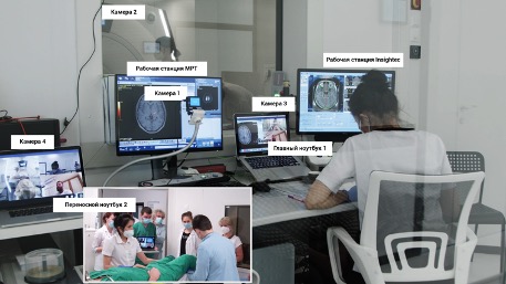Figure 5-17. Camera positions and view from the operating room Firstly, to ensure safety and constant visual monitoring of the patient’s condition, we needed to provide secure video streaming (in real-time) of the patient’s condition in the MRI room during treatment. Additionally, foreign experts were required to evaluate the results of neurological testing conducted by our team of neurologists to assess treatment effectiveness. The challenge was that due to the design features of the MRI room and the constant magnetic field, it was impossible to place cameras directly inside the room. We conducted 3 procedures using a webcam Microsoft, mounted on the ceiling of the MRI room. During the 3rd operation on MRI thermometry, intense noise began to occur, so the only solution was to remove the camera from the MRI room and install it in front of the observation window (camera 2 in Fig. 5.17). We streamed the video feed from the cameras into a video conference Microsoft Teams. A portable laptop was used during neurological tests MacBook Pro with a built-in camera (portable laptop 2 in Fig. 5.17)
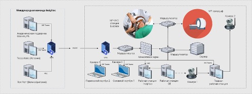Figure 17. Online Operating Room Infrastructure Secondly, it was necessary to organize the transmission of telemetry from the equipment InSightec to the international team of engineers so they could observe how the chosen treatment strategy aligns with the equipment settings and adjust them if necessary to achieve the desired outcome. No one in the world had such experience. After analyzing all available technologies, the most promising technologies were selected Virtual Network Computing (VNC) и Port Address Translation, with which we were able to organize secure remote connection sessions for experts located during treatment in the UK, Spain, Israel, and St. Petersburg (Fig. 5.18)
Thirdly, it was necessary to ensure the transmission of the patient’s MRI images so that the international team could assess the effectiveness of the treatment and adjust the strategy with us. Since the software and hardware system General Electric represents a fully closed system, we did not have the opportunity to connect to it directly or use additional technologies for intercepting and transmitting images (similar to converters HDMI в USB). To solve this task, we used a camera directed at the MRI monitor, and the video stream from the camera was transmitted to a video conference for all participants (camera 1 in Fig. 5.18). This image was not used for making clinical decisions but only provided the ability to manage settings on the console by the MRI-technician. The highest quality MRI image in real-time, as usual, was transmitted to the InSightec ExAblate 4000 (workstation InSightec in Fig. 5.18) according to the device protocol InSightec. Then the image was broadcast from the console InSightec protocol experts VNC. Microsoft Teams used as a platform for video conferencing among all participants (see Fig. 5.18). The choice of this technology was due to the corporate standard InSightec, and its functionality met all necessary requirements. We used 4 cameras: camera 1 for the MRI console, camera 2 for the MRI room, camera 3 for the neurosurgeon, camera 4 for neurological tests and temporary close-ups inside the MRI room. Portable laptop 2 with camera 4 was brought into the MRI room only when the MRI machine was not scanning (see Fig. 5.18)
Neurosurgeon, radiologist, and MRI-technician communicated with experts using the main laptop 1 (see Fig. 5.18). The workstation console InSightec thanks to the use of the protocol VNC allowed the neurosurgeon in Ufa to operate the MRgFUS device and enabled the use of the mouse cursor in collaboration with experts from the operating table manufacturer. The MRI workstation console was controlled by a radiologist and an MRI specialist.
Thus, solving these three tasks made it possible to ensure the necessary level of patient safety and conduct the world’s first telemedicine treatment using MRgFUS.
## Comparison of Surgery Outcomes Performed with Telemedicine Monitoring and Independently
In total, thalamotomy using the MRgFUS method under international online supervision was performed on 38 patients with tremor (11 patients with ET and 27 patients with PD), and independently we operated on 56 patients (16 patients with ET and 40 patients with PD) (Fig. 5.19). There were no significant age differences between patients in the specified groups.
In the ET group with telemedicine support, the procedure was successful in 10 (81.8%) out of 11 patients, while in the self-treatment group, all 16 patients (100%) were successful. No recurrence of tremor was recorded in either group. When evaluating the treatment results by CRST (Fig. 5.20) No statistically significant differences were found between the groups of self-treatment and treatment with telemedicine support
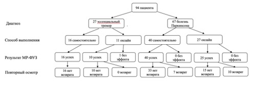Figure 5-19. Research flow diagram In the group with telemedicine support, the procedure was successful in 25 (92.6%) out of 27 patients, while in the self-treatment group, all 40 patients (100%) were successful. A partial recurrence of tremor within 1 year was observed in 10 (40%) out of 25 patients in the telemedicine support group, and in 7 (17.5%) out of 40 in the self-treatment group, with statistically significant differences ( p = 0,04 by the χ2 criterion). When comparing the condition of operated patients by UPDRS (part III – motor functions) it was found that the improvement achieved in the self-treatment group was statistically more significant ( p = 0,0026 by the Wilcoxon criterion) than in the treatment group with telemedicine control (Fig. 5.21)
 and treatment with telemedicine support ("Online") | VS Buzaev International MRgFUS Medical Treatment Centre")Figure 5-20 Results of MRgFUS application in patients with ET in self-treatment groups (“Solo”) and treatment with telemedicine support (“Online”) Thus, we were the first to demonstrate that remote expert monitoring during treatment with MRgFUS is effective and safe. For ET and PD, the treatment outcomes in the groups with remote telemedicine monitoring and independent treatment were generally comparable in terms of achieved improvement, complication rates, and long-term results (tremor recurrence). Interestingly, in the PD group, the percentage of symptom reduction was significantly higher in the independent treatment group than in the telemedicine monitoring group; this can be explained by the fact that during online training, experts were restricted by a strict protocol with clearly defined targets, whereas in the process of independent treatment, we had the opportunity to target other areas with MRgFUS not included in the manufacturer’s protocol (for more details, see Chapter 7)
 and treatment with telemedicine support ("Online") | VS Buzaev International MRgFUS Medical Treatment Centre")Figure 5-21. Results of MRgFUS application in patients with PD in the self-treatment group (“Solo”) and treatment with telemedicine support (“Online”) The method we developed for launching a telemedicine remote treatment program with MRI-guideded focused ultrasound under online supervision can be recommended for implementation by other centers in the process of mastering this technology. The world’s first international publication on this topic was presented by us in 2023 ([Buzaev et al., 2023](https://www.ncbi.nlm.nih.gov/pmc/articles/PMC10914008/)) and has already received very favorable feedback. Given the rapid development of the method and the emergence of new MRgFUS centers worldwide and in our country, the experience gained is very valuable and timely. Since various telemedicine approaches are currently in the spotlight, some of the developments and technological solutions we have made can be transferred from neurosurgery to other clinical specialties.
---
### [MRgFUS for Dystonias ](https://buzaevclinic.com/kniga-fokusirovannyj-ultrazvuk-pod-kontrolem-mrt-v-nevrologii/mr-fuz-pri-distoniyah/)
**Published:** November 2, 2024
**Author:** Rezida Galimova
**Content:**
# MRgFUS treatment of dystonia
## General Information
Dystonia is a clinical syndrome characterized by involuntary, arrhythmic twisting movements in various parts of the body, as well as dynamic changes in muscle tone, leading to the formation of pathological postures or posture-dependent changes (Levin et al., 2022). Dystonia is characterized by: stereotypical contractions in a specific muscle group (rotational, flexion, or extension pattern, etc.); increased hyperkinesis during voluntary movements and its change with posture changes; improved control over involuntary dystonic hyperkinesias with certain tactile or proprioceptive stimuli (corrective gestures); involuntary contraction of additional muscle groups that do not anatomically coincide with the primary dystonic movement (the phenomenon of “overflow dystonia”), usually observed at the peak of a dystonic spasm Geyer, Bressman, 2006; Sitburana et al., 2009; Albanese et al., 2019).
Dystonic syndrome develops gradually: initially, hyperkinesias occur only for a short time and/or only during certain actions (positions), but over time their duration increases, the severity of dystonia escalates, pathological postures appear, and sometimes contractures Albanese et al., 2019). There is also a gradual spread of dystonic manifestations to various regions of the body (in generalized forms). Some patients with dystonia may experience one or more periods of complete or partial remission lasting from several hours to several years in the early years of the disease.
A special manifestation is dystonic tremor (Illarioshkin, Ivanova-Smolenskaya, 2011). It typically has a frequency of 3–7 oscillations per second and is equally common in generalized, segmental, or focal/multifocal dystonia. Its clinical features are similar to the dystonic hyperkinesis characteristics discussed above, which significantly facilitates diagnosis. In particular, dystonic tremor is characterized by significant variability, asymmetry, and dependence on maintaining a specific posture, intensification during voluntary movements or attempts to resist dystonic spasm, weakening at rest, and the presence of corrective gestures.
According to etiology, the following are distinguished: 1) primary dystonia – all forms (usually genetic), where dystonia is the only symptom of the disease (may only be combined with tremor), with no laboratory changes or clear signs of pathology in neuroimaging studies (CT, MRI); 2) dystonia plus – forms characterized by a combination of dystonic syndrome with myoclonus, parkinsonism, or chorea, where clinical and laboratory results suggest certain neurochemical disturbances without signs of neurodegeneration (dopa-responsive dystonia, myoclonus-dystonia, etc.); 3) secondary dystonias – drug-induced forms and syndromes caused by acquired reasons and external factors (neurotrauma, stroke, neuroinfections, demyelinating processes, etc.). Dystonia can also be one of the manifestations of a large number of multisystem neurodegenerative diseases ( Albanese et al., 2019).
The classification of dystonia by the prevalence of hyperkinesis is also generally accepted:
• focal dystonia (involving one part of the body – oromandibular dystonia, CD, etc.);
• segmental dystonia (involving ≥2 adjacent body areas, for example, Meige’s syndrome – a combination of blepharospasm, oromandibular dystonia, and CD)
• multifocal dystonia (involving ≥2 distant body areas, such as blepharospasm and foot dystonia)
• hemidystonia (involving muscles on one side of the body, which usually clearly indicates the secondary nature of dystonia and the location of the pathological process in the contralateral hemisphere of the brain)
• generalized dystonia (involving muscles of one or both legs, the torso, and at least one other part of the body)
Hereditary forms of dystonia are distinguished (they usually debut in childhood and tend to generalize) and sporadic forms (more often manifest in adulthood as focal phenotypes). To date, more than 30 genes associated with the development of hereditary dystonias are known (Levin et al., 2022; Domingo et al., 2021).
The overall prevalence of dystonia may be 3–11 cases per 100,000 population for generalized forms and 30–60 cases for focal forms ( Steeves et al., 2012). Thus, within the framework of movement disorders, dystonia is second in prevalence only to ET and PD. Among the various types of dystonia, CD (focal form) is the most common – 4.98 per 100,000 population ( Steeves et al., 2012).
Dystonic syndromes significantly affect the quality of life: 25 to 50% of patients suffer from depression, and pain is one of the key complaints in 67–75% of patients. The deterioration in quality of life and impaired motor activity impact the professional activities of patients with dystonia, making the problem socially and economically significant ( Butler et al., 1998).
Treatment of dystonias is a challenging task. Medication therapy is highly effective only for a few forms: for instance, in dopa-responsive dystonia, small doses of levodopa have a dramatic effect, and in patients with ADCY5-with associated dystonia (dyskinesia), significant relief can be achieved with caffeine intake. Unfortunately, in the vast majority of cases of dystonia, such a favorable outcome from medication treatment is not expected. Usually, the first step involves attempts to prescribe anticonvulsants, benzodiazepines, anticholinergics, muscle relaxants, tetrabenazine, clozapine, etc., including in various combinations. Their intake may be accompanied by a certain (sometimes even persistent) symptomatic effect; however, most often, the results of such therapy are quite modest, and increasing the dosages of the drugs used leads to pronounced side effects (drowsiness, impaired coordination, general weakness, decreased mental performance, dry mouth, accommodation disorders, etc.). Particularly unfavorable for conservative therapy are generalized (dopa-unresponsive) forms of idiopathic dystonia.
In focal forms, the method of choice is the use of intramuscular injections of botulinum toxin ( Albanese et al., 2019), which in most cases allows for the complete or partial alleviation of dystonic spasms for an average period of 3 to 6 months. After this, re-administration of the drug is required. In addition to its peripheral action, botulinum toxin has a favorable effect on the central pathophysiology of dystonia by reducing afferentation from muscles involved in hyperkinesis (Levin et al., 2022). Cases of prolonged “therapeutic remission” have been described with regular administration of botulinum toxin, attributed to its beneficial effects on peripheral and central mechanisms of neuroplasticity. Repeat courses of botulinum therapy for patients with focal dystonias have been successfully used worldwide for over 40 years. However, in 6–14% of cases, botulinum toxin proves to be less effective. Treating patients with retrocollis and anterocollis, as well as with combined forms of CD, presents a particular challenge. Surgical treatment is indicated when medication is ineffective, and there is progression of segmental or generalized dystonia, or insufficient control of symptoms of focal dystonias.
## Surgical Treatment of Dystonias
The first operation for spasmodic torticollis dates back to the mid XVII century, when the German surgeon Isaak Minnius performed a transection of the sternocleidomastoid muscle. In XIX in the century, I.V. Buyalsky first performed the transection of the accessory nerve in the neck for spasmodic torticollis, later W.W. Keen carried out the crossing of the first 3 anterior cervical roots, and at the beginning of the 20th century O. Foerster developed a technique of posterior rhizotomy for such patients. The effectiveness of peripheral operations, which continued to improve, was undeniable; however, extensive denervation of the neck muscles leads to serious complications (flaccid paralysis of the muscles, sensory disturbances, vertebral artery injuries), up to fatal outcomes. Therefore, surgeries at the level of muscles, nerves, and roots, as well as on the conduction pathways of the spinal cord (such attempts were also made), are currently not used for dystonias of any localization and are of only historical interest (Tyurnikov et al., 2007)
The basis of modern surgical treatment for patients with dystonic syndromes is various types of functional stereotactic operations on the brain. The two most adequate interventions are considered to be destruction VIM and some other subcortical nuclei and, mainly, DBS with an impact primarily on the globus pallidus ( GPi). Destructive surgeries, widely used in clinical practice since the mid-20th century and having played a significant role in treating patients with severe, disabling forms of dystonia, have become c 1990-years largely giving way to the method DBS, primarily due to the safety of bilateral stimulation and the ability to perform (if necessary) fine, dynamic adjustment of the stimulation mode
First Results of Bilateral Stimulation GPi were published P. Coubes et al. (1999). In the authors’ clinic, an 8-year-old girl with severe primary generalized dystonia, bedridden and requiring constant sedation and mechanical ventilation, underwent surgery. Six months after the operation, the patient was able to attend a regular school. Currently, worldwide, the number of patients with generalized, segmental, and focal forms of dystonia successfully receiving treatment through DBS GPi, runs into thousands. Bilateral surgery c conducting DBS GPi until recently, remains the most radical and effective method of providing real help to patients with severe movement disorders, impaired walking, and self-care (Levin et al., 2022; Alterman, Snyder, 2007).
Thalamic DBS may be quite successful for dystonic tremor, especially for DYT1-form of the disease. In primary writing tremor, stimulation VIM accompanied by almost complete disappearance of tremor ( Racette et al., 2001). In some cases, such patients leave the generator turned off for most of the day and turn it on only when they need to write something.
There are also several recent reports in the literature about the successful application of DBS on the subthalamic nucleus area in the treatment of patients with primary (generalized, segmental, cervical) dystonia, tardive dystonia, dystonia-parkinsonism, isolated tongue dystonia ( Tisch, 2018; Liu et al., 2019; Wu et al., 2019; Li H. et al., 2021; Mercado-Pimentel et al., 2021). More detailed results of traditional functional stereotactic interventions for dystonia are presented in Chapter 1.
## Treatment of Dystonia with MRgFUS
The global experience of surgical treatment of dystonias using the new MRgFUS method remains limited. There are about 15 publications on this topic in the literature, presenting a preliminary assessment of the dynamics of the clinical condition of patients after stereotactic ablation of subcortical nuclei using the MRgFUS method, conducted in several clinics worldwide, based on relatively small groups of patients with various forms of dystonias. Japanese researchers can be considered pioneers in this field S. Horisawa и T. Taira.
The initial results of using MRgFUS for dystonias indicate that this method, in terms of effectiveness, apparently does not fall short DBS. The ventrooral nucleus of the thalamus was used as a target for ultrasound ablation ( VO) и PTT (campotomy), only in a few reports is there mention of targeting GPi. The main work is currently limited to focal dystonias, dystonia-parkinsonism, and dystonic tremor. According to several authors Golfrè Andreasi et al., 2024; Peters et al., 2024), Thalamotomy using the MRgFUS method appears to be a very promising and less invasive alternative DBS to eliminate dystonic tremor, however, opinions regarding the most appropriate target VIM, GPi, PTT and others) differ, requiring further accumulation of experience
In Table 8.1, comparative data from some recent publications are presented, focusing on the analysis of the effectiveness of surgical treatment of various forms of dystonia using MRgFUS and DBS.
Thus, the treatment of dystonias using the MRgFUS method is at an early stage of development, and practically every newly described series of cases and even individual cases of different forms of this clinically heterogeneous movement disorder subjected to MRgFUS are of significant interest.
**Table 8.1.** Global Experience in Surgical Stereotactic Treatment of Dystonia: Comparison of Recent Studies in MRgFUS and DBS.
| Form of dystonia | Link | Year | Method | Target | Result |
|---|---|---|---|---|---|
| Musician’s Dystonia | Horisawa et al., 2018 | 2018 | MRgFUS | VO | 90% improvement observed over 12 months |
| Hand dystonia | Horisawa et al., 2021б | 2021 | MRgFUS | VO | Improvement of 70% during a 12-month follow-up (complications: persistent dysarthria – 1, transient dysarthria – 3, transient facial paresis – 2, symptom recurrence after 3 months – 3) |
| Dystonic tremor | Golfrè Andreasi et al., 2024 | 2024 | MRgFUS | VIM | 53.8% improvement on the contralateral side during a 12-month observation period |
| Dystonic tremor | Peterset al.,2024 | 2024 | MRgFUS | VIM | Improvement of 41–61% during a 12-month observation period |
| X-dystonia-parkinsonism | Jamora et al., 2021 | 2021 | MRgFUS | PTT | Improvement of 40% during 12-month observation (in 2 patients – arm pain) |
| Generalized, cervical/segmental | Krause et al., 2020 | 2020 | DBS | GPi | 60% improvement observed over 8–16 years. |
| Isolated dystonia DYT1-positive and DYT1-negative) | Li J.M. et al., 2021 | 2021 | DBS | Subthalamic nucleus | Improvement of 50–60% with observation over 5–12 years. |
| Generalized, cervical, blepharospasm, post-stroke, dystonia in PD | Kamel et al., 2021 | 2021 | DBS | GPi | 65% improvement observed over 7 years |
| Secondary dystonia (cerebral palsy, post-hemorrhagic dystonia, post-anoxic dystonia, post-encephalitic dystonia, post-stroke dystonia) | Ozturk et al., 2021 | 2021 | DBS | GPi | Improvement of 40–60% observed over 29 months |
| Cervical dystonia | Blahak et al., 2021 | 2021 | DBS | GPi | Improvement 40–50% |
| Cranial dystonia | Ren et al., 2022 | 2022 | DBS | GPi | Improvement of 52–58% during 36 months of observation |
| Tongue dystonia | Wu et al., 2021 | 22021 | DBS | Subthalamic nucleus | 65% improvement observed over 7 years |
| Idiopathic dystonia | Li H. et al., 2021 | 2021 | DBS | Subthalamic nucleus | 90% improvement observed over 14 years. |
Designations: STN – subthalamic nucleus ## Personal Experience in Treating Patients with Dystonia Using MRgFUS
In Russia, the first stereotactic surgeries for patients with dystonia using the MRgFUS method were performed at the V.S. Buzaev International Medical Centre (Ufa) (Galimova et al., 2023; Galimova et al., 2022).
Treatment using the MRgFUS method was performed on 19 patients with various forms of dystonia. Among them were 13 patients with CD, 3 patients with myoclonus-dystonia, and 1 patient each with multifocal, tardive, and segmental dystonia. The following scales were used to assess the severity of the clinical picture: CRST, TWSTRS, UDRS, Burke-Fahn-Marsden Dystonia Rating Scale Burke–Fahn–Marsden Dystonia Rating Scale, BFMDRS) (Appendix 4)
We used various targets or combinations of targets ( PTT, VO, VIM, ZI, GPi), selecting them individually for each patient based on:
• described in the literature results of radiofrequency ablations and individual clinical cases of MRgFUS application in patients with dystonia, since for this condition there is currently no scientific basis for the advantages of any particular target
• clinical intraoperative assessment during trial reversible ultrasound interventions, which allow modeling the effect on a specific brain area and finding the most effective target for the patient
Targets were subjected to at least 2 exposures to ultrasound at temperatures above 55°C. The duration of operations ranged from 150 to 240 minutes, with energy variations from 20,096 to 35,731 J and temperature fluctuations in the range of 54–62°C C. During the trial sonications and final target selection, clinical focus was on the severity of dystonic symptoms (including dystonic hand tremor), assessment of muscle tone by palpation, and the patient’s sensations. Side effects were also monitored after each ultrasound exposure.
Given the diversity of phenotypes and the small number of operated patients with specific variants of dystonia, we present the obtained results not so much in a statistically generalized form, but as a series of clinical descriptions, some of which have global precedence
### Cervical dystonia
Cervical dystonia is the most common form of focal dystonia, with a prevalence of 4.98 per 100,000 population ( Steeves et al., 2012). Cervical dystonia is particularly difficult to treat with botulinum toxin due to the complex anatomy of the neck muscles and the polymorphism of clinical manifestations of the developing motor syndrome. Therefore, improving surgical approaches to the treatment of cervical dystonia is considered a very important task.
In our clinic (V.S. Buzaev International Medical Centre, Ufa), treatment using the MRgFUS method was conducted on 13 patients with movement disorders. The average age of the patients was 42 \[39; 53\] years. In all cases, the family history for dystonia or other movement disorders was negative. All patients with CD received several courses of botulinum toxin injections (more than 5) and were refractory to this type of therapy. The possibility DBS was rejected due to subjective aversion to the implantation of a mechanical device in the brain (patient refusal) or due to difficulty accessing medical centers that can provide continuous monitoring with selection and adjustment of stimulation parameters
As targets for MRgFUS in this category of patients, we used PTT, VO, ZI ((rese) or a combination of targets (Table 8.2). Since there are currently no clearly scientifically based standards and algorithms for the destruction of any single target in patients with movement disorders, the choice was made individually, taking into account the experience described in the literature with specific features of the clinical syndrome. The median time for treatment using the MRgFUS method was 117 \[79; 139\] min, median number of sonications – 12.0 \[11,0; 14,5\].
The average clinical follow-up period for patients was 13.3 ± 3.4 months, from July 2021 to April 2023. Before treatment with MRgFUS, the average total score for TWSTRS (severity assessment of diabetes mellitus) was 19.0 \[15,2; 22,2\], after treatment (in the last available observation period) – 5.5 \[4,0; 9,2\]. Thus, as a result of the surgery, patients achieved an improvement of 71.7 \[56,3; 77,8\]% (r = 0.0025 by the paired Wilcoxon test) (Fig. 8.1)
All patients underwent MRI of the brain immediately and 2 hours after completing treatment with MRgFUS to assess the resulting lesions and edema around the target points (Fig. 8.2)
Table 8.2. Characteristics of Operated Patients with Cervical Dystonia and MRgFUS Targets (Beginning) Table 8.2. Characteristics of Operated Patients with Cervical Dystonia and MRgFUS Targets (continued) After the procedure, 6 patients experienced mild side effects such as gait disturbances and postural instability, which persisted for 3 weeks. Two patients had pronounced logorrhea, which completely regressed within 1 month while taking quetiapine (25 mg/day). Two patients experienced memory decline 1 month after treatment with MRgFUS, followed by gradual recovery by the end of the first year of observation. Two patients showed changes in handwriting with the development of slight micrographia and gradual recovery during postoperative monitoring.
Figure 8.2. MRI of a patient with DM 2 hours after ablation PTT и VO on the right using the MRgFUS method in axial (a) and coronal (b) projections. Arrows indicate ablation foci. In one patient with severe head tremor, hyperkinesis returned 6 months after the operation. He underwent repeat treatment 9 months after the first procedure, and in the following 4 months, there was no recurrence of head tremor symptoms. It should be noted that none of the operated patients experienced a return of laterocollis or torticollis symptoms.
As examples, we provide a brief description of several clinical observations of patients with diabetes mellitus operated on using the MRgFUS method.
**Woman, 39 years old** , has been ill since 2006, when isolated head tremor first appeared, gradually intensifying. In 2012, a diagnosis of ET was made, and clonazepam 2 mg/day was prescribed. The patient noted a positive effect with the cessation of tremor when taking the medication, but the effectiveness of clonazepam gradually decreased, and its dosage could not be increased due to the side effect of drowsiness. In 2020, head tilt to the right appeared, and the diagnosis was revised in favor of idiopathic CD. Courses of botulinum therapy were started, initially with a positive effect, but their effectiveness gradually decreased and almost disappeared after 4 courses. Since 2021, she has been taking propranolol up to 120 mg/day, with some reduction in head tremor, but with a short-term and insufficient effect. The patient underwent treatment using the MRgFUS method: the targets were selected PTT и VO on the left, after the operation, a good effect was noted in the form of a significant reduction in head tremor hyperkinesis and the disappearance of laterocollis. However, after 3 months, there was a 70% recurrence of head tremor, while the symptoms of laterocollis were still absent. After 10 months, a repeat operation was performed with different targets – VIM on the right, ZI on the left. After repeated exposure to MRgFUS, a 90% regression of head tremor was achieved; the indicator for TWSTRS decreased from 4 to 0 points. The treatment was deemed effective.
**Man, 30 years old** , complaints of head tilt to the left and its tremor. Parents noticed episodic head tremor at 3 years old, and by 15, the head tremor intensified, becoming noticeable to others and the patient himself. A diagnosis of ET was established. At 23, left-sided laterocollis suddenly developed subacutely, and the diagnosis was revised in favor of focal idiopathic CD. Botulinum therapy was prescribed with significant improvement, and subsequently, botulinum therapy courses were repeated 5 times. The latest botulinum therapy courses were ineffective, and clonazepam was taken up to 4 mg/day with a slight positive effect. The patient underwent treatment with MRgFUS: after ablation PTT on the right, there was a subjective improvement of 85%. However, the patient had difficulty enduring the treatment – during the MRgFUS procedure, headache, nausea, and a single instance of vomiting occurred. Of the planned target points, it was only possible to perform the impact on PTT, but with a good result and a reduction in the total score for TWSTRS from 9 to 4. The effect lasts for 1 year, the treatment conducted is considered effective
**Woman, 32 years old,** dystonic head tremor with a tilt to the right. She has been ill since 2007 when episodic head twitching first appeared, gradually increasing in duration. Positive family history: head tremor is present in the older sister, head tilt in the father. In 2009, the patient was diagnosed with ET and prescribed propranolol, which she took for 1 month with little effect. In 2015, a diagnosis of CD was established. Clonazepam 0.5 mg/day was prescribed, but even at this initial dose, poor tolerance of the drug was noted, and it was discontinued. A course of botulinum therapy was conducted with significant improvement, and such courses were subsequently conducted regularly, but since 2021, their effect gradually decreased and shortened. Currently, she occasionally takes propranolol at a dose of 20–40 mg/day. The patient underwent treatment with the MRgFUS method: impact on the PTT и ZI on the left, VO crights. Achieved subjective improvement of 80%, indicator by TWSTRS decreased from 17 to 5 points. The effect lasts for 10 months, and the treatment is considered effective.
### Myoclonus-dystonia
Myoclonus-dystonia (myoclonic dystonia) is a rare form of dystonia that, according to the modern classification, belongs to the “dystonia plus” group. The prevalence of myoclonus-dystonia is approximately 0.2 per 100,000 population ( https://rarediseases. info.nih.gov/diseases/7139/myoclonus-dystonia). Idiopathic forms of myoclonus-dystonia (usually genetically determined) and secondary forms are distinguished
Among the secondary forms, post-hypoxic myoclonus-dystonia—Lance-Adams syndrome—should be mentioned. This is a chronic condition characterized by peculiar myoclonus-like hyperkinesias of the facial, limb, and trunk muscles, occurring in the weeks following brain hypoxia experienced by the patient (usually due to cardiac arrest) Freund et al., 2017). Lance–Adams syndrome – a very rare disease ( Zhu et al., 2018). In a study by the University of Ontario, it was noted that 12.5% of people who survived cardiac arrest had some moderate or severe neurological disorders, including Lance–Adams syndrome ( Stiell et al., 2009). Drug therapy for Lance–Adams syndrome is often ineffective. In the literature, there are isolated cases of application DBS, results of which are quite controversial in this category of patients (Ö ztürk et al., 2021).
In our clinic, treatment using the MRgFUS method was performed on 3 patients with myoclonus-dystonia, one of whom had post-hypoxic myoclonus-dystonia (Lance–Adams syndrome). Below are brief descriptions of the clinical observations.
**Male, 73 years old** , right-handed, has been ill for 10 years. At the first visit to the neurologist, symptoms of segmental dystonia were identified, involving hyperkinesis of the left arm, left-sided torticollis, and myoclonus in the face, neck, arms, and mouth. Treatment with clonazepam at a dose of 4 mg/day was ineffective, and higher doses were intolerable due to unacceptable sedative effects. Levodopa test was negative. Family history negative. Initial overall severity assessment of dystonia according to UDRS made up 38 points, according to TWSTRS – 22 points, on the Unified Myoclonus Rating Scale ( Unified Myoclonus Rating Scale, UMRS) – 84 points. Treatment was conducted using MRgFUS with targeting PTT и VIM on the right. On the first day after treatment, the subjective regression of symptoms was 65%, and upon examination after 3 months, it was already 85%. Indicators for UDRS decreased from 38 to 29 points, according to TWSTRS – c 22 up to 19 points, according to UMRS – from 84 to 22 points.
**Woman, 47 years old** , with generalized persistent idiopathic myoclonus-dystonia. Onset in early adulthood, with manifestations of right-sided laterocollis and torticollis, as well as involvement of the trunk and lower limbs in the form of postural tremor, resting tremor, and myoclonus of both legs. MRI-guided focused ultrasound was applied to areas GPi и PTT on the left, the subjective regression of symptoms was 50%. The total score for UDRS decreased from 45.5 to 27.5, according to TWSTRS – from 25 to 14, by CRST – from 34 to 21. The patient is satisfied with the result of the 1st stage of treatment; dynamic monitoring continues and the 2nd stage is planned.
**Woman, 55 years old** , a case of post-hypoxic myoclonus (Lance–Adams syndrome). Severe generalized myoclonus and aphonia developed 8 weeks after traumatic asphyxia in 2019. The patient could not stand and required assistance with care and feeding. Clonazepam, levetiracetam, pregabalin, normokinesin, and levodopa in maximum tolerated doses were ineffective. Consultations were held in several medical centers regarding the possibility of conducting DBS, but the operation was declined due to the lack of global experience in treating patients with Lance–Adams syndrome using this method. Upon examination, the patient exhibited mild spontaneous myoclonic jerks in the right hand. Action myoclonus occurred with every active movement in each limb. Stimulus-induced myoclonus was detected upon tactile stimulation of the right limbs. Due to pronounced negative myoclonus in the legs, the patient required assistance to stand up.
Pallidothalamotractotomy using MRgFUS was performed in January 2022. We selected as the target PTT, since, according to our previous experience in treating dystonia, the impact on this structure is characterized by a favorable safety and efficacy profile. After the surgery, the total score on the UDRS decreased from 49 to 29, by BFMDRS – from 58 to 40, according to UMRS – from 100 to 60. During treatment with MRgFUS, the patient began to communicate with simple words. After treatment, the severity of myoclonus in the right limbs decreased to a mild degree; the intensity of negative myoclonus in the legs decreased, allowing the patient to stand, leaning on a walker or stationary support with both hands, and walk several meters with the help of one person. She could hold objects with each hand. However, the patient retained a range of diverse post-hypoxic neurological symptoms, many of which cannot be eliminated with MRgFUS. Despite documented improvement, the patient’s subjective satisfaction with the achieved result was minimal, raising complex ethical questions about patient selection for such essentially palliative treatment in the future.
We conducted a database search PubMed и Google Scholar by the following keywords: « Lance Adams syndrome», «myoclonus dystonia», «MRgFUS», «focused ultrasound». At the time of writing this chapter, we did not find any cases in the literature of using MRgFUS for the treatment of myoclonus-dystonia, and our experience Galimova et al., 2022) turned out to be the only publication on this topic
### Other Dystonias (Multifocal, Segmental, Tardive)
We provide brief descriptions of individual cases of other forms of dystonia successfully operated on by us using the MRgFUS method
**Male, 57 years old** , with multifocal progressive idiopathic dystonia (without family history), involving the distal parts of the arms, masticatory muscles, and right-sided lateroretrocollis. Onset at age 31, the disease is progressing. Has repeatedly received courses of botulinum therapy with limited effect. Treatment with MRgFUS was carried out in stages, with the points of impact selected PTT и VO on the left at the 1st stage of treatment VO on the right – at the 2nd stage. During the treatment, symptom regression was subjectively assessed by the patient at 60%. Indicator by BFMDRS before treatment was 40 points, after treatment – 26 points, the indicator for TWSTRS – 29 and 15 points respectively, the indicator for CRST – 46 and 25 points, respectively
**Male, 51 years old** , with craniocervical idiopathic dystonia (without family history) with adult-onset and dystonia manifestations more in the left limbs, as well as right-sided laterocollis. In the first stage of treatment, ablation was performed using MRgFUS PTT on the left with a regression of symptom severity subjectively by 50%; score indicators for BFMDRS decreased c 20 up to 5, per UDRS – from 34 to 13, according to TWSTRS – c 17 up to 6. The patient experienced a return of symptoms after 3-4 months, after which the second stage of treatment was conducted: the points of impact were selected GPi и PTT on the right, resulting in the total score for UDRS decreased from 49 to 15.
**Male, 39 years old,** with tardive orofacial dystonia combined with left-sided laterocollis and torticollis, retrocollis. MRgFUS was performed on PTT и VO on the right VO on the left. As a result, the total score for UDRS decreased from 20 to 6, according to BFMDRS – c 20 up to 6, by TWSTRS – from 27 to 9; subjectively, immediately after the operation, improvement was 55%, and after 5 months – already 90%. It should be noted that within 1 week after the operation, the patient experienced symptoms of logorrhea, which resolved after 1 month.
Thus, our experience indicates that MRgFUS is an effective and safe method for treating patients with various forms of dystonia. If necessary, a repeat non-invasive procedure can be successfully performed in patients who did not achieve optimal clinical effect after the first MRgFUS treatment.
Assessment of head tremor fixed in a stereotactic frame
When treating patients with dystonia (less commonly with other diseases), an important aspect is the objective assessment of head tremor. The challenge is that during treatment with MRgFUS, the patient’s head is fixed with a stereotactic frame, preventing movement during the procedure. While hand tremor control during the operation is easily performed visually using standard tests, assessing the effectiveness of head tremor treatment is complicated due to its rigid fixation.
Existing methods for assessing head tremor fixed in a stereotactic helmet do not meet modern requirements for MRI compatibility, as they affect image quality, interfere with precise surgical procedures due to interference from the electrical wiring system, or make the operation impossible or even dangerous ( Kreiner, 2003). H.J. Kreiner (2003) proposed a method for determining head movements based on recording responses from elements of the stereotactic helmet or the patient’s neck muscles caused by spontaneous head movements; it was suggested to use strain gauges attached to the support elements of the stereotactic helmet, transmitting signals via electrical wires to the interface and operator monitor area. However, this method of data transmission is accompanied by interference, degrading the output on modern MRI scanners, making its application impossible. Additionally, strain resistors require relatively high forces for measurement, indicating low sensitivity of the device. Thus, there is a need to improve the design for head fixation in the MRgFUS device to accurately and safely assess head tremor during surgery.
To ensure MRI compatibility, it is necessary to use strain sensors or gauges that do not contain electronic components and ferromagnetic materials, as they are placed directly in the MRI machine’s operating area. We have proposed a new device for determining spontaneous head movements of the patient in real-time with a high sampling rate and the ability to graphically visualize head movements of the patient, rigidly fixed by a stereotactic helmet. We proposed using a piezo-optical transducer, which can be installed on the structural elements of a standard stereotactic helmet or its mounting elements during MRgFUS treatment. It is known that piezo-optical transducers used for measuring deformations (stresses) have the highest sensitivity compared to others.
The device we developed for detecting spontaneous head movements of a patient using a piezo-optical transducer allows for the recording of stresses on a highly sensitive element caused by elastic deformations of the rod, enabling the determination of movements of the patient’s fixed head. It consists of a sensor integrated into the body of a support screw, optical pathways in the form of optical fiber, and a signal processing interface. The sensor consists of a screw-shaped body with a photoelastic element installed. The body and elements of the device are made from non-ferromagnetic materials such as polymers (e.g., polyamide), non-ferrous metals and their alloys (e.g., duralumin or brass), as well as other types of carbon. This allows the device to be used in the MRI machine’s operational area. Three fiber optic cables are connected to the device, which are routed from the control room into the MRI machine’s magnetic field area. One optical cable strand delivers light from an LED to the sensor, and then the signal goes from the sensor through two optical strands to a dual-area photodetector, returning to the control room from the MRI exposure area. In the control room, outside the MRI machine’s magnetic field, the fiber optic cables are connected to an analog-to-digital converter, which is connected via an interface to a computer. The measured values are transmitted through the interface to a personal computer, where they are graphically visualized in real-time and saved using special software.
Our device differs from the proposed one H.J. Kreiner and taken as a prototype because it does not contain magnetic elements and electrical components, this allows it to be used in the MRI machine’s operating area. It enables real-time graphical visualization of the patient’s head movements, rigidly fixed by a stereotactic helmet, with a high sampling rate (≥5000 measurements per second)
Fig. 8.3. Variant of the stereotactic helmet used with the option to assess head tremor. Possible sensor placement locations on the head fixation device are shown Fig. 8.4. Screws for head fixation on support posts Sensors and/or detectors can be attached to one or several structural elements of the stereotactic frame and/or its mounting elements (Fig. 8.3). In the frame used, four support posts have special screws for head fixation (Fig. 8.4). A transducer or sensor is installed on one of the posts (Fig. 8.5), which registers elastic deformations from the pressure of the head on one of the support screws. The presence of tremor is visualized by the output signal from the optical sensor, which registers stresses from elastic deformations on the structural element of the stereotactic helmet. The light beam from the light source, located in the operator’s area, travels through the optical fiber to the sensor’s sensitive element and returns to the photodetector with a magnitude proportional to the stress magnitude from elastic deformations on the structural element of the stereotactic helmet caused by head tremor. The amplitude of the output signal (transmitted by the sensor to the monitoring system for visual display) is used to assess the severity of head tremor hyperkinesis.
Figure 8.5. Placement of the sensor on one of the support posts Figure 8.6. Manufactured and assembled sensor for the project. Figure 8.7. Head tremor measurement kit with sensor attachment to the stand. Figure 8.8. Software with tremor visualization. In some model variants, it is possible to use multiple sensors installed on both the structural elements of the stereotactic helmet or its mounting elements, as well as on the neck muscles involved in head tremor. Visual information is displayed from the sensors independently and in parallel, allowing for more precise monitoring of the patient’s condition and minimizing the possibility of error during the treatment procedure.
The application of this method does not require any structural or technological changes to the existing equipment for treatment using MRgFUS, and the sensor is installed reliably through a mechanical method without using glue. Establishing a connection with the operator does not require complex, costly work, eliminates possible magnetic and electrical interference, and does not require expenses for shielding the data transmission line to reduce the level of “noise”.
In Fig. 8.6, the sensor manufactured and assembled for the project is shown, in Fig. 8.7, the entire set for measuring head tremor with the sensor attached to the stand and corresponding devices is presented. In Fig. 8.8, a screenshot of the software with tremor visualization is provided.
The device we developed and tested for assessing head tremor, fixed in a stereotactic frame, proved to be very useful and informative during MRgFUS procedures. Considering the possibilities for its further modernization, the device can also be used in other areas of modern stereotactic neurosurgery
---
### [Authors ](https://buzaevclinic.com/kniga-fokusirovannyj-ultrazvuk-pod-kontrolem-mrt-v-nevrologii/avtorskij-kollektiv-monografii-fokusirovannyj-ultrazvuk-pod-kontrolem-mrt-v-nevrologii/)
**Published:** April 2, 2024
**Author:** Rezida Galimova
**Content:**
# Authors of the monograph
## The monograph is dedicated to the treatment of Parkinson’s disease, essential tremor, dystonias, and a number of other neurological disorders

Rezida Maratovna Galimova
PhD, neurosurgeon [V.S. Buzaev International Medical Centre](https://buzaevclinic.com) (Ufa),
assistant professor of the Department of Neurosurgery and Medical Rehabilitation with the IDPO course at BSMU
- [WhatsApp](https://wa.me/79677406099)
- [VK](https://vk.com/rezidagalimova)
[](https://buzaevclinic.com/wp-content/uploads/mrgfus-book-illustrations/Illarioshkin-scaled.jpg)
[Illarioshkin Sergey Nikolaevich ](https://neurology.ru/o-centre/expert/illarioshkin-sergey-nikolaevich.html)– Academician of the Russian Academy of Sciences, Doctor of Medical Sciences, Professor, Deputy Director for Research at the Federal State Budgetary Scientific Institution “Research Center of Neurology”, Director of the Brain Institute at the Federal State Budgetary Scientific Institution “Research Center of Neurology”, Head of the Department of Neurology at the N.A. Semashko Scientific and Educational Institute of Clinical Medicine of the Federal State Budgetary Educational Institution of Higher Education “Russian University of Medicine” of the Ministry of Health of Russia, President of the National Society for the Study of Parkinson’s Disease and Movement Disorders (Moscow)

Nabiullina Dinara Ilgizovna
PhD
Chief Physician and Neurologist at Intelligent Neurosurgery Clinic, Ltd [V.S. Buzaev International Medical Centre](https://buzaevclinic.com) (Ufa)
- [WhatsApp](https://wa.me/79297577095)
- [Telegram](https://t.me/n_dinara)
- [VK](https://vk.com/dinabiullina)

Safin Shamil Makhmutovich – Doctor of Medical Sciences, Head of the Department of Neurosurgery and Medical Rehabilitation with a course of IDPO FSBEI HE [“Bashkir State Medical University”](https://bashgmu.ru) of the Ministry of Health of Russia (Ufa)
Akhmadeeva Gulnara Nailevna – PhD, assistant professor of the Department of Neurology at the [Bashkir State Medical University](https://bashgmu.ru) of the Ministry of Health of Russia, neurologist at the “Intelligent neurosurgery clinic, Ltd” [V.S. Buzaev International Medical Centre (Ufa)](https://buzaevclinic.com)
Gushcha Artem Olegovich – Doctor of Medical Sciences, Professor of the Russian Academy of Sciences, Head of the 1st Neurosurgery Department at the Institute of Clinical and Preventive Neurology of the FSBSI “[Research Center of Neurology](https://neurology.ru/)” (Moscow)
Sidorova Yulia Alexandrovna – neurologist at the Intelligent Neurosurgery Clinic, Ltd [V.S. Buzaev International Medical Centre](https://buzaevclinic.com) (Ufa)
Teregulova Dinara Ravilievna – PhD, neurologist at the Intelligent Neurosurgery Clinic, Ltd [V.S. Buzaev International Medical Centre](https://buzaevclinic.com) (Ufa)
Tyurnikov Vladimir Mikhailovich – PhD, Senior Researcher of the 1st Neurosurgical Department of the Institute of Clinical and Preventive Neurology of the FSBSI “[Research Center of Neurology](https://neurology.ru/)” (Moscow)
---
### [General Principles of the MRgFUS Method ](https://buzaevclinic.com/kniga-fokusirovannyj-ultrazvuk-pod-kontrolem-mrt-v-nevrologii/obshchie-osnovy-metoda-mr-fuz/)
**Published:** November 2, 2024
**Author:** Rezida Galimova
**Content:**
# General Principles of the MRgFUS Method
By clinical medicine standards, the MRgFUS method can be considered relatively “young,” but the preliminary outcomes of its 15-year development and remarkable achievements in treating nervous system diseases are quite impressive. Before presenting the main capabilities, results, practical experience, and prospects of using MRgFUS in neurology, it seems appropriate to provide a brief analysis of the general principles of using ultrasound for therapeutic purposes.
## History of Surgery with Focused Ultrasound
Ultrasound refers to mechanical vibrations and waves in elastic media in the frequency range (20,000 Hz – 10 10 Hz). Low-frequency ultrasound (2×10 4 – 105 Гц), средних (10 5 – 107 Гц) и высоких частот (10 7 – 1010 Hz). In medicine, it is customary to distinguish low-intensity ultrasound (0.125–3.0 W/cm 2) and high intensity (from 3.0 W/cm 2) (Reznikov et al., 2015).
Еще в конце XVI century Italian Lazzaro Spallanzani suggested that bats use inaudible sounds for orientation in the dark, but in practice, the use of ultrasound began, as is often the case, with military purposes: in 1916, submarines started using sonar to detect enemy ships (Reznikov et al., 2015). Also, in the first half of the 20th century, the medical application of ultrasonic waves began. The first mentions of ultrasound in medicine appear in 1918, and of the therapeutic significance of ultrasonic waves in 1939, when they began to be used for the treatment of inflammatory diseases of muscles and joints Patzold, 1918; Pohlman et al., 1939).
The use of focused ultrasound waves for intracerebral ablation was first described J.G. Lynn et al. (1942). Later F.J. Fry (1958) developed a complex device for its time with 4 piezoelectric transducers for the safe ablation of intracranial tumors by performing a craniotomy to create a “window” for transmitting acoustic waves. Since then, for more than half a century, similar attempts to use ultrasonic radiation as the “ideal tool” for brain impact have been made repeatedly, however, despite some successes ( Zubiani, 1951; Lindstrom, 1954; Fry, Meyers, 1962; Hynynen et al., 1993), until the late 1990s, the main problem had not been solved—the passage of ultrasound through the skull bones. All operations were performed after trepanation of the skull without opening the dura mater, whereas the passage of focused ultrasound through the skull bones caused a significant reduction in ultrasound strength and heating of the bone. This problem was solved using multiple synchronized sources of ultrasonic radiation, which were evenly placed on a hemisphere around the head; none of them caused significant heating of the skull and brain tissues along the beam path (Tyurnikov, Gushcha, 2016). Additionally, each source had a controller that adjusted the phase shift so that the ultrasonic waves from all sources reached the target point simultaneously and acted briefly Hynynen, Jolesz, 1998; Clement et al., 2000). As a result, the heating occurred with such a high gradient that just 3 mm from the target point, the effect of the ultrasound was negated, and the tissue temperature did not increase. To reduce the heating of the skin and skull bones, the space between the helmet with radiation sources and the patient’s head, limited by an elastic membrane, was filled with circulating cold water.
Fig. 2.1. Focused ultrasound system ExAblate 4000 (InSightec), integrated with an MRI scanner Other technical improvements have been proposed (Tyurnikov, Gushcha, 2016). For instance, while the initial systems of focused ultrasound used ultrasound imaging for treatment, which has limited targeting accuracy and does not allow real-time temperature measurement at the target, the combination of focused ultrasound with MRI has significantly improved the control of the ablation focus and enabled precise thermometry to assess the degree of impact Hardy et al., 1994).
The procedure parameters began to be adjusted (in the intervals between focused ultrasound pulses) based on the obtained MRI image. Temperature monitoring in the destruction zone provides the method with maximum efficiency and safety ( Hynynen et al., 1993; Ricke, Butts Pauly, 2008).
The result of many years of work by numerous researchers was the system ExAblate 4000 (InSightec), consisting of a helmet with 1024 piezoelectric elements, which can be used to exert thermal effects on the brain with very high precision by focusing ultrasound waves at a frequency of 650 kHz (Fig. 2.1). It is integrated into a single complex with an MRI scanner of 1.5 or 3 Tesla and allows optimal direction of acoustic energy to a strictly localized target, as well as control of heating temperature and thermal dose at the target point. Thus, the precision of ablation with high-intensity focused ultrasound is ensured firstly by intraoperative management through MRI scanning and real-time thermometry, and secondly by using a phased array antenna to control the ultrasound beam.
Pilot therapeutic procedures using MRgFUS in 2009 were described in patients with neuropathic pain, but the first approved indication for clinical use of MRgFUS in 2016 was ET ( Martin et al., 2009; Osterholt et al., 2020; Abe et al., 2021). In 2018, the use of the method for the tremor form of Parkinson’s disease was approved ( Martínez-Fernández et al., 2020; Perlmutter, Ushe, 2020; Gallay et al., 2021; Lin et al., 2021), and later for a whole range of other diseases. To this day, movement disorders remain the leading area of application for MRgFUS in neurology.
Table 2.1 presents the main historical milestones in the development of MRgFUS technology and its application for the treatment of movement disorders.
**Table 2.1** The history of the development of MRgFUS technology and its application for the treatment of movement disorders
| 1880 | P. Curie | Discovered the piezoelectric effect, which led to the creation of ultrasonic transducers. | Jagannathan et al., 2009 |
|---|---|---|---|
| 1935 | J. Gruetzmacher | Developed a piezoelectric focusing emitter | Gruetzmacher, 1935 |
| 1938 | M.R. Pohlman | Described the therapeutic effect of ultrasound waves. | Jagannathan et al., 2009 |
| 1942 | J.G. Lynn, T.J. Putnam | Described the effect of focused high-intensity ultrasound HIFU) and conducted the first experimental work on animals, resulting in destruction sites | Lynn et al., 1942; Jagannathan et al., 2009 |
| 1949 | L.M. Rosenberg | Developed focusing systems in our country. | Rosenberg, 1949, 1967 |
| 1949 | LG. Leksell | First used ultrasound for brain ablation. | Bradley, 2009 |
| 1958 | F.J. Fry, W.J. Fry | Developed an ultrasound device with 4 transducers focusing high-intensity ultrasound. For the first time, applied focused ultrasound on the open human brain through trephination holes. | Fry, 1958 |
| 1984 | O.S. Adrianov | Described reversible neuron suppression using ultrasound | Adrianov and others, 1984a, b |
| 1993 | K. Hynynen et al. | First proposed combining focused ultrasound treatment with MRI. | Hynynen et al., 1996 |
| 2001 | Company Insightec | Registered the first MRgFUS device – ExAblate. | Jagannathan et al., 2009 |
| 2009 | A.E. Magara et al. | Treated the first 10 patients with PD using the MRgFUS method. | Magara et al., 2014 |
| 2013 | W.J. Elias et al. | Conducted the first pilot study of the application of the MRgFUS method in patients with ET | Elias et al., 2013 |
| 2016 | W.J. Elias et al. | Completed the first randomized study of the application of the MRgFUS method in patients with ET. | Elias et al., 2016 |
| 2016 | FDA | MRgFUS method approved FDA for the treatment of ET | FDA News Release, 2016 |
| 2017 | Registration certificate for medical device No. RZN 2016/5230 | The MRgFUS method is approved for use in the Russian Federation. | Roszdravnadzor, 2017; Galimova et al., 2021 |
| 2018 | FDA | The MRgFUS method is approved FDA for the treatment of the tremor phenotype of PD | Park et al., 2019 |
| 2018 | S. Horisawa et al. | The first treatment using MRgFUS was performed on a patient with musician’s dystonia. | Horisawa et al., 2018 |
| 2020 | R. Martínez-Fernández et al. | Conducted the first randomized study to evaluate the effectiveness of MRgFUS treatment for Parkinson’s disease. | Martínez- Fernández et al., 2020 |
| 2021 | C.I. Morin et al. | Conducted an assessment of the effectiveness and safety of staged bilateral intervention in patients with ET using MRgFUS BEST-FUS). | Iorio-Morin et al., 2021 |
| 2021 | R. D. Jamora et al. | Conducted the first treatment X-linked myoclonus-dystonia | Jamora et al., 2021 |
Designations: FDA – U.S. Food and Drug Administration (Quality Control Management
U.S. Food and Drug Administration (FDA) ## Mechanisms of Focused Ultrasound Impact on the Body
Focused ultrasound, in the most general sense, is a technology that allows the concentration of ultrasound effects at a specific point in a particular organ. This enables the induction of various effects in tissues, which has therapeutic potential for many clinical conditions ( Foley et al., 2013). The control of the focus area when using this technology is carried out in real-time using a special thermometry program during MRI (Galimova et al., 2020)
Physical factors determining the impact of focused ultrasound on tissues can be divided into thermal and mechanical. Typically, in practice, they are combined with each other, although they can have isolated effects. The result of the physical impact of ultrasound is always a certain biological effect, which depends not only on the power, duration, and mode (continuous or pulsed) of exposure but also on the specific tissue (target). For example, muscle and bone react differently to ultrasound due to their different acoustic parameters.
**Thermal exposure** Continuous delivery of acoustic energy increases the temperature at the focal point of the waves inside the human body ( Al-Bataineh et al., 2012). The increase in temperature is due to the absorption of energy from ultrasound waves in a viscous medium, as well as the scattering of ultrasound as it passes through mechanical inhomogeneities in the tissue. The magnitude and duration of the temperature increase are referred to as the “thermal dose” delivered to the tissue. Ultrasound energy can be used to create a low level of hyperthermia lasting from minutes to hours (local hyperthermia) ( Jang et al., 2010). On the contrary, short and localized exposure to high temperatures destroys tissues by denaturing proteins and forming coagulative necrosis ( Webb et al., 2011).
As a rule, to achieve the effect of thermal coagulation of brain tissue, an ultrasound energy intensity of about 1000 W/cm² is used (Kholyavin, 2019). Since part of the ultrasound energy is lost during its passage to the target point and calculating the amount of heat delivered to the intracerebral target is usually impossible, the main parameter used to regulate the size of the destruction focus is the temperature in the exposure zone. It has been established that raising the temperature at a certain point in the brain to 57°C leads to complete cell death within 1 second, and at a temperature of 54°C C – within 3 seconds Schlesinger et al., 2017). Ultrasound heating of tissue above 65–70° C not recommended, as in this case there may be dilation of capillaries with the formation of less dense necrosis and an increased risk of tissue hemorrhages (Kholyavin, 2019)
**Mechanical impact.** High-intensity ultrasound pulses lead to significant pressure changes in the target tissue, which, in turn, cause numerous effects—from vibration to cavitation ( Jang et al., 2010; Uchida et al., 2012).
The most significant mechanical effect of focused ultrasound is cavitation – the oscillation of gas bubbles as ultrasound waves pass through tissues ( Uchida et al., 2012). These dissolved gases can be either introduced microbubbles (for example, to open the blood-brain barrier (BBB) – see chapter 9) or microbubbles created by the ultrasound itself due to negative pressure ( Chen et al., 2009; Deng, 2010). If the transmitted energy is sufficiently high, the rapid and intense pulsation of microbubbles leads to their collapse (non-stationary or collapsing cavitation), the emergence of areas with a sharp local increase in pressure, temperature rise, and tissue destruction. Low-energy ultrasound leads to stable cavitation, causing transient damage to cell membranes and increasing their permeability.
The cavitation process is less controllable compared to the thermal effect of ultrasound exposure (Kholyavin, 2019). Reducing the frequency of ultrasound lowers the energy intensity threshold at which cavitation occurs. Thus, by increasing the frequency of ultrasound exposure, the risk of cavitation can be reduced or avoided. The introduction of microbubbles (used as a contrast agent in ultrasound imaging) allows for more controllable, stable cavitation at relatively low ultrasound power.
The constant time-averaged force acting on an object in an acoustic field is called the acoustic radiation force. This force arises from the transfer of motion from the sound field to the object ( Rooney, Nyborg, 1972). Using ultrasound, it is possible to create tissue microflows in biological objects ( Karazapryanov, Titianova, 2016). Radial forces acting on cells in the field of ultrasound waves are more complex: they are associated with the “standing wave” effect and allow particles to be collected in lines. This is used to determine, for example, the erythrocyte sedimentation rate or to displace contrast agents to the walls of blood vessels in laboratory animals (Gurbatov, Klemina, 2011; Dayton et al., 1999).
The power of acoustic radiation is the primary physical mechanism leading to phenomena such as radial torque, acoustic streaming, acoustic levitation, and acoustic fountain effects. Radiation torque causes the rotation of symmetrical particles, while asymmetrical particles may predominantly rotate in an orientation corresponding to the acoustic field. An acoustic field propagating in a fluid medium can induce a bulk flow of liquid, known as acoustic streaming. The flow rate depends on several factors—absorption coefficient, speed of sound, kinematic viscosity, intensity, beam geometry, and nonlinear propagation ( Dalecki, 2004). The induction of fluid flow in an acoustic field, caused by the impact of ultrasonic waves, can trigger apoptosis processes responsible for the formation of delayed cell destruction. Additionally, the mechanical impact of ultrasound on cell membranes affects the functioning of ion channels and thus participates in the proposed neuromodulatory effect of transcranial ultrasound (Kholyavin, 2019)
By combining biological effects, various results can be achieved. The most commonly used is the thermal effect of tissue destruction and protein denaturation ( Webb et al., 2011; Al-Bataineh et al., 2012). Increase in temperature to a relatively low (42° C) and maintaining it for several minutes can increase blood supply and drug absorption in the target region without irreversible tissue damage ( Wang et al., 2010; Thanou, Gedroyc, 2013). Mechanical effects are used to destroy regions (histotripsy), as well as to deliver drugs to the target and create pores with ultrasound (sonoporation) ( Liang et al., 2010). Stable cavitation creates an acoustic stream that increases fluid flow in the cellular environment. This acceleration of flow can help open pores and direct drug molecules into the cell Wu et al., 2002; Collis et al., 2010). Focused ultrasound can reversibly increase the permeability of the vascular wall (especially when interacting with administered microbubbles), allowing chemicals to pass through it and penetrate the surrounding tissue Deng, 2010). A similar effect was used for the temporary opening of the BBB and the delivery of various compounds to the brain ( Tung et al., 2011; Park et al., 2012).
Thus, the impact of focused ultrasound on body tissues through interconnected thermal and mechanical factors leads to a variety of biological effects, such as thermocoagulation, histotripsy, modulation of cell functions, thrombolysis, sonoporation, increased permeability of histohematological barriers, etc., which are used in various methods of treatment and diagnosis of human diseases.
### Modern Capabilities and Features of MRgFUS Impact on the Brain
As mentioned above, in neurology and neurosurgery, the use of focused ultrasound is particularly challenging due to the need to pass through the skull bones, which absorb and reflect a significant amount of ultrasonic energy, disrupting the focusing process. To overcome this effect, it has been proposed to use transducers with a large number of high-energy sources. Circulation of cool water around the patient’s head helps prevent thermal damage to the skin, and the hemispherical design of the helmet itself allows for more transmitters and proper distribution of thermal energy.
Another issue with transcranial passage of ultrasound beams is their significant aberration. The uneven thickness of the skull and the inner plate of the cranial bones, combined with the high speed of sound waves within bone tissue, leads to the defocusing of ultrasound beams ( Hynynen et al., 2006). To solve this problem, a computerized multichannel phased array sensor in the form of a hemisphere is currently used. The direction of each beam from the transducer is controlled by computer calculations and adjusted for varying skull thickness based on CT scanning. In combination with acoustic modeling, this allows phase adjustment to focus the waves on a small area ( Hynynen et al., 2006).
If we summarize the main technological innovations that contributed to the practical implementation of the MRgFUS method for brain surgeries, we can highlight 5 fundamental solutions implemented in the setup ExAblate (Kholyavin, 2019):
1\) the presence of a large number of ultrasound sources (1024 sources) evenly distributed around the patient’s skull, directing ultrasound waves convergently into the target area of the brain
2\) guiding the ultrasound beam to target points in the brain using an MRI scanner integrated with a device generating ultrasound energy
3\) preliminary CT scan of the patient’s brain to create a virtual spatial model of the skull and verify the thickness of its bones, as well as phase-amplitude correction for each ultrasound source (using large phased-array ultrasound transducers and adaptive focusing methods). This allows effective compensation for the defocusing of ultrasound energy caused by waves passing through the skull bones;
4\) conducting MRI thermometry during the therapeutic session to obtain temperature maps of the patient’s brain, ensuring real-time temperature control at the target site. The temperature after processing MRI images is measured by the shift in proton resonance frequency using fast gradient echo sequences, and the exposure volume is determined using T2-weighted fast spin echo sequences ( Hynynen et al., 1996);
5\) conducting test exposures (test sonications) at the target point in the brain with heating in the temperature range of reversible tissue damage (41–45° C) under the control of the patient’s neurological status. Short-term heating to the specified temperatures leads to the functional deactivation of the target area without causing tissue necrosis
Thanks to points 1–3, precise targeting of the intracerebral target is ensured, and points 4–5 allow for feedback between the impact and the treatment result. This makes it possible to adjust the focus of the impact during the treatment session if necessary, achieving the maximum therapeutic effect without adverse side effects. When the optimal position of the target in the desired zone is established, confirmed by the effect of trial (reversible) sonications, decisive heating to a temperature of 51–64° is performed C, what leads to the formation of a therapeutic ablation focus. One ultrasound pulse lasts several seconds, and the controlled destruction of brain tissue is achieved with high precision (the average error is no more than 0.50–0.75 mm). Considering the various sizes of target structures in different situations of functional stereotaxis, a therapeutic session can result in a destruction focus with a diameter of 2–3 to 10 mm.
The ability to perform trial, reversible effects on the brain using ultrasound at sub-therapeutic intensity levels while simultaneously assessing the patient’s response is a significant advantage of the MRgFUS method compared to standard stereotactic radiofrequency ablation or the use of a gamma knife. Trial sonication, which does not cause irreversible tissue damage, allows for the desired clinical effect (e.g., suppression of tremor) to be observed or warns of possible complications (paresis, dysarthria). This ensures high safety of the method and creates conditions for achieving better results. Moreover, adjusting the position of the target in the brain does not increase the invasiveness of the operation, as is observed in invasive stereotactic interventions, where changing the target position requires inserting an electrode into the brain along a new trajectory.
In studies on biological models, a clear demarcation of the coagulation necrosis focus from the surrounding tissue was identified, with no tissue damage outside the impact zone (Kholyavin, 2019 Cohen et al., 2007). On the MRI of the brain of patients in mode T2-weighted images immediately after treatment in the target area show 3 concentric zones: a hypointense zone I in the center of the lesion, surrounded by a hyperintense zone II, which, in turn, is surrounded peripherally by a slightly hyperintense zone III. Zones I и II represent necrotic tissue and areas of cytotoxic edema, whereas the zone III reflects the perifocal edema of the surrounding intact brain tissue. At the boundary of the zones II и III a thin hyperintense rim can be observed, caused by the accumulation of hemosiderin. Zone III disappears within 1–7 days, whereas the zones I и II evolve into a cyst of round or oval shape within 1–4 weeks. It has been established that the diameter of the area II and the size of the destruction in the target zone located in the gray matter of the brain, 1 day after exposure, corresponds to the size of the 51° isotherm contour C according to MRI thermometry data conducted during the treatment session (Kholyavin, 2019; Bond, Elias, 2018). For this condition, the exposure should be at a temperature not lower than 51° C must last more than 3 seconds. For targets located in the area of the brain’s conductive pathways (white matter), which are more resistant to thermal damage, exposure at the specified temperatures must be repeated 4 times. This is explained by the dense arrangement of axons and their protection by the myelin sheath ( Magara et al., 2014).
Thus, the main advantage of MRgFUS technology compared to other types of functional impacts on the brain is obvious – the absence of the need for a craniotomy. When treating with MRgFUS, there is no risk of infection or hemorrhagic complications, no implants in the brain tissue, and no need for programming and constant monitoring of the stimulator’s condition (as is the case with DBS). This procedure does not require a sterile operating room or anesthesia (which allows for constant contact with the patient during the treatment session and necessary adjustments to the focus for maximum therapeutic effect) and is essentially performed on an outpatient basis. A clear result is immediately visible and achieved in one session. It is important to note that most possible side effects (usually mild) occur directly during the procedure and generally resolve on their own within a few days (Galimova et al., 2020; Gallay et al., 2018). All the features of MRgFUS significantly reduce the duration of patients’ stay in medical facilities and ease the postoperative recovery period overall.
It is no coincidence that the analysis of the economic component revealed a statistically significantly higher efficiency of using MRgFUS technology compared to DBS and stereotactic radiosurgery ( Ravikumar et al., 2017). The projected costs for therapy using MRgFUS were significantly lower compared to the aforementioned methods, despite the high cost of the equipment and consumables used (membrane and gel for sealing the fluid cooling the patient’s head, screws for its fixation, etc.), as well as the fact that the procedure occupies the MRI scanner for 3–6 hours. Summarizing the experience gained in various clinics, it can be concluded that the economic advantages of the MRgFUS method are achieved due to the absence of:
• prolonged hospitalization
• anesthetic management
• severe complications
• implantation of devices into the body and their subsequent replacement;
• the need for repeated visits to adjust the generator parameters
Alongside the undeniable advantages of MRgFUS, like any other method, it has its limitations and drawbacks (Tyrnikov, Gusha, 2016). In particular, the procedure cannot be performed on patients with thick skull bones or well-developed spongy bone tissue of the skull, previously installed intracerebral implants, foreign bodies in the brain, etc. Additionally, the system has limitations in generating high temperatures at points more than 5 cm away from the intercommissural line. In the work W.S. Chang et al. (2015) for these reasons, during thalamotomy using MRgFUS, 28% of patients could not complete the operation. A certain disadvantage of MRgFUS is the inability to conduct MER, as well as micro and macrostimulation during the operation – according to some authors, macrostimulation is considered more controllable and informative compared to trial sonication ( Palur et al., 2002).
Thanks to all the above-mentioned advantages, the MRgFUS technology, having made significant progress over 15 years, has secured a strong position in the treatment of patients with chronic progressive CNS diseases that are indications for stereotactic interventions on the brain. It is one of the proven highly effective methods for treating movement disorders (ET, PD, dystonia, Holmes tremor), successfully used in patients with neuropathic pain of central and peripheral origin. Clinical experience is gradually accumulating in the treatment of obsessive-compulsive disorder, depression, epilepsy, and some neuro-oncological syndromes using MRgFUS. Early-stage clinical research is underway in the application of MRgFUS for temporary opening of the BBB (targeted delivery of chemotherapeutic agents to tumor tissue, introduction of stem cells or genetically engineered constructs into target brain areas, increasing the availability of antibodies to pathological proteins in neurodegenerative diseases), sonothrombolysis in patients with ischemic stroke, neuromodulation, treatment of hydrocephalus, and more. (Kholyavin, 2019; Mainprize et al., 2016; Baek et al., 2022; Samuel et al., 2023). The most significant of these developments are discussed in detail in the following chapters.
---
### [Eduard Izrailevich Kandel ](https://buzaevclinic.com/kniga-fokusirovannyj-ultrazvuk-pod-kontrolem-mrt-v-nevrologii/eduard-izrailevich-kandel/)
**Published:** April 2, 2024
**Author:** Rezida Galimova
**Content:**
# Eduard Izrailevich Kandel
Eduard Izrailevich Kandel was born on August 14, 1923, in Odessa in the family of an employee. He started school in Moscow, where his parents moved in 1930. In 1940, Eduard became a student at the 1st Moscow Medical Institute. After the war began, he combined his studies with working as a paramedic at the Moscow ambulance station, and in 1942 he evacuated with his family to Tomsk, where he continued his studies at the medical institute. In March 1943, Eduard Kandel returned to Moscow, and in October 1944, he graduated with honors from the Faculty of Medicine of the 1st Moscow Medical Institute. He was immediately drafted into the army and served as a military doctor on the 3rd Belorussian Front. He was awarded the medals “For Military Merit” and “For the Capture of Königsberg”.
Laureate of the USSR State Prize Professor E.I. Kandel Immediately after demobilization in 1946, E.I. Kandel entered the postgraduate program at the N.N. Burdenko Institute of Neurosurgery of the USSR Academy of Medical Sciences. After successfully defending his PhD thesis, the young doctor and scientist was sent for one year to the Ministry of Health of Kyrgyzstan, after which he returned to Moscow and became a junior researcher at the Institute of Neurosurgery, engaging in various surgical activities and research. A troubling episode was his sudden dismissal during the height of the “doctors’ plot,” but in mid-1953 justice prevailed, and Eduard Izrailevich was reinstated. At the Institute of Neurosurgery, Eduard Izrailevich distinguished himself as a talented, original researcher and surgeon. He quickly became a senior researcher and senior surgeon in the rehabilitation clinic. Laureate of the USSR State Prize, Professor E.I. Kandel studied tumors of the optic thalamus, explored the possibilities of using ganglioblockers in neurosurgery, and developed issues of vascular neurosurgery (Likhterman, 2013)
E.I. Kandel performs a stereotactic surgery on a patient with Parkinson’s disease. After a long internship in England in 1950, Eduard Izrailevich found his true calling and fully devoted himself to a completely new field of science that inspired him in the best foreign clinics. This field is functional and stereotactic neurosurgery. He quickly mastered the new technology and was the first in the Soviet Union to perform selective destruction of subcortical structures using the stereotactic method for the treatment of parkinsonism, dystonia, and other extrapyramidal diseases. Thus, E.I. Kandel became a recognized pioneer of this direction in our country.
(a) (b) Stereotactic calculations on intraoperative ventriculography. a – Ventriculography with target point calculations. b – E.I. Kandel (right) and V.V. Peresedov perform stereotactic calculations in the operating room. To the fullest extent, Eduard Izrailevich showcased his talent at the Institute of Neurology of the USSR Academy of Medical Sciences, where he transferred in 1965 at the invitation of the institute’s director, N.V. Konovalov. Here, he organized and led a new neurosurgical department, investing much effort and passion into equipping it with the most advanced equipment of that time. For performing stereotactic operations, he developed and implemented the method of cryodestruction using liquid nitrogen ( Kandel, 1965). This method was constantly improved (with the participation of scientists from the Institute of Neurology of the USSR Academy of Medical Sciences and the Institute of Physical Problems of the USSR Academy of Sciences, several models of cryoinstruments were created) and was successfully applied not only in various extrapyramidal pathologies but also in brain tumors (transnasal stereotactic cryohypophysectomy and others), vascular malformations, pain syndromes, epilepsy. A system for calculating the target point during stereotactic operations based on ventriculograms was developed. Professor E.I. Kandel, together with his closest student V.V. Peresedov, operates: visual control of the course of stereotactic intervention on the brain on the screen of an electro-optical converter. An authorial stereotactic complex—the Kandel-Peresedov apparatus—was created, earning universal recognition. Throughout his life, E.I. Kandel acquired and passionately promoted new experiences in the surgery of various movement disorders, wrote classic monographs and guides, and nurtured his own school of functional neurosurgeons. His major work “Functional and Stereotactic Neurosurgery” Kandel, 1989), translated into English and published in 1989 by the publishing house Springer («Functional and stereotactic neuro surgery»), became an event in the global medical community
Operated by Professor E.I. Kandel: visual control of the stereotactic intervention on the brain on the screen of the electronic-optical converter Eduard Izrailevich, as an erudite scientist well-acquainted with the latest literature and the best clinics in Europe and America, always had a keen sense for promising directions in neurosurgery. His department at the Institute of Neurology became renowned for its pioneering research on stereotactic exclusion of brain aneurysms, stereotactic removal of intracerebral hematomas using the “Archimedes screw,” cryosurgery of brain tumors, pain syndromes, and the introduction of mathematical methods in the diagnosis and prognosis of hemorrhagic strokes (in collaboration with the famous mathematician, Academician of the USSR Academy of Sciences I.M. Gelfand). Together with I.A. Ivanova-Smolenskaya and scientists from the Institute of Control Problems of the USSR Academy of Medical Sciences, he proposed an original method for quantitative and qualitative assessment of tremor using spectral analysis of the electromyography envelope. Eduard Izrailevich had the ability to inspire people with his ideas and established close cooperation with scientists from the USSR Academy of Sciences and the Academy of Medical Sciences, understanding that the most groundbreaking ideas and technologies are born at the intersection of sciences.
(a) (b) Cryosurgical device. a – internal structure. b – the cannula of the cryosurgical device is inserted using a stereotactic apparatus into the specified subcortical structure (moment of surgery before the start of local freezing) In 1985, E.I. Kandel, as part of a team of authors, was awarded the USSR State Prize for the development of vascular neurosurgery issues.
Monograph by E.I. Kandel ” Functional and Stereotactic Neurosurgery» (Springer, 1989).In the early 1980s, Professor E.I. Kandel became the chairman of the Moscow Society of Neurosurgeons and remained in this honorary position until the end of his life. Thanks to his knowledge of English, Eduard Izrailevich often spoke at international neurosurgical forums (which was a great rarity in the USSR at that time), conducted extensive correspondence, gradually gaining worldwide recognition. As a natural result, he was elected vice-president of the World Federation of Neurosurgical Societies.
M. Zhvanetsky, E. Kandel, A. Mironov. V. Smekhov and E. Kandel. According to the witty expression of Professor L.B. Likhterman, Eduard Izrailevich Kandel was “the most sociable person among neurosurgeons.” He was friends with Iosif Kobzon (whose sister he was married to), Andrei Mironov (whom he unsuccessfully tried to save in a Riga clinic in August 1987 after the actor suffered a massive subarachnoid hemorrhage), Vladimir Vysotsky, Veniamin Smekhov, and other celebrities. He was well-known in literary and artistic circles, always receiving invitations to premieres, openings, and anniversaries (Highest Title, 1994). His opinion was respected. For example, it was E.I. Kandel who gave the name to the remarkable film “Belarusian Station” during the filming stage (Likhterman, 2013). V. Vysotsky dedicated his famous song to him, the lyrics of which began like this:
> He was a surgeon, even a neurosurgeon,
> Brain specialist,
> At the Congress in Rio de Janeiro
> In front of him, everyone was small fry… etc.
>
> V. Vysotsky
In the collection of E.I. Kandel, there are lines gifted to him by Alexander Galich, whom Eduard Izrailevich greatly assisted during a difficult time for the poet:
> Dr. Kandel and Others!
> No good without evil…
> Your neurosurgery –
> It’s just a miracle!
> Sleep, dear people –
> Healing is near!
> Dr. Kandel and Others –
> I bow to you deeply!
>
> Alexander Galich
Eduard Izrailevich was a passionate collector, proudly introducing his friends and colleagues to a rare collection of antique medical books, stamps, and lamps. Visitors to his famous office were always amazed by the vast research archive with hundreds of articles, folios, photographs, drafts for future books, etc., which he kept in perfect order (Highest Title, 1994)
It’s amazing how diverse this vibrant person could be in life: emotional, explosive, intense, and at the same time a thoughtful, unhurried conversationalist, a tough leader (who easily raised his voice and was often quite harsh with his employees!) and simultaneously a lover of life with a great sense of humor and the ability to make friends. The main thing is that he, as a truly creative person, left a deep and unique mark in neurosurgery.
---
### [Selection and Preparation of Patients ](https://buzaevclinic.com/kniga-fokusirovannyj-ultrazvuk-pod-kontrolem-mrt-v-nevrologii/otbor-i-podgotovka-pacientov/)
**Published:** November 2, 2024
**Author:** Rezida Galimova
**Content:**
# Selection and Preparation of Patients
As with other high-tech treatment methods, a crucial condition for the successful application of MRgFUS is the proper selection of patients, with careful consideration of all indications, contraindications, risk/benefit ratio, and prognosis. Since the main experience with MRgFUS in neurology today involves ablative functional interventions for movement disorders, this chapter will primarily focus on the principles of selecting this particular category of patients.
## Stages of Patient Selection for Treatment Using MRgFUS
In 2004 T.Ferguson и G.Frydman introduced the term “electronic patient” to describe people who are well-informed about their illness (sometimes even more than their treating doctors) and actively participate in making decisions about their health and medical care, using available information online. A survey of more than 670 patients who sought treatment at our center over 3 years using MRgFUS showed that 79% of them found information independently while searching for new treatment methods through various electronic resources. Usually, such inquiries come from patients with a long history of the disease and an already established diagnosis.
During the initial contact with a patient seeking MRI-guided focused ultrasound, it is necessary to ensure the accuracy of the diagnosis and the adequacy of the ongoing therapy. If conservative therapy was insufficient (in terms of medication dosages, duration of intake) or not entirely correct, the patient is referred for treatment to a neurologist specializing in movement disorders. If, despite adequate therapy, the patient continues to experience severe, disabling symptoms, they proceed to the next stage—an in-depth examination (in-person or online) at a specialized MRI-guided focused ultrasound center.
### Features of Using Telemedicine Technologies in Patient Selection
Telemedicine is the use of electronic communication technology for the purpose of providing remote medical care. In the Russian Federation, telemedicine consultations are conducted based on Article 36.2 of the Federal Law dated 29.07.2017 No. 242-FZ. Research on interactive telemedicine technologies in real-time using videoconferencing is actively developing for many neurological diseases. For patients with movement disorders, this is particularly relevant, considering their limited mobility, the progressive nature of the disease, and often the lack of qualified specialists in movement disorders in certain localities ( Schneider, Biglan, 2017; Espay et al., 2019). A sharp increase in attention to the possibilities of telemedicine occurred in connection with the pandemic COVID-19 and the movement restrictions it causes ( Srinivasan et al., 2020).
For the purpose of remote selection of patients for MRgFUS at the neurosurgery clinic of the V.S. Buzaev International Medical Centre (Ufa), we have developed a methodology for conducting neurologist consultations via telemedicine. The corresponding algorithm is presented below.
At the very beginning, the doctor meets with the patient and any accompanying relatives, confirming the patient’s consent for others to be present during the consultation. Complaints and medical history, including family history, are collected (this is significant, for example, in patients with ET). Special attention should be given to the medications being taken, as surgical treatment is the method of choice only for patients refractory to medication therapy. For example, “medication-resistant tremor” is defined as persistent disabling tremor that remains despite attempts to use at least two standard drugs (propranolol, primidone) in sufficient therapeutic doses. It is important to timely identify possible contraindications for the use of MRgFUS (Table 4.1)
**Table 1 .** Contraindications for the use of MRgFUS in patients with ET and PD
| **Contraindications** | **Explanations and clarifications** |
|---|---|
| Inability to perform MRI | Implanted metallic devices incompatible with MRI; weight restrictions; allergy to contrast agents; severe claustrophobia that cannot be overcome. |
| Presence of implants in the brain or on the skull bones | Shunting systems; electrodes; plates; artificial dura mater; clips |
| Obstacles for the passage of ultrasound waves | Presence of areas in the brain, bones, and skin that have increased ultrasonic absorption capacity in the projection of the proposed paths of ultrasound waves (cystic-gliotic changes, changes in the projection of a removed shunting system, scars on the skin surface, etc.) |
| Presence of CNS diseases | Brain tumors, aneurysms, etc. |
| Presence in the medical history of certain diseases during the previous year | Acute cerebrovascular accidents of ischemic or hemorrhagic nature; seizure episodes. |
| Taking anticoagulants | Over the past 2 weeks. |
| Unstable cardiovascular diseases and severe arterial hypertension | Unstable angina, regardless of medication; documented myocardial infarction within the previous 6 months, congestive heart failure with ejection fraction < 40%; unstable ventricular arrhythmia; uncontrolled atrial arrhythmia; diastolic blood pressure > 100 mmHg while taking medication |
| Chronic kidney failure | Severe renal impairment with glomerular filtration rate <30 mL/min/1.72 m 2 and/or undergoing dialysis treatment |
| Infectious disease | Acute phase |
| Presence of certain conditions in medical history | Abnormal bleeding; hemorrhages; coagulopathy. |
| Pregnancy | At any stage. |
| Alcohol and other psychoactive substance abuse | Taking medications affecting the CNS in the past 6 months |
| Contrast agent administration within the previous 24 hours | Diagnostic studies (MRI, CT, ultrasound, X-ray examination) using contrast agents. |
| Increased skull bone density | Bone density coefficient of the skull less than 0.35 (± 0.05) according to CT data |
| Personal characteristics of patients | The patient cannot or does not wish to maintain the required stationary position during treatment (approximately 2 hours) and cannot communicate independently with the staff during therapy. |
### Neurological examination using telemedicine technologies
This section presents a minimal set of tests for assessing neurological status, feasible and sufficient to make a conclusion based on the results of an online examination (tests selected based on our 3-year experience). An online consultation takes an average of 60–80 minutes. The patient requires an assistant to help with trials and tests to assess the severity of tremor and other neurological symptoms.
**Assessment of cranial nerves** During the discussion with the patient about their medical history, speech fluency is assessed, along with any disorders such as dysarthria, dysphonia, or aphasia. Eye movements are evaluated: the patient is asked to look up–down–right–left plus convergence, with a short pause in each position. Facial symmetry is assessed: the patient is asked to close their eyes tightly, raise their eyebrows, puff out their cheeks, and purse their lips. Neck mobility is evaluated: the patient is asked to turn their head right and left, tilt their head right and left. The condition of the tongue, extended from the mouth, is assessed for fasciculations and tremor at rest; the patient is also asked to move the tongue side to side to evaluate the range of motion.
**Indirect assessment of muscle strength, muscle rigidity, and bradykinesia** Initially, it is necessary to assess the symmetry of muscle mass in the arms and legs. For a basic assessment of the symmetry of volume and movement speed, the patient should be asked to perform a full range of movements with their arms and legs. A bradykinesia test is conducted: clenching and unclenching the fist, pronation-supination of the hand, tapping the toe on the floor.
**Assessment of coordination and gait.** Evaluated: rapid alternating hand movements – from object to nose; patient’s normal gait, walking on toes and heels; patient’s ability to stand in the Romberg test
**Tremor assessment** Patient examination at rest: hands should rest relaxed on the armrests of the chair or on the knees in a neutral position between pronation and supination. The patient is asked to subtract 7 from 100 aloud (assessment of the presence or absence of tremor at rest and during this test). Resting tremor is assessed on a scale from 0 to 4 (0 – no tremor; 1 – slight tremor, may be intermittent; 2 – moderate amplitude tremor, may be intermittent; 3 – significant amplitude tremor; 4 – severe disabling tremor)
Examination of the hands while maintaining posture: the patient is asked to extend their arms forward, spreading their fingers. Then the patient is asked to slowly bend their arms at the elbows and perform slow pronation-supination of the forearm, noting changes in tremor and the presence of a kinetic component. To objectify the assessment of postural tremor, the patient can be asked to hold a glass of water alternately in outstretched hands. Postural tremor is assessed on a scale from 0 to 4, similar to the severity gradations of tremor oscillations for resting tremor mentioned above. To identify positional increases in tremor amplitude, it is important to ask the patient to place their hands in different positions (Fig. 4.1)
Examination of hands during movement (detection of kinetic tremor): the patient is asked to touch their nose with their index finger.
You can also ask the patient to alternately lift a glass of water with their right and left hand and bring it to their mouth. Some patients experience an increase in tremor intensity with slight arm loading. In this test, the intentional component of the tremor and dysmetria are evaluated separately. Kinetic tremor is assessed on a scale from 0 to 4, similar to the severity gradations of oscillatory tremors for resting tremor mentioned above.
Fig. 4.1. In the photo, a neurologist conducts tests with a patient via video call with various hand positions to identify positional enhancement of tremor **Letter evaluation** In patients with ET, handwriting is characterized by large, sweeping movements and tremors, while in patients with PD, it primarily features micrographia (with or without tremor oscillations)
The study of resting tremor, postural tremor, and kinetic tremor is also conducted in the lower limbs in a lying position. Postural tremor is assessed using the Barré test, and kinetic tremor of the legs is evaluated during the heel-knee test. All three types of tremor are assessed using the aforementioned scale from 0 to 4.
When assessing tremor in the limbs, it is essential to conduct provocative tests to identify the functional component of the tremor.
Via video call, it is also possible to determine axial tremor (tongue tremor at rest and postural, voice tremor, facial muscle tremor, head tremor at rest while lying down and postural while sitting, trunk tremor at rest while lying down and postural while sitting), assess the presence and severity of laterocollis, retrocollis, torticollis. All types of tremor are also assessed on a scale from 0 to 4.
To evaluate the spiral and line drawing test, a standard form is sent to the patient in advance, which they must print out. During the online appointment, the doctor instructs the patient on how to perform these tasks: the drawing is done without resting the hand (Fig. 4.2)
The water pouring test from glass to glass and the water carrying test to the mouth with a teaspoon are also evaluated. All research results are entered by the doctor into the clinical tremor assessment scale ( Clinical Rating Scale for Tremor, CRST), parts 1 and 2.
Selected patients are re-examined by a neurologist on the eve of treatment with MRgFUS.
Figure 4.2. Part of the scale CRST (drawing spirals) by the patient during in-person and online examination We conducted a separate study in patients with ET for sub-
claims that the online approach is identical to an in-person examination and does not
yields to it for the purposes of selection for treatment using
MRgFUS. During the online examination, the median total score for
CRST 40.5 \[33,2; 46,8\], when conducting an assessment in these same
patients at in-person examination – 41 \[32; 47\]. In Fig. 4.3 presented
comparison of patient group data indicators: statistically significa-
no significant differences detected ( p > 0,005 by the Wilcoxon criterion)
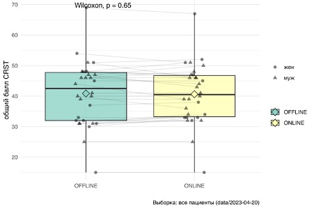Figure 4.3. Scale Indicators CRST in patients with ET during in-person and online examinations To confirm that the online approach is identical to an in-person examination when assessing tremor on the scale CRST, we conducted a test for equivalence *non–inferiority*. We used t-criterion for comparing average scores CRST two groups, assuming equal deviations. According to the statistical analysis conducted, no differences were found between the compared groups (p=0.58). In the next stage of the analysis, we accepted a threshold of 5 points as an allowable error and formulated an alternative hypothesis: the medians of the total score on the scale CRST «online” and “offline” examinations differ by more than 5 points (this wording is softer as it allows for not only equivalence but also errors in the range of 0–5 points). As a result, the value obtained was p<0,04 that allows us to reject the alternative hypothesis and conclude that the online examination differs from the in-person examination by no more than 5 points
Thus, it was established that the assessment of ET severity obtained through telemedicine technologies can be an adequate clinical measure for evaluating tremor during remote medical consultations.
### Computed tomography
In the second stage of selection, the question of the technical feasibility of performing the MRgFUS procedure is addressed, specifically the assessment of the ultrasound conductivity of the skull bone tissue. For this, the patient is referred for a CT scan of the skull, and the study must be conducted according to special rules.
The MRgFUS console software allows for the calculation of bone density only with strict adherence to a special CT protocol. Compatible protocols with the program General Electric (BONEPLUS), Toshiba (FC30), Canon (FC30). The enhancement filter cannot be used for planning UEO, Contrast. Images are analyzed raw in format FC30 RAW. As a rule, no enhancing filters are applied to CT scans intended for use in MRgFUS procedures. CT is performed with a uniform slice thickness of 0.625–1.25 mm with a 0 mm interval and a resolution of 512 × 512. The series should cover the entire skull, not limited to the target area. The upper slice is always above the skull (in the air), and the last slice reaches the base of the skull. The slices are oriented so that the axial CT images align with the plane AC–PC and perpendicular to the midline.
The patient sends their CT images via a secure communication channel, which are then uploaded to the MRgFUS console. The software allows for the calculation of the bone tissue ultrasound conductivity coefficient Scull Score) \[423\] using the built-in program (Figure 4). The higher the indicator Scull Score, the better the permeability of bone tissue for ultrasound. A coefficient of 0.35 is considered suitable for treatment using MRgFUS: if the bone density is ≥0.35, it is considered technically feasible to perform the procedure.
In principle, even with a dense skull (for example, with “borderline values of the indicator Scull Score) A neurosurgeon has certain technical capabilities during MRgFUS to achieve the necessary temperature at the target point and obtain an acceptable clinical effect by increasing energy, power, frequency, and duration of sonication. However, this is often associated with the occurrence of pain syndrome, which is difficult for patients to endure.
If the bone ultrasound conductivity density coefficient in a patient is below 0.35, treatment is currently considered impossible: even the maximum parameters of MRgFUS cannot guarantee heating at the target point. For such situations, we, together with a team of endocrinologists, have developed a special method to improve bone tissue permeability. Endocrinologists have created a program to change bone density using alendronic acid-based medications taken over 6 months. At the time of writing this monograph, 8 patients in our Center have undergone or are continuing this course of treatment, and two of them successfully underwent MRgFUS after completing the course.
Figure 4.4. Calculation of the ultrasound conductivity coefficient of bone tissue ( Scull Score).### Preparatory stage immediately before performing MRgFUS
To ensure the safety of the MRgFUS procedure, all patients at this stage are required to undergo the following standard examinations (before arriving at the center): complete blood count (no more than 10 days); urinalysis (10 days); blood biochemistry (glucose, protein, creatinine, urea, bilirubin, alanine aminotransferase, aspartate aminotransferase) (10 days); blood test for blood type and Rh factor; blood test for human immunodeficiency virus (6 months) HBs-antigen (6 months), hepatitis C (6 мес) COVID-19 (72 Wassermann reaction (6 months); coagulogram (10 days); electrocardiography (10 days); duplex scanning of brachiocephalic arteries, veins of the lower extremities (10 days); examination by a general practitioner at the place of residence with obtaining a conclusion (10 days). If deviations are detected, patients are additionally consulted by a cardiologist/therapist and endocrinologist at our center to correct somatic pathology and ongoing treatment. In our patient group, this necessity arose in 37% of cases.
For the final decision on the possibility of treatment using MRgFUS, the patient is examined by a neurologist and a neurosurgeon. To objectively assess the condition of patients, a wide range of the standard scales listed below is used.
• Universal scales for any pathologies: MoCA (Montreal Cognitive Assessment – Montreal Cognitive Assessment (MoCA) Nasreddine et al., 2005), HADS (Hospital Anxiety and Depression Scale – Hospital Anxiety and Depression Scale Zigmond, Snaith, 1983), SF-36 (The 36-Item Short Form Health Survey – Short Health Status Survey of 36 Items Ware et al., 1994), MADRS (Montgomery–Åsberg Depression Rating Scale – Montgomery–Åsberg Depression Rating Scale Montgomery, Åsberg, 1979), ESS (Epworth Sleepiness Scale – Epworth Sleepiness Scale) Johns, 1991), Beck Depression Inventory ( Beck et al., 1961).
• Scales for Assessing Tremor Hyperkinesis in Patients with ET and PD. The most commonly used scale for assessing tremor is CRST; at the same time, a general assessment is conducted as well as an assessment for each side of the body separately ( Fahn et al., 1988; Khandhar, Marks, 2007). In ET, it is also advisable to use scales TETRAS (The Essential Tremor Rating Assessment Scale – scale for assessing the severity of essential tremor) Elble, 2016) и QUEST (Quality of Life in Essential Tremor questionnaire – Quality of Life Questionnaire for Essential Tremor Tröster et al., 2005).
• In PD, the “gold standard” is the use of UPDRS (part I–IV and the total score, as well as the separate score for the operated and non-operated sides) ( Movement Disorder Society Task Force on Rating Scales for Parkinson’s Disease, 2003), Hoehn and Yahr functional scale Hoehn, Yahr, 1967) и PDQ-39 (Peto et al., 1998).
• In patients with dystonia, depending on the form (CD, myoclonus-dystonia, segmental dystonia), the clinical condition is advisable to determine by UDRS (Unified Dystonia Rating Scale – Unified Dystonia Rating Scale) ( Comella et al., 2003), BFMDRS (Burke–Fahn–Marsden Dystonia Rating Scale – Burke-Fahn-Marsden Dystonia Rating Scale Burke et al., 1985), UMRS (Unified Myoclonus Rating Scale – Unified Myoclonus Rating Scale) ( Frucht et al., 2002), TWSTRS (Toronto Western Spasmodic Torticollis Rating Scale – Western Toronto Spasmodic Torticollis Rating Scale Comella et al., 2015), as well as on CRST (Fahn et al., 1988).
The duration of a neurologist’s examination using scales necessary for a specific patient is 1.5–2 hours. During the joint consultation of a neurologist and a neurosurgeon, in addition to clarifying the indications for MRgFUS, the patient is given a detailed explanation of the treatment stages, sequence of manipulations, possible sensations during their execution, and other questions. Then, an examination by a cardiologist (therapist) of our center is conducted. After completing all the specified procedures, a final decision on the surgery is made collegially.
Preparing a patient for MRgFUS involves discontinuing antiplatelet therapy 7 days prior, new generation anticoagulants 48 hours prior, and warfarin until the international normalized ratio normalizes. A few hours before MRgFUS, the patient’s symptomatic medications (anti-Parkinson’s, anti-tremor, etc.) are also discontinued. It is recommended that the patient refrain from eating for at least 6 hours and drinking for at least 4 hours before undergoing MRgFUS.
On the day of the procedure, the patient arrives at the center with a relative or close person 2 hours before the start of treatment (fasting). A nurse inserts a peripheral venous catheter, applies compression stockings, and provides nausea and vomiting prevention (ondansetron 4 mg 30 minutes before the procedure, metoclopramide 10 mg intravenously may be administered to patients with ET). To prevent thrombotic complications, elastic stockings are applied. Due to the duration of the procedure (2–3 hours), we recommend using a diaper during the operation. Blood pressure is monitored in all patients, and glucose levels are monitored in patients with diabetes. Sixty minutes before the installation of the neurosurgical frame, the nurse applies lidocaine cream (if there is no allergic reaction) to the patient’s skin at the anticipated frame attachment sites.
Figure 4.5. An MRI-technician assembles a phantom for daily quality control. ### Equipment calibration and application of the stereotactic frame
While the neurosurgeon and nurse prepare the patient on the morning of the surgery, the MRI diagnostics doctor, together with the MRI-technician, calibrates the MRgFUS machine before each operation. To do this, they perform a procedure on a special mannequin DQA. This procedure allows us to determine that the equipment is operating without errors, the thermal spot is formed at the correct size and at the right point. It is also important to ensure that the membrane to be used on the patient is defect-free. Figure 4.5 shows how the MRI-technician secures the mannequin inside the MRgFUS helmet.
The patient’s head is completely and thoroughly shaved 30 minutes before the treat-
nia. The necessity of thorough preparation is due to the fact that
what air bubbles that remain at the hair roots prevent
due to the possibility of cavitation during ultrasound.
On the scalp, four stereotactic fixation points are marked with a marker
stereotactic frame. Local anesthesia of the fixation points of the stereotactic frame was performed with ropivacaine.
The stereotactic frame for head immobilization is fixed to the skull with screws at four points. We recommend securing it in the supraorbital area on the right and left, as well as in the projection of the occipital protuberances at the back as low as possible, so that during treatment the surface area of the cranial part of the skull is as wide as possible; this allows a greater number of ultrasound waves to penetrate the cranial cavity and reach the necessary temperature indicators during the procedure.
Then place it on the head as low as possible (above the frame screws)
ется только что проверенная при помощи DQA Elastic disposable
silicone membrane (Fig. 4.6). This membrane contains a coil-
coils for MRI, and it is necessary to ensure that the coils are correctly
function. At the ends of the coils, deformations are eliminated even with
outer side. The membrane should fit snugly against the skin
covers to prevent water leaks. It is adjusted to fit dif-
Head measurement of the patient: for this, in the central part of the membrane, there is a-
Circular markings are used, cutting along which with scissors the fragment-
Elements of the membrane, you can change its size.
After this, the patient is placed on the table and is ready for the procedure.
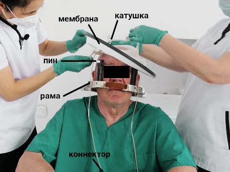Figure 4.6. The neurosurgeon and the operating nurse are applying the membrane.
---
### [Principles of Organizing an MRgFUS Center ](https://buzaevclinic.com/kniga-fokusirovannyj-ultrazvuk-pod-kontrolem-mrt-v-nevrologii/principy-organizacii-centra-mr-fuz/)
**Published:** November 2, 2024
**Author:** Rezida Galimova
**Content:**
# Principles of Organizing an MRgFUS Center
To achieve optimal treatment results using MRgFUS technology, the proper functioning of the relevant medical center is extremely important. This concerns both specialized equipment and the organization of the operating room, as well as the coordinated actions of the entire team of specialists, including training in this complex technique.
## Equipment
Neurosurgical focused ultrasound device for the treatment of neurological disorders ( ExAblate, InSightec) is a specially designed helmet with a phased array transducer containing 1024 ultrasound elements. The helmet is mounted on a table and equipped with a manually operated positioning system integrated with MRI (see Fig. 2.1). The MRgFUS procedure itself is performed inside the MRI machine. To date, it has been demonstrated that the MRgFUS procedure can be safely and effectively performed using 1.5 and 3 T devices, allowing for high-resolution intraoperative imaging thanks to a special MRI coil ( Iacopino et al., 2018). Preoperative and intraoperative MRI scanning of the brain allows for precise calculation of target coordinates and assessment of the overall view of the area of interest. In our country, the focused ultrasound table ExAblate 4000 (InSightec, v. 7.0.404) compatible with MRI equipment from the company General Electric, in other countries, MRI machines are also used Siemens.
The focused ultrasound system consists of several components: a patient table with an integrated helmet; stereotactic frame; workstation; water system for cooling the patient’s head surface; equipment cabinet; electronic systems unit ( front-end). The workstation is a computer with a monitor installed in the MRI control room and used by the neurosurgeon to connect the ultrasound device with the MRI and conduct treatment. During treatment, the standard MRI table is replaced with a patient table equipped with an integrated ultrasound wave transducer, a system for securing the stereotactic frame and positioning, which connects to the water and front-end Blocks.
Block front-end and the equipment cabinet serves as the electronic control center for the entire system operation – from performing sonications and maintaining proper water circulation to monitoring the workstation.
The water system is installed in the technical room and provides degassing, circulation, and cooling of the water that fills the space between the patient’s head and the transducer; the cooled circulating water in the circuit is necessary to prevent excessive heating of the skin surface and to prevent burns.
During the procedure, the patient is conscious, and after each ultrasound exposure (sonication), the functional effect of the manipulation is assessed. This allows the interdisciplinary team of specialists (neurosurgeons, neurologists, neuroradiologists) to refine and confirm the correct target and determine the necessary number of sonications. Trial low-power sonications are used to assess the thermal map, confirming the absence of overheating areas outside the impact zone and ensuring beam focus on the target point. After this, the amount of energy delivered is gradually increased. At this stage, the patient, who remains conscious throughout the procedure, undergoes an assessment of neurological status and clinical condition after each sonication, real-time thermography, and anatomical verification based on MRI data. After neuroimaging and clinical confirmation of the target (neurological improvement without side effects), the power of the exposure and, accordingly, the temperature at the target point are increased (heating from 55 to 60°C), which causes actual tissue damage ( Fishman, 2017; Health Quality Ontario, 2018).
Unlike stereotactic ablative radiosurgery, MRgFUS does not involve irradiation of surrounding tissues, allowing for the avoidance of potential side effects (such as edema of brain structures not adjacent to the target point, aseptic meningitis, actinic keratosis, alopecia, etc.). The procedure is performed under conditions of short-term hospitalization or day hospital care ( Fasano et al., 2018; Weidman et al., 2019; Valentino et al., 2020).
Our experience treating over 200 patients over 3 years shows that, considering possible clinical situations (including emergencies), it is advisable to always have the following additional equipment on hand: electric suction, defibrillator, oxygen concentrator, electrocardiography machine with attachment straps; electric trimmer and razor; Ambu bag valve mask; sharp scissors (for membrane adjustment); glucometer; blood pressure monitor; video camera
The main necessary consumables for performing MRgFUS are as follows: a) for equipment preparation: phantom for daily quality control ( daily quality assurance, DQA), a tank of clean filtered water for filling the water system of the MRgFUS, a special detergent for cleaning the water system of the MRgFUS; b) for the patient: special membrane InSightec (see below), a set of peripheral venous catheters, compression stockings, diapers, skin marker, etc.; b) medications: resuscitation kit and kit for treating anaphylactic shock, antihypertensive, antiemetic, decongestant medications, non-steroidal anti-inflammatory drugs, 5% glucose solution, crystalloid fluids; c) materials for patient protocol and intraoperative testing: MRI-guideded focused ultrasound safety checklist, spiral test and letter sheets, pencil or pen, plastic bottle with water level, plastic spoon, toothpick or non-magnetic plastic skewer for sensitivity testing
Among the consumables for the patient, a special membrane deserves a separate description InSightec, mandatory when performing treatment using the MRgFUS method. It is an elastic silicone membrane placed on the patient’s head to isolate the space between the transducer and the scalp using cool degassed water. The membrane functions as a full-fledged MRI coil, achieved by special rings installed on the right and left, allowing high-quality visualization of brain structures during treatment planning and enabling the creation of a thermal map during the procedure. The membrane adapts to the patient’s head size by adjusting the circumference of the inner ring. After positioning the patient on the focused ultrasound table, the membrane forms multiple folds that should be smoothed out. If it is impossible to eliminate the folds, they must be marked during planning as areas through which ultrasound waves do not pass due to the large number of air bubbles in these formations.
We have proposed additional methods for objectively assessing the effectiveness and safety of the MRgFUS procedure. Many patients note the greatest severity of tremor when performing certain actions, often related to professional activities. For example, for a patient serving in a law enforcement agency who had difficulties shooting a pistol, we created a special pistol mock-up with a laser pointer to assess the effect of the procedure (Fig. 3.1). For a patient whose work involved training locksmiths to use tools, we used plastic children’s toys such as adjustable wrenches, screwdrivers, and nuts for testing. For a patient who worked as a seamstress before the illness, we used thin toothpicks to simulate needles. This kind of experience is important in managing patients with kinesia-specific forms of movement disorders.
Special device-model with laser guidance for assessing forearm and hand tremor during the MRgFUS procedure. A particular challenge is the assessment of head tremor: this task cannot be solved in the usual way during the MRgFUS procedure due to the fixation of the patient’s head in a stereotactic frame. To assess head tremor oscillations in such situations, we developed a special device described in Chapter 8.
## Team of specialists providing treatment using MRgFUS
The main team providing treatment using MRgFUS includes a neurosurgeon, neurologist, neuroradiologist, MRI-technician, and nurse.
The neurosurgeon bears full responsibility for the safe and correct execution of the entire treatment using MRgFUS, the coordinated work of the formed team, and the proper operation of the equipment. In many centers, a mandatory requirement for admission to perform MRgFUS is specialization in functional neurosurgery.
An important member of the team is a qualified neurologist specialized in movement disorders. The neurologist conducts the initial selection of patients, adjusts (if necessary) medication therapy, determines pharmacoresistance, and assesses the patient’s clinical condition using scales. During each stage of MRgFUS, the neurologist evaluates the neurological status and, together with the neurosurgeon, determines the course of further treatment. Additionally, the neurologist continues to monitor the patient in the postoperative period, overseeing the medication regimen, prescribing rehabilitation activities, and assessing the patient’s condition dynamics, including using special scales.
The neuroradiologist also participates in organizing treatment throughout its duration: initially determines the skull bone density coefficient using CT scanning, conducts brain MRI before the MRgFUS stage for surgery planning, collaborates with the neurosurgeon to develop a treatment plan in terms of safety and effectiveness, provides necessary intraoperative imaging during repeated sonications, and performs follow-up MRIs at various times after the procedure.
A universal nurse with procedural and dressing skills prepares the patient for treatment (insertion of a venous catheter, shaving hair, setting up a neurosurgical frame and placing the membrane together with the neurosurgeon, positioning the patient on the focused ultrasound table). During the MRgFUS procedure, the nurse administers medications as prescribed by the neurologist, measures blood pressure, and, if necessary, measures blood glucose levels, etc.
The MRI-technician assists the neuro-radiologist at every stage – in determining the patient’s skull density ratio, performing MRI for surgery planning, conducting quality control before the start of the MRgFUS procedure (using a phantom DQA) and so on
Additional team members who may be involved at various stages if necessary: therapist, cardiologist, endocrinologist, anesthesiologist, clinical psychologist, psychiatrist, etc.
## Patient, Relatives, and the Concept of the “Open Operating Room”
Optimal results of MRgFUS can be achieved through coordinated work and a full understanding of the technical aspects of the procedure not only by all team members but also by those close to the patient. The presence and involvement of loved ones at all stages of treatment help form the right expectations from the intervention, objectively assess its outcomes, and maintain the necessary mindset of the patient.
During the procedure, constant contact with the patient is necessary. According to the equipment instructions, for the safety of the procedure, the patient must be given an emergency stop button. However, this is not enough; additional adequate instruction is very important. The patient should know in which cases to press the button and which sensations are normal and should not cause concern.
Common sensations experienced by patients during MRgFUS that should not hinder the continuation of treatment: dizziness, nausea, mild pain, taste sensations (bitterness)
Sensations and symptoms requiring cessation of sonication:
• severe headache
• new neurological symptoms arising during intraoperative monitoring (coordination disorders in limbs, sensory deficit, etc.);
• vomiting
The patient should feel safe and be able to communicate with the team while conscious throughout the procedure. This will help avoid interruptions in sonications and achieve both better results in terms of effectiveness and a smaller edema size in the impact area in terms of safety.
To create favorable conditions for the effective conduct of the procedure, we proposed the concept of an “open operating room.” The control room is separated by a glass partition with blinds. With the patient’s consent, the room can be made well-visible by placing chairs for the patient’s relatives behind the glass. Relatives are allowed to enter the control room, express their concerns, and offer help. If the patient has poor tolerance for being alone in the MRI room for an extended period, we allow relatives to be nearby and hold the patient’s hand during the procedure. Since there are no incisions, this treatment does not require sterile conditions, and loved ones can freely be near the patient. Feeling supported, the patient in the operating room is less focused on internal sensations and tolerates the treatment more easily, avoiding unnecessary pressing of the stop button. At the same time, relatives, participating in the course of treatment, feel additional responsibility for the patient’s postoperative care.
An advantage of having relatives nearby is the ability to discuss further treatment strategies if necessary. This is very helpful in complex situations where standard MRI-guided focused ultrasound achieves an incomplete clinical effect (e.g., partial regression of tremor), but further treatment in an attempt to improve the result carries a certain risk of developing neurological deficits. The patient and relatives receive the most complete information, see and evaluate the achieved effect, and can consciously, in close contact with the medical team, participate in making a balanced decision about the advisability and possibility of continuing the treatment. According to our data, discussion in the format of such an “expanded open consultation” may be required in 1/3 of cases of functional operations using MRI-guided focused ultrasound. Based on our experience, this approach increases patient and family satisfaction with the course and results of treatment even with a suboptimal clinical effect.
The presented concept of an “open operating room” implies high requirements for the preparation of a qualified team of specialists performing treatment using MRgFUS.
---
### [Introduction ](https://buzaevclinic.com/kniga-fokusirovannyj-ultrazvuk-pod-kontrolem-mrt-v-nevrologii/vvedenie/)
**Published:** April 2, 2024
**Author:** Rezida Galimova
**Content:**
```
100-anniversary of the birth
Eduard Izrailevich Kandel
dedicated to
```
# Introduction
Over the past 30 years, the treatment of nervous system diseases, primarily extrapyramidal movement disorders, using approaches of functional stereotactic neurosurgery has reached a qualitatively new level. The active implementation of the method of deep brain stimulation deep brain stimulation, DBS), the refinement of traditional destructive stereotactic surgeries and the emergence of non-invasive methods for precision ablation of cerebral targets have dramatically changed the face of modern neurology. Today, it is difficult to imagine managing patients with Parkinson’s disease (PD), essential tremor (ET), dystonias, and many other conditions without these familiar surgical technologies, which help overcome severe disability and significantly improve patients’ quality of life
It would not be an exaggeration to say that one of the most striking innovations in this rapidly developing field of neuroscience has been the development of the MRI-guided focused ultrasound (MRgFUS) method. It represents a unique alternative to traditional ablative functional interventions. The MRgFUS method allows for non-invasive targeted impact on a specific point deep within the brain by focusing hundreds of ultrasound beams simultaneously directed at the target from an external spherical emitting device. And if the artistic symbol and emblem of stereotactic neurosurgery is traditionally considered to be the image of Wilhelm Tell hitting an apple on a boy’s head with a crossbow (illustration), then MRgFUS can probably be compared to another weapon—a bundle of invisible “magic arrows” from popular computer games, inevitably striking the intended target. Dedicated to the 100th anniversary of the birth of Eduard Israilevich Kandel
Wilhelm Tell. Engraving XIX века. The method of directed ultrasound impact on the brain has a long history, but it was implemented at the modern technological level 15 years ago: the first operation using MRgFUS was performed in 2009. Today, the method is mainly used in patients with extrapyramidal pathology. Thanks to the official approval of the U.S. Food and Drug Administration, the European Medicines Agency, Roszdravnadzor, and other authorities, the MRgFUS method is now becoming increasingly widespread worldwide. If in 2018, the milestone of the 1000th patient was reported, by today the total number of patients with ET and PD treated with the MRgFUS method has already exceeded 15,000. And this is far from the limit. For example, a very indicative fact: in 2021, for the first time in the U.S., the number of ET patients operated on using MRgFUS exceeded the number of patients with this diagnosis referred for DBS. The total number of operational MRgFUS installations in different countries worldwide in 2024 exceeded 150. In the Russian Federation, 4 clinics already have such installations (2 in Moscow, and 1 each in Saint Petersburg and Ufa), and there is no doubt that their number will gradually increase.
The obvious advantages of MRgFUS over other methods of stereotactic interventions on the brain are the absence XIX century. (From: Collected Works of Schiller in Translations by Russian Writers. Edited by S.A. Vengerov. St. Petersburg: Brockhaus-Efron; 1901. Vol. 1. 480 p.) incisions, trepanation of the skull, anesthesia, implantation of foreign bodies, and the associated various complications. MRI-guideded focused ultrasound does not require a sterile operating room, and the clinical result of the intervention can be assessed directly during the operation. The effect of ultrasound ablation performed by the MRgFUS method can be preliminarily modeled during trial sonications at lower temperatures, with the temperature regime at the target point being controlled in real-time. All this allows for maximum precise targeting and thus achieving an optimal response to treatment. The safety of the procedure and the ability to model the clinical effect have led to the reintroduction of bilateral ablations for ET and other diseases in functional stereotactic neurosurgery, which were considered unacceptable for many years due to the risk of severe complications.
The pioneer of MRgFUS in our country was the V.S. Buzaev International Medical Centre in Ufa, whose specialists successfully performed the first operation using this technology on a patient with PD on May 5, 2020. This work is conducted under the scientific and methodological guidance and in close cooperation with the Research Center of Neurology (Moscow). To date, the total number of patients operated on at the V.S. Buzaev International Medical Centre with various central nervous system (CNS) diseases is approaching 200, with a number of our proposed innovations and developments having global priority. We present this extensive experience in the current monograph and hope it will be very useful for all our colleagues interested in mastering the MRgFUS method or interested in issues related to the treatment of movement disorders.
It is symbolic that the monograph on MRgFUS is being published just a few months after the scientific medical community solemnly celebrated the 100th anniversary of the founder of functional and stereotactic neurosurgery in our country and one of the leaders in this field worldwide, the creator and long-time head of the neurosurgical department of the Neurology Research Institute of the USSR Academy of Medical Sciences (now the Scientific Center of Neurology), Professor Eduard Israilevich Kandel. A true innovator in neurosurgery, he left a bright creative legacy. That is why we dedicate the first publication in our country on this breakthrough, innovative stereotactic technology to his memory.
The authors express deep gratitude for the continuous support in their work to Doctor of Medical Sciences [Igor Vyacheslavovich Buzaev,](https://buzaev.ru) professor of the Department of Hospital Surgery at the Federal State Budgetary Educational Institution of Higher Education “[Bashkir State Medical University](https://bashgmu.ru)” of the Ministry of Health of Russia, one of the founders of the [Intelligent Neurosurgery Clinic, Ltd at the V.S. Buzaev International Medical Centre](https://buzaevclinic.com). The method of treating patients using focused ultrasound without incisions, pain, and anesthesia inspired him in 2017: it resonated with the ideas that his father, [Vyacheslav Stepanovich Buzaev](https://buzaev.ru/patient/faq/post-my-father-buzaevvs/) – Doctor of Medical Sciences, founder of the interventional radiology school in the Republic of Bashkortostan, who performed the first coronary angiographies, coronary artery stenting, emergency endovascular treatment of acute myocardial infarction in the republic and transitioned from open to minimally invasive cardiovascular surgery, had advocated throughout his life. Seeing a technology that combines humanity, progressive medical development, and a change in the philosophy of medical care, I.V. Buzaev made every effort to open the first MRgFUS center in the Russian Federation. This monograph was written with his assistance.
The authors express special gratitude to Ildar Dinarovich Khalfin, who believed in the potential of MRgFUS technology, became one of the founders of our center, and continues to make a significant contribution to its development, the expansion of the geographical reach of patients, and staff training. Many years ago, faced with the inability to help a loved one with a movement disorder, he took on the challenge of opening the first MRgFUS clinic in Russia, where the dream of alleviating severe suffering became possible!
---
### [We treat together with charitable foundations and VHI ](https://buzaevclinic.com/prices/partners/)
**Published:** September 21, 2024
**Author:** Igor Buzaev
---
### [News ](https://buzaevclinic.com/news/)
**Published:** May 25, 2021
**Author:** Aleksej Savelev
---
### [Whole-body MRI ](https://buzaevclinic.com/services/expert-mri-imaging/mri-all-body/)
**Published:** March 27, 2023
**Author:** Aleksej Savelev
**Content:**
# Whole-body MRI. What is this examination and why is it performed?
Whole-body MRI or oncosearch is a method based on combining MRI data of the main areas of the human body, performed in modes most informative for detecting oncological diseases.
**This method is exclusively aimed at detecting oncological diseases and secondary changes in the human body.**
## ADVANTAGES
An undeniable advantage is that this method does not involve radiation exposure for the patient, which is especially important for individuals who need to undergo the procedure multiple times.
This diagnostic method is not a substitute for targeted studies of specific areas of the human body, which focus on a detailed examination of a particular area to identify any structural or morphological changes. This The examination is prescribed as an additional diagnostic tool for patients with cancer suspicion, rather than as a primary or preventive measure.
When performing a whole-body MRI, the condition of the following organs and systems is assessed for the presence of neoplasms (from 0,5-1,0 cm in diameter):
- Brain and spinal cord
- Soft tissues of the neck, supra- and infraclavicular zones, axillary areas.
- Organs of the Chest – Lungs, Mediastinum
- Abdominal organs – liver, pancreas, spleen
- Retroperitoneal organs – kidneys, adrenal glands, retroperitoneal soft tissues
- Pelvic organs in women – uterus, ovaries, bladder
- Male pelvic organs – prostate gland, seminal vesicles, bladder
- Skeleton bones in scanning areas
Do not assess the condition:
- Larynx and Pharynx
- Thyroid and Parathyroid Glands
- Mammary glands
- Esophagus, stomach, and intestines
- Bones and soft tissues of the upper limbs
- Bones and soft tissues of the lower extremities, below the knee joints
## Preparation
No special preparation is required for a full-body MRI. It is advisable to avoid eating 5-6 hours before the examination, exclude foods that cause gas formation from the diet for a day, and moderately fill the bladder before tomography for better visualization.
## Conducting Research
The method involves placing the patient in a high-voltage magnetic field (safe for the human body). The examination takes about 2 hours and is performed by attaching special magnetic coils to the patient, which are positioned from the head to the lower limbs, allowing visualization of organs and tissues at all these levels.
IMPORTANT!
During the examination, **intravenous administration of a contrast agent** may be required if an oncological process is suspected and when searching for metastases.
An extended examination of a specific area may be required during the examination if an oncological process is suspected.
Confirm the final cost of the examination when scheduling by phone.
You need to have with you Referral for a full-body MRI from an oncologist or another specialist
In case of detecting pathological changes that require detailed assessment to clarify their nature, the patient may need further targeted diagnostic examination.
After a whole-body MRI, an oncologist consultation is necessary based on the results of the study.
---
### [Publication Archive ](https://buzaevclinic.com/archive/)
**Published:** September 13, 2024
**Author:** Igor Buzaev
---
### [MRI of the heart ](https://buzaevclinic.com/services/expert-mri-imaging/heart-mri/)
**Published:** November 7, 2023
**Author:** Aleksej Savelev
**Content:**
# MRI of the Heart: Advantages, Features, Contraindications
## **What is cardiac MRI?**
Magnetic Resonance Imaging (MRI) is an important tool in heart examination, helping doctors obtain highly detailed images of this organ in a completely non-invasive and safe manner for the patient. The study allows for identifying issues and determining treatment strategies, especially in cases where other imaging methods do not provide the necessary information.
## **What are the advantages of cardiac MRI?**
Multiplanar non-invasive imaging and the ability to study with contrast agent administration allow for the most accurate assessment of the heart and vascular anatomy. The absence of ionizing radiation makes MRI an excellent choice for examining children, pregnant women, and patients requiring periodic monitoring.
## **How is an MRI of the heart performed?**
The study is conducted in the clinic, does not require hospitalization, and lasts about 60 to 90 minutes. The operator conducting the study will be in the adjacent room at the computer. It is necessary that your clothing does not have metal objects—they may be attracted to the magnet or distort the image. Before the study begins, a catheter will be inserted into a vein in your arm for the administration of a contrast agent—gadolinium.
## **What diseases can cardiac MRI diagnose?**
Myocarditis and pericarditis – cardiomyopathies – heart defects – heart tumors – amyloidosis and hemochromatosis – ischemic heart disease – aortic diseases This is by no means a complete list of conditions where cardiac MRI can assist the doctor and patient. Cardiac MRI is an important tool for monitoring the heart health of athletes and people actively engaged in sports.
## **What are the contraindications for cardiac MRI?**
Contraindications for MRI include metallic or electrical implants, devices, or foreign bodies. Most modern electronic devices implanted in the heart are safe for MRI. Heart valve prostheses, joint prostheses, and intravascular stents do not limit the examination. Nailya Rifkhatovna Mukhamadeeva, cardiologist, therapist, studied cardiac MRI at the A.N. Bakulev Center for Cardiovascular Surgery
---
### [MRI of the spine ](https://buzaevclinic.com/services/expert-mri-imaging/mri-neck-spine/)
**Published:** September 21, 2021
**Author:** Aleksej Savelev
**Content:**
# MRI of the cervical spine (C-spine)
MRI of the cervical spine: T2-STIR-sag
MRI of the cervical spine: T2-sag
MRI of the cervical spine: T2-MERGE-axial
Magnetic resonance imaging of the cervical spine without contrast agent
#### Rating:
#### Reviews [2GIS](https://2gis.ru/ufa/search/%D0%BA%D0%BB%D0%B8%D0%BD%D0%B8%D0%BA%D0%B0%20%D0%91%D1%83%D0%B7%D0%B0%D0%B5%D0%B2%D0%B0/firm/70000001044251429/55.983612%2C54.753242/tab/reviews) [Yandex](https://yandex.ru/search/?lr=172&text=%D0%BA%D0%BB%D0%B8%D0%BD%D0%B8%D0%BA%D0%B0+%D0%B1%D1%83%D0%B7%D0%B0%D0%B5%D0%B2%D0%B0+%D1%83%D1%84%D0%B0&src=suggest_B) [Google](https://www.google.com/search?gs_ssp=eJzj4tVP1zc0TCuoKiipSEsyYLRSNagwMU6xNLY0NTU3TDExNUw1tjKoMDZKMTGzNE0yTzRLS0lMSfOSvbDrwu4LOy7sBeJdFzYoXNh4sfnC9gsbLmy9sOnCBgDV6CUR&q=%D0%BA%D0%BB%D0%B8%D0%BD%D0%B8%D0%BA%D0%B0+%D0%B1%D1%83%D0%B7%D0%B0%D0%B5%D0%B2%D0%B0&oq=%D0%9A%D0%BB%D0%B8%D0%BD%D0%B8%D0%BA%D0%B0+%D0%B1%D1%83%D0%B7%D0%B0%D0%B5%D0%B2%D0%BE&aqs=chrome.1.69i57j46i13i175i199j0i13j0i8i10i13i30.2750j0j15&sourceid=chrome&ie=UTF-8#)
#### What you will receive
Medical report with recommendations
We will record the MRI data on DVD-disk. On the disk envelope, we will indicate QR-CODE, which will allow you to download the study description one day after the examination.
For an additional fee, we can print the research results on film.
#### Who needs this service?
Indications to MRI of the cervical spine:
- pains in шее
- limitations in neck and head mobility
- instability of blood pressure, hearing and vision impairments
- clarification of the diagnosis “vegetative-vascular dystonia”
- dizziness
- suspicion of cervical spine osteochondrosis, presence of spinal canal stenosis, hernias, tumors, or metastases
- control over treatment process for the established diagnosis
#### Who cannot receive this service? Contraindications for MRI:
- Presence of electronic medical devices in the body (pacemaker, etc.)
- Presence of metallic elements in the area of study (prostheses, clips, fragments)
- Inadequate patient behavior (panic attack, psychomotor agitation)
- State of drug or alcohol intoxication
- Inability to remain still during the examination (due to severe pain or other reasons)
- The necessity of continuous resuscitation measures (artificial respiration, etc.)
#### How we do it
MRI **does not require anesthesia or special preparation**.
Before the procedure, the MRI-technician will check you with a special device to ensure there are no metals in your body or clothing that react to the magnetic field. Then, you will be placed on the MRI table and given instructions. You will be given a button in your hand, which can instantly stop the examination if pressed. Then, the examination protocol, consisting of several programs, will be initiated.
If necessary, additional research programs may be implemented after consultation with you to clarify the obtained data.
#### What is included in the service?
The research protocol in different clinics can vary significantly. MRI is always a compromise between the time spent on the examination and its quality.
We use all recommended programs to ensure the highest quality of research, allowing for a detailed examination of the brain without missing pathological changes.
An important aspect is the research step – thickness “sections “, which we will obtain as a result of the research. The smaller the step and the more slices, the more detailed we can assess the condition of the tissues.
Our MRI protocol for the cervical spine includes the following programs:
*Program / slice thickness*
T2 3 D sagittal 2.2 мм
T1 or T1 FLEX/DIXON sagittal 3 мм
T2 STIR sagittal 3 мм
T2\* MERGE axial 3 мм
---
#### How long will it take?
The basic MRI protocol for the cervical spine will take about 30 minutes.
#### How else can this be done?
You can assess the condition of the cervical spine based on symptoms during a neurologist’s examination.
Hardware diagnostic methods will help the doctor make an accurate diagnosis.
In addition to MRI, CT (computed tomography) and ultrasound can be performed.
But it should be understood that CT uses gamma radiation, the dose of which needs to be strictly controlled; and ultrasound provides much less informative results.
#### You may need
- Neurologist Consultation
- Neurosurgeon Consultation
- Second expert opinion (description of MRI studies conducted in other clinics)
---
### [Payment for Services ](https://buzaevclinic.com/payment/)
**Published:** May 31, 2023
**Author:** Aleksej Savelev
**Content:**
# Payment for Services at the V.S. Buzaev International Medical Centre®
To pay for the service using a smartphone, you have the following options:
## Payment directly at our medical center
Regular payment through a cash terminal with receipt issuance.
## Promo cвязьБанк
Follow the payment [link](https://3ds.payment.ru/cgi-bin/ecomm_payment_ref_processor?id=3469255&rnd_hash=a9c3be0771c5e2512583aa62a409421252d8e6b00ddd4ee61db8acdb2bb5d831), enter the amount, and complete the payment.
Pay via PSB
## Fast Payment System
Follow the payment [link](https://buzaevclinic.com/payment/fast-payment/), enter the amount, and complete the payment.
[Pay via SBP ](https://buzaevclinic.com/payment/fast-payment/)
---
You can check the prices for services on the [Price List](https://buzaevclinic.com/prices/) page
[Offer ](https://buzaevclinic.com/wp-content/uploads/2023/06/oferta_02.pdf)
---
### [Sustainable Development ](https://buzaevclinic.com/values/sustainable-development-goals/)
**Published:** September 23, 2021
**Author:** Igor Buzaev
**Content:**
# We contribute to the development of humanity
We believe in a bright future and strive to make it better for everyone. Here’s how we work every day to help you and your loved ones:
## Health and Well-being
The focus of our attention is ensuring a healthy lifestyle and promoting patient well-being.
## Quality Education
We help you and your children receive quality education.
- In 2024, we established the V.S. Buzaev Award and fund residencies for young doctors.
- Our book on focused ultrasound [is distributed for free](https://buzaevclinic.com/kniga-fokusirovannyj-ultrazvuk-pod-kontrolem-mrt-v-nevrologii/).
- We conduct lectures for patients and schoolchildren, have obtained a license for educational activities, and are developing training programs.
## Decent Work and Economic Growth
We create new jobs and offer rewarding work to the best specialists in the field of medicine.
## Equality of Opportunities
We believe that every person deserves equal opportunities for career growth. [We develop effective individuals who strive to continuously learn and work.](https://buzaevclinic.com/team/this-is-the-way/)
## Innovations and Infrastructure
We implement advanced technologies to improve the quality of medical care.
- In 2020, we began using MRgFUS in Russia and the post-Soviet countries.
- In 2020, we were the first in the world to implement MRgFUS using international medical proctoring (remote online surgery)
- In 2021, we treated dystonia with MRI-guided focused ultrasound among the first 10 cases described worldwide.
- In 2022, we were among the first three clinics in the world to treat hypothalamic hamartoma using MRgFUS.
- In 2022, we performed the world’s first simultaneous bilateral surgery with using MRgFUS for essential tremor
- In 2022, we conducted the first treatment of neuropathic pain in Russia using MRgFUS.
- In 2022, we became one of the first in the world and the first in Russia to begin treating the akinetic-rigid form of Parkinson’s disease using MRgFUS.
## Reducing Inequality
Thanks to medical tourism, we create accessible medicine to help people worldwide receive quality treatment. Our specialists have performed MRI-guided focused ultrasound therapy for patients with tremor and Parkinson’s disease from [more than 25 countries](https://buzaevclinic.com/global/countries/) where this technology is not yet available.
## Sustainable Cities and Communities
We believe that quality medical care makes our city better and safer. That’s why we opened our center here in Ufa, to be closer to our family and friends. We feel immense gratitude to the residents of Ufa, our schools, and [university](https://bashgmu.ru/) who helped us become who we are. Now we want to give back by creating a new medical asset for Bashkortostan. Our center attracts smart and talented people who want to live and work in our beautiful city.
## Responsible Consumption and Production
We care about our planet and strive to reduce waste. We recycle paper and were among the first to switch to electronic document management, including for medical records. We also stopped using disposable shoe covers, even though it costs us more in the short term.
## Combating Climate Change
We are actively participating in the fight against climate change. For example, we have planted over 50 trees near our medical center.
## Partnership
We are open to collaboration and believe that together we can make the world a better place in the interest of sustainable development of innovations. Among our partners, there are many wonderful organizations that share our values and aspirations.
---
### [Price List ](https://buzaevclinic.com/prices/catalogue/)
**Published:** September 16, 2024
**Author:** Igor Buzaev
**Content:**
# Price List. Catalog of Prices and Services at V.S. Buzaev International Medical Centre
After scheduling a medical service, the administrator will definitely contact you the day before your visit to the clinic to confirm the appointment. In medicine, schedules can unexpectedly shift for various patient-related reasons, in which case the administrator will offer another convenient time or date for you.
Please answer the call from the clinic!
An unconfirmed appointment may be rescheduled or canceled.
Services can be paid for at the clinic by card, transfer, or using [FPS (Fast Payment Service).](https://buzaevclinic.com/payment/fast-payment/)
## Prices for the treatment of movement disorders with MRI-guided focused ultrasound
[Information on prices, special offers, and discounts for MRgFUS treatment of tremor, Parkinson’s disease, and dystonia here. ](https://buzaevclinic.com/prices/mrgfus-tremor-parkinsons-treatment/)
## Discounts and Special Offers
[Information on discounts and special offers for other services here ](https://buzaevclinic.com/prices/promo/). This does not concern the treatment of movement disorders, Parkinson’s disease, tremor, and dystonia with MRI-guided focused ultrasound.
---
### [Legal section ](https://buzaevclinic.com/legal-compliance/)
**Published:** September 28, 2024
**Author:** Igor Buzaev
**Content:**
# Compliance with legal requirements
Information required for placement on medical organizations’ websites in accordance with Russian Federation legislation
## Information about the Institution
V.S. Buzaev International Medical Centre (Intelligent neurosurgery clinic, Ltd) “Intelligent Neurosurgery Clinic “**,** abbreviated name — Intelligent neurosurgery clinic, Ltd
Registered trademark “V.S. Buzaev International Medical Centre “
[About the Legal Entity ](https://buzaevclinic.com/legal-compliance/legal-entity/)
[Partner Map ](https://buzaevclinic.com/wp-content/uploads/2024/03/karta-partnera.pdf)
[Presentation about the center ](https://buzaevclinic.com/downloads/vs-buzaev-international-medical-centre-presentation-rus.pdf)
[Information about Medical Professionals ](https://buzaevclinic.com/team/), involved in providing medical services, about their level of professional education and qualifications
## Medical Activities
**License** for conducting medical activities [No. Л041-01170-02/00324442](https://roszdravnadzor.gov.ru/services/licenses?qrguid=94f81ddbad5d3860143442249ea825f9) FROM 05.11.2020
## Higher authorities
[Addresses and phone numbers ](https://buzaevclinic.com/legal-compliance/authorities/)
## Regulatory documents of government authorities
[Regulatory Documents Governing Medical Activities ](https://buzaevclinic.com/legal-compliance/legislation-medicine/)
## Internal documents of Intelligent neurosurgery clinic, Ltd “Intelligent neurosurgery clinic, Ltd “
[Data Protection ](https://buzaevclinic.com/wp-content/uploads/2022/11/%D0%97%D0%B0%D1%89%D0%B8%D1%82%D0%B0-%D0%BF%D0%B5%D1%80%D1%81%D0%BE%D0%BD%D0%B0%D0%BB%D1%8C%D0%BD%D1%8B%D1%85-%D0%B4%D0%B0%D0%BD%D0%BD%D1%8B%D1%85-11.22.pdf)
[Public Offer ](https://buzaevclinic.com/wp-content/uploads/2023/06/oferta_02.pdf)
## Educational Activities
[License ](https://islod.obrnadzor.gov.ru/rlic/details/5f5777f4-d58e-c5a9-1391-a43fe493d265/) L035-01198-02/01042757 dated 01.02.2024 for conducting educational activities.
[Materials on Compliance with Regulatory Requirements for Educational Activities ](https://buzaevclinic.com/legal-compliance/education-documents/).
## Website and Personal Data Processing
[Personal Data Processing Policy ](https://buzaevclinic.com/legal-compliance/privacy-policy/) on the website [https://buzaevclinic.com](https://buzaevclinic.com/)
---
### [Our Specialization ](https://buzaevclinic.com/conditions/)
**Published:** December 18, 2023
**Author:** Aleksej Savelev
**Content:**
# Our Specialization
## Nervous Diseases
[Tremor ](https://buzaevclinic.com/conditions/tremor/)
[Essential tremor ](https://buzaevclinic.com/conditions/tremor/essential-tremor/)
[Parkinson’s Disease ](https://buzaevclinic.com/conditions/parkinsons-disease/)
[Dystonia ](https://buzaevclinic.com/conditions/dystonia/)
[Neuropathic pain ](https://buzaevclinic.com/conditions/neuropathic-pain/)
[Migraine ](https://buzaevclinic.com/conditions/headache-facial-pain/myths/)
Idiopathic dizziness
[Hypothalamic Hamartoma ](https://buzaevclinic.com/conditions/hypothalamic-hamartoma/)
## [Heart Diseases ](https://buzaevclinic.com/services/heart-team-cardiology-vascular-surgery/)
Hypertensive disease
Atherosclerosis
Coronary heart disease
Angina pectoris
Arrhythmia
Atrial fibrillation (AFib)
We also perform lipid metabolism correction (for elevated cholesterol levels, high-density lipids, etc.), conduct ECG, echocardiography, and ultrasound diagnostics of major vessels.
## Congenital Heart Defects
Patent ductus arteriosus
Atrial septal defect
Ventricular septal defect
## Endocrinological
Obesity (metabolic syndrome)
Diabetes mellitus
[Reduced thyroid function (hypothyroidism) ](https://buzaevclinic.com/conditions/endocrinology-hormones/hypothyroidism/)
Hyperthyroidism
## Gastroenterological Diseases
Diagnosis of the esophagus, stomach, colon (video-EGD, videocolonoscopy), detection of Helicobacter pylori, consultation with a gastroenterologist
## Urological Diseases
Consultations are conducted by a neuro-urologist-andrologist, with possible cystoscopy, uroflowmetry, and ultrasound examinations.
## Gynecological Diseases
## Diseases of the Joints and Spine
We can offer taping PRP-therapy, magnetotherapy, shockwave treatment, introduction of Alloplant biomaterials into paravertebral points and joint cavities to relieve chronic inflammation
## Childhood Diseases
Pediatrician appointment, children’s preventive vaccinations
[Sign up ](tel:+78003008462)
---
### [List of Research Publications ](https://buzaevclinic.com/research/publications/)
**Published:** September 17, 2024
**Author:** Igor Buzaev
---
### [Personal Data Policy ](https://buzaevclinic.com/legal-compliance/privacy-policy/)
**Published:** December 19, 2023
**Author:** Aleksej Savelev
**Content:**
# Privacy Policy of Personal Data on the Website
## Privacy Policy for Users’ Personal Data on the Website buzaevclinic.com
## What information do we collect?
We collect the information you provide by filling out the feedback form on our website.
**At the same time, we may ask you to specify:**
- Your name
- Your phone number
However, you can visit our site anonymously.
The site may use cookie. You can disable them in the security settings of any browser ( Safari, Firefox, Chrome).
## How do we use the collected information?
Any information you provide to us may be used to call you back and clarify order details.
We do not sell, trade, or provide your public or confidential information to other companies without your consent, except in cases:
- **Email and phone for application processing:** We can use this information for company newsletters, updates about our services, promotions, etc.
## **Do we provide information to third-party companies?**
We may share your information with trusted companies that service our site, as we have entered into confidentiality agreements with them.
We may disclose your information if required by law, our company’s regulations, or to protect the rights, property, or safety of others.
## **We store your personal data indefinitely.**
## **You can withdraw your personal data by submitting a written statement.**
## On-line privacy policy
This privacy policy applies only to information collected through our website and does not apply to information obtained by other means.
## Your consent
By using our website, you agree to this privacy policy.
## Changes in Privacy Policy
If we make changes to our privacy policy, we will post them on this page and update the date of the last modification.
**Date of last modification: January 17, 2020**
---
### [List of Main Abbreviations ](https://buzaevclinic.com/kniga-fokusirovannyj-ultrazvuk-pod-kontrolem-mrt-v-nevrologii/acronyms/)
**Published:** November 2, 2024
**Author:** Rezida Galimova
**Content:**
# List of Main Abbreviations
| BP | Parkinson’s disease |
|---|---|
| YOUR | visual analog scale |
| GABA | g-gamma-aminobutyric acid |
| BBB | blood-brain barrier |
| CT | computed tomography |
| KUPCT | ultrasound conductivity coefficient of bone tissue |
| MRI | magnetic resonance imaging |
| MRgFUS | MRI-guideded focused ultrasound |
| MRT | microelectrode recording |
| SPECT | single-photon emission computed tomography |
| PET | positron emission tomography |
| ЦД | cervical dystonia |
| CNS | central nervous system |
| ЭТ | essential tremor |
| CRST | Clinical Rating Scale for Tremor (clinical tremor rating scale |
| DBS | deep brain stimulation (deep brain stimulation) |
| DQA | daily quality assurance (daily quality control) |
| GPi | globus pallidus internus (internal segment of the globus pallidus) |
| HADS | Hospital Anxiety and Depression Scale (Hospital Anxiety and Depression Scale |
| MADRS | Montgomery–Åsberg Depression Rating Scale (Montgomery–Åsberg Depression Rating Scale |
| MDS | International Parkinson and Movement Disorder Society (International Parkinson and Movement Disorder Society |
| MoCA | Montreal Cognitive Assessment (Montreal Cognitive Assessment Scale |
| PDQ-39 | The 39-Item Parkinson’s Disease Questionnaire (Questionnaire for Parkinson’s Disease (39 items) |
| PTT | pallidothalamic tract (pallidothalamic tract) |
| QUEST | Quality of Life in Essential Tremor Questionnaire (Quality of Life Questionnaire for Essential Tremor |
| SF-36 | The 36-Item Short Form Health Survey (short health status survey of 36 items) |
| TWSTRS | Toronto Western Spasmodic Torticollis Rating Scale (Western Toronto Spasmodic Torticollis Rating Scale |
| UDRS | Unified Dystonia Rating Scale (Unified Dystonia Rating Scale |
| UPDRS | Unified Parkinson’s Disease Rating Scale (Unified Parkinson’s Disease Rating Scale |
| VIM | ventral intermediate nucleus of the thalamus (ventral intermediate nucleus of the thalamus) |
| VOP | ventral oral posterior nucleus of the thalamus (ventro-oral posterior nucleus of the thalamus) |
| ZI | zona incerta (undefined zone) |
---
### [Conclusion ](https://buzaevclinic.com/kniga-fokusirovannyj-ultrazvuk-pod-kontrolem-mrt-v-nevrologii/zaklyuchenie/)
**Published:** April 2, 2024
**Author:** Rezida Galimova
**Content:**
# Conclusion
The experience gained in recent years with MRgFUS for extrapyramidal and other nervous system disorders shows that this technology represents more than just a new method of functional therapeutic impact on the brain based on a stereotactic approach. Essentially, thanks to MRgFUS, we are now talking about erasing the boundary between conservative and surgical neurology. This significantly expands the possibilities of providing effective assistance to various categories of patients with severe, often disabling movement, sensory, and behavioral disorders.
As discussed in the monograph, MRgFUS represents, on one hand, a certain alternative DBS (taking into account the existing limitations for macroelectrode brain stimulation due to its invasive nature, surgical risks, the need for constant monitoring of the stimulator’s operation mode, etc.), and on the other hand, a qualitatively new level of ablative stereotactic neurosurgery. For example, trial reversible ultrasound interventions allow modeling the effect on a specific area of the brain and finding the most effective target for a particular patient VIM, PTT, VO, ZI and others – in isolation or in combinations). Positive experience has been gained worldwide and in our clinic with effective two-stage treatment, i.e., safe repeat surgeries when symptoms return in patients with ET, PD, dystonias. The application of several developed methodological techniques (asymmetry of created lesions, timely cessation of impact on the second side with sufficient antitremor effect, symptom control during trial sonications) has breathed new life into the idea of bilateral stereotactic ablations in ET and potentially in other movement disorders. Our results indicate that such operations using MRgFUS can be safe both in staged and simultaneous execution, but this requires strict adherence to all requirements and adequate consideration of existing risks, indications, and contraindications.
It can be expected that the use of MRgFUS for the treatment of various movement disorders will gradually expand. Simultaneously, methodological foundations and indications for the use of ablative high-intensity MRgFUS modes will be refined for non-movement neurological disorders—pain syndromes, epilepsy, affective disorders resistant to conservative therapy, hydrocephalus, thrombotic ischemic stroke for the purpose of artery recanalization, etc. The initial results of such interventions look promising, although significant additional experimental and multicenter randomized clinical studies are still required here.
A promising variety of MRgFUS is the use of low-intensity ultrasound exposure. It allows for temporary opening of the BBB for the treatment of neurodegenerative and other CNS diseases and also lays the foundation for implementing a fundamentally new mode of neuromodulation in practice. This demonstrates the significance of MRgFUS not only for clinical practice but also for fundamental neurology and neurophysiology.
Thus, despite its youth, the MRgFUS method has already firmly established itself and confidently looks to the future.
---
### [Educational Activity Documents ](https://buzaevclinic.com/legal-compliance/education-documents/)
**Published:** September 25, 2024
**Author:** Igor Buzaev
**Content:**
# Documents on Educational Activities
## Section Content
- [Main Information ](#01)
- [Structure and Governance of the Educational Organization ](#02)
- [Documents ](#03)
- [Material and Technical Support and Equipment of the Educational Process ](#06)
- [Paid Educational Services ](#07)
- [Financial and Economic Activities ](#08)
- [Vacancies for Admission (Transfer) of Students ](#09)
- [Accessible Environment ](#10)
- [International Cooperation ](#11)
## General Information
1. Full name of the educational organization:
Intelligent Neurosurgery Clinic, Ltd. Abbreviated name: Intelligent neurosurgery clinic, Ltd.
2. Date of establishment: July 26, 2019
3. Founders of the organization: Buzaev Igor Vyacheslavovich, Khalfin Ildar Dinarovich.
4. There are no branches or representative offices of the educational organization.
5. Location of the educational organization, location implementation of educational activities under additional professional programs internship location, final assessment location: 450059, Republic of Bashkortostan,
Ufa, Rikhard Zorge St. 17/4, office 13 (2nd floor)
6. Operating hours: Monday — Saturday
7. Working hours:
Monday — Friday: 9:00 — 21:00
Saturday: 10:00 — 18:00
8. Contact phone:
[+7 (800) 300-84-62](tel:+78003008462)
9. Email address:
<help@tremor.care>
10. Web page:
[https://buzaevclinic.com/education](https://buzaevclinic.com/education/)
The structure of the official website of an educational organization in the information and telecommunication network “Internet,” as well as the format for presenting information by the educational organization that is mandatory for placement on the Site, are determined in accordance with [Article 29 of the Federal Law “On Education in the Russian Federation” from 29.12.2012 № 273-Federal Law ](https://db-nica.ru/documents/regulations/sveden/fz-ob-obrazovanii-273.pdf).
## Structure and Governing Bodies of the Educational Organization
Educational Organization Department of Continuing Education Intelligent Neurosurgery Clinic, Ltd.
All structural units are located at: 450059, Republic of Bashkortostan, Ufa, Rikhard Zorge St. 17/4, Office 13.
**Administration** General Director Rezida Maratovna Galimova
Deputy General Director Saveliev Alexey Borisovich
HR Specialist Gilchik Galina Borisovna
Regulations on Administration — [open ](https://8hnh35p8oe.a.trbcdn.net/wp-content/uploads/2023/10/1-%D0%9F%D0%BE%D0%BB%D0%BE%D0%B6%D0%B5%D0%BD%D0%B8%D0%B5-%D0%BE%D0%B1-%D0%B0%D0%B4%D0%BC%D0%B8%D0%BD%D0%B8%D1%81%D1%82%D1%80%D0%B0%D1%86%D0%B8%D0%B8-DS.pdf)
**Academic Department (including student admissions, organization of the educational process):** Position About the Educational Department, Organization of the Educational Process, and Admission Organization — [open ](https://8hnh35p8oe.a.trbcdn.net/wp-content/uploads/2023/10/2-%D0%9F%D0%BE%D0%BB%D0%BE%D0%B6%D0%B5%D0%BD%D0%B8%D0%B5-%D0%BE%D0%B1-%D0%BE%D1%80%D0%B3%D0%B0%D0%BD%D0%B8%D0%B7%D0%B0%D1%86%D0%B8%D0%B8-%D1%83%D1%87%D0%B5%D0%B1%D0%BD%D0%BE%D0%B3%D0%BE-%D0%BE%D1%82%D0%B4%D0%B5%D0%BB%D0%B0-DS.pdf)
## Documents
1. [Organization Charter ](https://8hnh35p8oe.a.trbcdn.net/wp-content/uploads/2023/10/%D0%A3%D1%81%D1%82%D0%B0%D0%B2.pdf).\*
2. [License ](https://islod.obrnadzor.gov.ru/rlic/details/5f5777f4-d58e-c5a9-1391-a43fe493d265/) L035-01198-02/01042757 dated 01.02.2024 for conducting educational activities
3. [Internal Regulations for Students ](https://8hnh35p8oe.a.trbcdn.net/wp-content/uploads/2023/10/3-%D0%9F%D1%80%D0%B0%D0%B2%D0%B8%D0%BB%D0%B0-%D0%B2%D0%BD%D1%83%D1%82%D1%80%D0%B5%D0%BD%D0%BD%D0%B5%D0%B3%D0%BE-%D1%80%D0%B0%D1%81%D0%BF%D0%BE%D1%80%D1%8F%D0%B4%D0%BA%D0%B0-%D0%BE%D0%B1%D1%83%D1%87%D0%B0%D1%8E%D1%89%D0%B5%D0%B3%D0%BE%D1%81%D1%8F-%D1%81%D0%BB%D1%83%D1%88%D0%B0%D1%82%D0%B5%D0%BB%D1%8F-DS.pdf)\*
4. [Internal Work Regulations ](https://8hnh35p8oe.a.trbcdn.net/wp-content/uploads/2023/10/%D0%9F%D0%92%D0%A2%D0%A0.pdf)\*
5. Collective agreement (if available)
6. Self-Assessment Report Results (published at the end of the academic year)
7. Prescriptions of bodies exercising state control (supervision) in the field of education (if available)
8. Local regulatory acts of the educational organization ([Order on the approval of local regulatory documents](https://8hnh35p8oe.a.trbcdn.net/wp-content/uploads/2023/10/%D0%9F%D1%80%D0%B8%D0%BA%D0%B0%D0%B7-%D0%BE-%D0%9B%D0%9D%D0%90-S.pdf)) on the main issues of organizing and implementing educational activities:
- [Order on the Establishment of the Department of Continuing Education ](https://8hnh35p8oe.a.trbcdn.net/wp-content/uploads/2023/10/%D0%9F%D1%80%D0%B8%D0%BA%D0%B0%D0%B7-%D0%BE-%D1%81%D0%BE%D0%B7%D0%B4%D0%B0%D0%BD%D0%B8%D0%B8-%D0%9E%D0%94%D0%9E-S.pdf);
- [class schedule ](https://8hnh35p8oe.a.trbcdn.net/wp-content/uploads/2023/10/5-%D0%A0%D0%B5%D0%B6%D0%B8%D0%BC-%D0%B7%D0%B0%D0%BD%D1%8F%D1%82%D0%B8%D0%B9-DS.pdf) students
- [forms, frequency, and procedure for current academic performance monitoring and interim assessment of students ](https://8hnh35p8oe.a.trbcdn.net/wp-content/uploads/2023/10/6-%D0%9F%D0%BE%D0%BB%D0%BE%D0%B6%D0%B5%D0%BD%D0%B8%D0%B5-%D0%BE-%D1%84%D0%BE%D1%80%D0%BC%D0%B0%D1%85-%D0%BF%D0%B5%D1%80%D0%B8%D0%BE%D0%B4%D0%B8%D1%87%D0%BD%D0%BE%D1%81%D1%82%D0%B8-%D0%B8-%D0%BF%D0%BE%D1%80%D1%8F%D0%B4%D0%BA%D0%B5-%D1%82%D0%B5%D0%BA%D1%83%D1%89%D0%B5%D0%B3%D0%BE-%D0%BA%D0%BE%D0%BD%D1%82%D1%80%D0%BE%D0%BB%D1%8F-%D1%83%D1%81%D0%BF%D0%B5%D0%B2%D0%B0%D0%B5%D0%BC%D0%BE%D1%81%D1%82%D0%B8-%D0%B8-%D0%BF%D1%80%D0%BE%D0%BC%D0%B5%D0%B6%D1%83%D1%82%D0%BE%D1%87%D0%BD%D0%BE%D0%B9-%D0%B0%D1%82%D1%82%D0%B5%D1%81%D1%82%D0%B0%D1%86%D0%B8%D0%B8-%D0%BE%D0%B1%D1%83%D1%87%D0%B0%D1%8E%D1%89%D0%B8%D1%85%D1%81%D1%8F-DS.pdf);
- [procedure and grounds for admission, transfer, expulsion, and reinstatement of students ](https://8hnh35p8oe.a.trbcdn.net/wp-content/uploads/2023/10/4-%D0%9F%D0%BE%D0%BB%D0%BE%D0%B6%D0%B5%D0%BD%D0%B8%D0%B5-%D0%BE-%D0%BF%D0%BE%D1%80%D1%8F%D0%B4%D0%BA%D0%B5-%D0%BF%D1%80%D0%B8%D0%B5%D0%BC%D0%B0-%D0%BF%D0%B5%D1%80%D0%B5%D0%B2%D0%BE%D0%B4%D0%B0-%D0%B8-%D0%BE%D1%82%D1%87%D0%B8%D1%81%D0%BB%D0%B5%D0%BD%D0%B8%D1%8F-DS.pdf);
9. [Regulations on Paid Services educational services ](https://8hnh35p8oe.a.trbcdn.net/wp-content/uploads/2023/11/%D0%9F%D0%BE%D0%BB%D0%BE%D0%B6%D0%B5%D0%BD%D0%B8%D0%B5-%D0%BE-%D0%BF%D0%BB%D0%B0%D1%82%D0%BD%D1%8B%D1%85-%D0%BE%D0%B1%D1%80%D0%B0%D0%B7%D0%BE%D0%B2%D0%B0%D1%82%D0%B5%D0%BB%D1%8C%D0%BD%D1%8B%D1%85-%D1%83%D1%81%D0%BB%D1%83%D0%B3%D0%B0%D1%85.pdf).
10. [Standard Contract Form for the Provision of Paid Educational Services with Legal Entities. ](https://8hnh35p8oe.a.trbcdn.net/wp-content/uploads/2023/11/%D0%A2%D0%B8%D0%BF%D0%BE%D0%B2%D0%B0%D1%8F-%D1%84%D0%BE%D1%80%D0%BC%D0%B0-%D0%B4%D0%BE%D0%B3%D0%BE%D0%B2%D0%BE%D1%80%D0%B0-%D0%BD%D0%B0-%D0%BE%D0%BA%D0%B0%D0%B7%D0%B0%D0%BD%D0%B8%D0%B5-%D0%BF%D0%BB%D0%B0%D1%82%D0%BD%D1%8B%D1%85-%D0%BE%D0%B1%D1%80%D0%B0%D0%B7%D0%BE%D0%B2%D0%B0%D1%82%D0%B5%D0%BB%D1%8C%D0%BD%D1%8B%D1%85-%D1%83%D1%81%D0%BB%D1%83%D0%B3-%D1%81-%D1%8E%D1%80.%D0%BB%D0%B8%D1%86%D0%B0%D0%BC%D0%B8.docx)
11. [Standard contract form for the provision of paid educational services with individuals. ](https://8hnh35p8oe.a.trbcdn.net/wp-content/uploads/2023/11/%D0%A2%D0%B8%D0%BF%D0%BE%D0%B2%D0%B0%D1%8F-%D1%84%D0%BE%D1%80%D0%BC%D0%B0-%D0%B4%D0%BE%D0%B3%D0%BE%D0%B2%D0%BE%D1%80%D0%B0-%D0%BD%D0%B0-%D0%BE%D0%BA%D0%B0%D0%B7%D0%B0%D0%BD%D0%B8%D0%B5-%D0%BF%D0%BB%D0%B0%D1%82%D0%BD%D1%8B%D1%85-%D0%BE%D0%B1%D1%80%D0%B0%D0%B7%D0%BE%D0%B2%D0%B0%D1%82%D0%B5%D0%BB%D1%8C%D0%BD%D1%8B%D1%85-%D1%83%D1%81%D0%BB%D1%83%D0%B3-%D1%81-%D1%84%D0%B8%D0%B7-%D0%BB%D0%B8%D1%86%D0%B0%D0%BC%D0%B8.docx)
12. Education
13. **Implemented Educational Programs**
14. **Additional Professional Development Program**
15. «”Current Issues in the Diagnosis and Treatment of Parkinson’s Disease”
16. The program “Current Issues in the Diagnosis and Treatment of Parkinson’s Disease” for adults over 18 years old has a clinical-diagnostic focus, a specialized nature, and includes disease diagnosis, pharmacological foundations of drug treatment, staging of the disease, and the development of major non-motor and movement complications of Parkinson’s disease, principles of neurosurgical treatment, and algorithms for choosing treatment strategies. The sequence of topics is determined by the logic of disease progression in an individual. The program is aimed at deepening knowledge in the field of neurology, developing additional skills in disease diagnosis, and teaching the basic principles and algorithms of diagnosis and treatment.
17. Implementation period — throughout the calendar year, completion of the educational program — 1.5 months.
18. The workload of training is 36 academic hours.
19. Class schedule – 6 academic hours per week.
20. Form of study – full-time, with partial leave from work.
21. Language of instruction – Russian.
22. Category of trainees – resident doctors, neurologists, therapists (doctors with higher professional education in one of the specialties: “General Medicine”, “Physician”, confirmed by a diploma)
23. Number of students in the group — up to 20 people.
24. Program Topic Titles:
25. Brief Basics of the Pathogenesis of Parkinson’s Disease
26. Diagnosis of PD
27. Medication Treatment for Parkinson’s Disease: Main Antiparkinsonian Drugs
28. Parkinson’s Disease Treatment Strategy
29. Non-motor symptoms and their correction
30. Surgical Treatment of Parkinson’s Disease, MRgFUS
31. Prevention, rehabilitation
32. The program is implemented in person.
33. Guidance. Teaching (Research and Teaching) Staff
34. **Head of the Educational Organization:**
35. [Nabiullina Dinara Ilgizovna ](https://buzaevclinic.com/team/doctors/dinara-nabiullina-neurologist/),
36. CEO, Chief Physician, Neurologist, PhD
37. тел. +7 929 757-70-95, email: nabiullina.dinara@yandex.ru
38. [Savelyev Alexey Borisovich ](https://buzaevclinic.com/team/alumni/aleksei-savelev/), Deputy General Director, PhD.
39. [Buzaev Igor Vyacheslavovich ](https://buzaevclinic.com/team/doctors/igor-buzaev/), Business Development Director, Cardiovascular Surgeon, Doctor of Medical Sciences
40. [Kachemaeva Olga Valeryevna ](https://buzaevclinic.com/team/doctors/olga-kachemaeva-neurologist/). Neurologist, PhD, Associate Professor of the Department of Neurology at BSMU.
41. [Mukhamadeeva Nailya Rifkhatovna ](https://buzaevclinic.com/team/doctors/nailia-mukhamadeeva-cardiology/ "Mukhamadeeva Nailya Rifkhatovna "). Cardiologist, therapist, PhD.
42. Nurmukhametova Svetlana Rafailovna. Neurologist-epileptologist, PhD. Leading expert on epilepsy in the Republic of Bashkortostan.
43. Branches and representative offices of the educational organization are absent.
## Material and Technical Support and Equipment for the Educational Process
Address (location) of the building, structure, facility, premises:
450059, Republic of Bashkortostan, Ufa, Rikhard Zorge St. 17/4, Office 13.
Non-residential **premises** . Total area 803.0 sq.m Training room 54.7 sq.m
Lease Agreement for Non-Residential Premises No. 3 dated January 1, 2020
**Availability of an e-learning system:** educational portal [https://edu.](http://www.edu.imcb.ru/)[buzaevclinic](https://edu.buzaevclinic.ru/)[.ru](http://www.edu.imcb.ru/)
**Availability of an electronic student enrollment system:** +
**Equipping lecture halls with multimedia tools and interactive learning tools:** multimedia projector BT-audio system, iMac 27”– 2 шт. iMac 21”– 2 шт. Macbook i5 – 3 шт.
**Availability of a high-speed corporate computing network providing access to the electronic information and educational environment:**
Intranet Wi-Fi 5ГГц, configured external access to educational resources through own VPN and to the portal [https://edu.](http://www.edu.imcb.ru/)[buzaevclinic](https://edu.buzaevclinic.ru/)[.ru](http://www.edu.imcb.ru/) – via the Internet using individual login and password
**Availability of server equipment**
**for the functioning of the electronic information and educational environment:**
Server based on Linux OS.
**Availability of tools for employees to create, store, deliver, and use electronic educational resources:**
MyOffice Package, Package for Creating Educational Courses iSpring Suite.
**Student Nutrition** – not provided in the educational institution
There is a dining option on the 1st floor of the building, in a restaurant business establishment.
**Health Protection Conditions for Students:**
- provision of primary health care in accordance with the legislation in the field of health protection
- promotion and training in healthy lifestyle skills, occupational safety requirements
- prevention and prohibition of smoking, consumption of alcoholic and low-alcohol beverages, beer, narcotic drugs, and psychotropic substances
- ensuring the safety of students during training
**Access to information systems and information and telecommunication networks**
In the classroom of the Educational Organization, it is possible to connect to the wireless network ” wi-fi» for access to the “Internet” network. Access to the wireless network ” wi-fi» is carried out using a login and password.
**Electronic resources** , to which access is provided for students:
- [Ministry of Science and Higher Education of the Russian Federation ](https://minobrnauki.gov.ru/) (https://minobrnauki.gov.ru/);
- [Ministry of Education of the Russian Federation ](https://edu.gov.ru/) (https://edu.gov.ru/);
- [Federal Portal “Russian Education” ](http://www.edu.ru/)» (http://www.edu.ru/);
- [Information System “Single Window Access to Educational Resources” ](http://library.isu.ru/ru/resources/edu_resources/window_edu.html);
- [Unified Collection of Digital Educational Resources ](http://school-collection.edu.ru/) (http://school-collection.edu.ru/);
- [Federal Center for Information and Educational Resources ](http://fcior.edu.ru/)
- [Proprietary educational portal with login and password access. ](http://edu.imcb.ru/)
## Paid Educational Services
Preschool, primary general, basic general, and secondary general education programs are not implemented.
## Financial and Economic Activities
| at the expense of federal budget allocations | 0% |
|---|---|
| at the expense of the budgets of the subjects of the Russian Federation | 0% |
| at the expense of local budgets | 0% |
| on contracts for the provision of paid educational services | 100% |
## Vacant places for admission (transfer) of students
- number of vacant places for admission (transfer) funded by federal budget allocations – none
- number of vacant places for admission (transfer) funded by the budget allocations of the constituent entities of the Russian Federation – none
- number of vacant places for admission (transfer) at the expense of local budget allocations – none
- number of vacant places for admission (transfer) funded by individuals and/or legal entities – **the number will be specified after the completion of the licensing of educational activities**
## Accessible Environment
The educational process is regulated by the requirements of federal state educational standards and the Federal Law of 29.12.2012 N 273-Federal Law “On Education in the Russian Federation”
The educational institution has a specialized classroom equipped for the comfortable stay and learning of individuals with disabilities, including those with impairments. Inclusive education is implemented through joint learning in one study group of students with disabilities, students with impairments, and students without such limitations. The institution has facilities specially equipped for practical classes that meet accessibility requirements for individuals with disabilities and impairments.
**Libraries** , adapted for use by people with disabilities and those with limited health capabilities, are not available
Sports facilities adapted for use by people with disabilities and those with limited health capabilities are not available.
## International Cooperation
The educational organization has no existing or planned agreements with foreign and/or international organizations in the field of education and research. There are no educational programs that have received international accreditation.
---
### [Leave a review? ](https://buzaevclinic.com/feedback/gratitude/)
**Published:** November 21, 2023
**Author:** Aleksej Savelev
**Content:**
# Would you like to leave a review?
## Support our philosophy and our approach to work!
Leave a positive review on specialized platforms
A positive review will help others find us to receive quality assistance.
[AboutDoctors ](https://prodoctorov.ru/ufa/lpu/68879-medicinskiy-centr-im-buzaeva/)
[Yandex ](https://yandex.ru/maps/org/mezhdunarodny_meditsinskiy_tsentr_imeni_v_s_buzayeva/180692492432/?ll=55.983673%2C54.753254&z=14)
[2GIS ](https://2gis.ru/ufa/firm/70000001044251429)
[SberHealth ](https://ufa.docdoc.ru/clinic/meditsinskiy_tsentr_im_v_s_buzaeva)
[НаПоправку ](https://napopravku.ru/ufa/clinics/meditsinskij-tsentr-im-buzaeva/otzyvy/)
[ДокТу ](https://doctu.ru/ufa/clinic/medicinskij-centr-im-buzaeva)
[VKontakte ](https://vk.com/buzaevclinic)
[Google Maps](https://goo.gl/maps/FDtHFQdhufGtKuSc6)
## Any suggestions?
If you are critical and have suggestions or comments for us, it would be much more constructive if you speak personally with the center’s founders or the director.
[Contact the creators of the V.S. Buzaev International Medical Centre ](https://buzaevclinic.com/feedback/contact-directors/)
---
### [Are you satisfied with the treatment at our clinic? ](https://buzaevclinic.com/feedback/)
**Published:** November 21, 2023
**Author:** Aleksej Savelev
**Content:**
# Are you satisfied with our clinic?
[](https://buzaevclinic.com/feedback/gratitude/)
[](https://buzaevclinic.com/feedback/contact-directors/)
---
### [Surgery for Tremor ](https://buzaevclinic.com/services/tremor-mrgfus-treatment/essential-tremor-surgery/)
**Published:** August 19, 2022
**Author:** Igor Buzaev
**Content:**
# Surgery for Tremor
Schematic illustration of how tremor treatment with focused ultrasound works MRI-guided focused ultrasound
## New in Russia
procedure for patients
with tremor, movement disorders and dystonias
[Sign up ](tel:+78003008462)
### What does it mean to live with trembling hands?
Drink from a glass or insert a key into a lock, write a text message, make a call, get dressed, brush your teeth, sign a document… Such everyday activities become difficult or even impossible for patients with essential tremor because their hands, legs, or head shake.
Tremor is the involuntary shaking of one or more parts of the body. There are a number of diseases that cause this movement disorder, such as Parkinson’s disease.
Another condition accompanied by shaking is essential tremor. It most often affects the hands, less frequently the legs, head, or voice. Essential tremor is a common condition, occurring in 0.3 to 22% of people depending on the region and its inhabitants. In about 25% of cases, tremor leads to a change of job, early retirement, or disability.
### How to help people with essential tremor?
But the diagnosis of “Essential Tremor” is not a sentence. Such people can be helped.
> Patients with essential tremor often experience severe psychological stress, and in severe forms of the disease, even the simplest daily activities may become impossible, significantly reducing quality of life. It is sad to see how society, by labeling a person with shaking hands, makes life even more difficult for someone with tremor. The exact causes of this disease are still unclear; research indicates that the trigger may be dysfunction in certain nerve cells in brain areas such as the thalamus. [Now my childhood dream has come true, where helping people with trembling hands is possible without incisions, by targeting the responsible areas of the thalamus ](https://youtu.be/AGoMGWEGXCk?si=rJ1QC_LRGYuos9UM)
>
> Neurosurgeon at the V.S. Buzaev International Medical Centre®, Rezida Maratovna Galimova.
Treatment of Essential Tremor and Dystonia with Ultrasound
MRI-guided focused ultrasound helps eliminate essential tremor, dystonia, and neuropathic pain.
## Methods of Treating Patients with Essential Tremor, Movement Disorders, and Dystonias
Until the last decade, various medications and deep brain stimulation surgery were used as standard treatments for essential tremor ( DBS), gamma knife treatment and radiofrequency thalamotomy
**Conservative treatment** Tremor is often not adequately controlled with medication. Drug therapy can be associated with side effects such as dizziness and, quite often, may not help.
**Deep Brain Stimulation ( DBS).** Deep brain stimulation is a neurosurgical procedure in which electrodes are implanted into the nuclei or pathways of deep brain structures and stimulated with alternating electrical current. This allows for a significant reduction in tremor. The advantage of the method is that it can treat both sides of the body simultaneously, but like any invasive surgery, deep brain stimulation carries risks such as infections or brain hemorrhage, and the stimulator needs to be replaced every few years.
**Thalamotomy with Gamma Knife** Thalamotomy with a gamma knife allows targeting ionizing radiation beams at the area suspected of causing tremor and irradiating it. The main disadvantage of the method is that the target area is determined by anatomical landmarks. Deviation of target nuclei positions from typical anatomy occurs in up to 40% of cases. If the irradiation is aimed at an innocent area, the effect becomes unpredictable. Complications may increase over several months due to the gradual death of brain cells in the irradiated zone. Cases of death after such treatment have even been described in the literature.
**Radiofrequency ablation** Radiofrequency ablation is a neurosurgical procedure in which electrodes are inserted into the brain under anesthesia. The patient is then awakened, tests are conducted to find the area responsible for the tremor, and it is cauterized using radiofrequency exposure, similar to a microwave. The main drawback of the procedure is brain damage during electrode insertion.
**Treatment with MRI-guided focused ultrasound** At the V.S. Buzaev International Medical Centre (Russia), an innovative method for treating tremor and involuntary movements of the hands, legs, and head using MRI-guided focused ultrasound (MRgFUS) has been introduced for the first time in Russia.
Comparison of Treatment Methods for Trembling Hands Presented [ in the table ](#pros-and-cons-mrgfus-dbs-surgery-of-tremor) at the end of this page
[Sign up ](tel:+78003008462)
## MRgFUS MRgFUS, Neuravive treatment, MRI-guided focused ultrasound. What is this magical method for treating tremor?
With MRI-guided focused ultrasound, patients with essential tremor can be treated effectively and gently. A new method for treating essential tremor and tremor-dominant Parkinson’s syndrome has been developed by the company Insightec and is labeled CE, that is, approved in Europe, approved FDA in the USA, and now approved by Roszdravnadzor and available in Russia. To date, approximately 5000 patients worldwide have been successfully treated with its help.
First, the safety and effectiveness of treating tremor with MRI-guided focused ultrasound are related to the fact that there is no need to open the skull and penetrate the brain with instruments.
Secondly, one of the incredible advantages of this method is that it allows the precise targeting of brain areas suspected of causing hand tremor with an accuracy of 0.1 mm using ultrasound. Ultrasound has a remarkable property; it can “stun” brain cells for 2-3 minutes. This allows the doctor to simulate and check what will happen if this specific area of the brain is turned off in a particular person. This helps avoid impairing brain functions, such as numbness or speech disorders, in the treatment of tremor. The doctor can determine whether targeting this specific area will help in combating hand tremor.
## MRI-guided focused ultrasound. Technology on the verge of a miracle
A helmet with a thousand ultrasound emitters is placed on the patient’s head. These ultrasound sources can be controlled individually. The ultrasound waves, like an optical magnifying glass, are focused on a millimeter-sized area of the brain.
At the target point, the energy delivered by the ultrasound heats the tissue. At low intensities, this temporarily stuns brain cells and allows for verification of targeting accuracy. At high energies, the ultrasound effect permanently disables the nerve cells responsible for tremor.
https://youtu.be/O3XEFzstU1c
During treatment, the patient is awake and does not require anesthesia. Doctors communicate with the patient and assess their sensations and neurological state. In most cases, tremor improves immediately after treatment, but on one side. The V.S. Buzaev International Medical Centre (Russia) is one of the most innovative medical centers in Europe. Currently, such operating rooms are not available in many European countries.
New Surgery. A film by Igor Buzaev about a new surgical method.
## Indications for the use of MRgFUS in the treatment of patients with essential tremor\*
1. Age over 22 years
2. Confirmed diagnosis of essential tremor and refractoriness to drug therapy (propranolol or primidone)
3. Absence of contraindications for performing a CT scan of the skull before therapy to determine bone density
4. No contraindications for MRI.
5. No pathological changes in the projection of the thalamus and subthalamic nucleus on brain MRI
6. The patient’s ability to undergo a therapy session without sedation or with minimal sedation.
7. The patient’s ability to understand and perform motor tests during the procedure and activate the button to stop sonication Stop Sonication).
\* — [Therapy of Movement Disorders Using the Focused ](https://buzaevclinic.com/wp-content/uploads/2020/05/Maket-bulletin1_20.pdf)[MRI-guideded focused ultrasound ](https://buzaevclinic.com/wp-content/uploads/2020/05/Maket-bulletin1_20.pdf)[Recommendations for Neurologists on Patient Selection ](https://buzaevclinic.com/wp-content/uploads/2020/05/Maket-bulletin1_20.pdf).
R.M. Galimova, S.N. Illarioshkin, I.V. Buzaev, O.V. Kachemaeva
## How to get treatment for hand tremors?
The attending physician, if modern and well-read, should inform patients with essential tremor about the treatment possibilities using MRgFUS and can refer them to our center. We also support the presence of the attending physician and the patient’s relatives during the procedure.
After a patient contacts us from anywhere in Russia or the world, we conduct an online consultation. During this, we determine whether treatment with focused ultrasound is suitable or if there are contraindications for our patient.
[Book an online consultation ](https://buzaevclinic.com/telemedical-consultations/)
## Are there any disadvantages of MRgFUS?
During any treatment, patients must be informed about the effectiveness and possible risks. [On our YouTube videos posted on the channel ](https://www.youtube.com/c/mrfustreatmenttremor/videos) with examples before and after treatment
First, there is a small chance that the tremor may return months or even years after treatment or may not improve at all. According to the literature, among patients with essential tremor, such cases are less than 5%.
Secondly, there is a slight risk of temporary or permanent muscle weakness, instability, loss of sensation, numbness, or tingling in the fingers or other parts of the body.
Thirdly, although the treatment can reduce tremor, it is important to understand that this procedure, like any other surgical methods, does not cure the underlying disease, nor does it prevent the deterioration or progression of the disease.
During the procedure, short periods of 10-20 seconds of ultrasound activation are conducted. At this moment, patients usually do not feel anything, but some may experience nausea, pain, or other sensations. These sensations disappear after the ultrasound is turned off.
MRI-guided focused ultrasound treatment — the latest non-invasive method to assist patients
with extrapyramidal movement disorders in Parkinson’s disease, patients with essential tremor, cervical dystonias are now available to residents of Russia and nearby countries
[Book an online consultation ](https://buzaevclinic.com/telemedical-consultations/)
### Table. Pros and Cons of Different Surgeries for Essential Tremor
| | Conservative | DBS | RFA | gamma knife | MRgFUS |
|---|---|---|---|---|---|
| Efficiency | <90% | >90% | >90% | <90% | >90% |
| Incisions and Holes in the Skull | No | there is | there is | No | No |
| Damage to areas not involved in tremor by radiation | No | No | No | Yes | No |
| Introduction of instruments through brain structures | No | there is | there is | No | No |
| Foreign bodies implanted in the brain | No | there is | No | no | No |
| The need for a repeat surgery to replace a depleted stimulator | No | there is | No | No | No |
| Possibility of Trial Target Determination Before Treatment | | after inserting the electrode into the brain | after inserting the electrode into the brain | No | there is a possibility without incisions and instruments |
| Risk of hemorrhage | No | there is | there is | extremely low | extremely low |
| Risk of suppuration | No | there is | there is | No | theoretically possible at the point of contact between the helmet and the skin, but no cases have been described in the literature |
| Anesthesia | no anesthesia | Awakening from Anesthesia During Surgery for Functional Tests | Awakening from Anesthesia During Surgery for Functional Tests | no anesthesia | no anesthesia |
| Average time in hospital | | 7 days | 7 days | less than a day | less than a day |
| In the intensive care unit after surgery | | maybe | maybe | extremely low probability | extremely low probability |
| Possibility of Bilateral Treatment | | possibly simultaneously | avoid due to possible loss of speech | No | possibly in stages, there are simultaneous publications |
Read more
[Essential Tremor. All About the Condition. ](https://buzaevclinic.com/conditions/tremor/essential-tremor/)
[Questions, answers, and useful articles ](https://buzaevclinic.com/f-a-q/)
[About Parkinson’s Disease ](https://buzaevclinic.com/conditions/parkinsons-disease/)
---
### [Vacancies ](https://buzaevclinic.com/team/careers/)
**Published:** December 27, 2022
**Author:** Aleksej Savelev
---